Upper Extremity Trauma Wrist
•Download as PPSX, PDF•
0 likes•101 views

lecture done by Prof. Ken Schreibman, PhD/MD Professor of Radiology for more radiology lecture plez visit : https://radiologymadeeasy.com/

Recommended
More Related Content
What's hot
What's hot (20)
Similar to Upper Extremity Trauma Wrist
Similar to Upper Extremity Trauma Wrist (20)
More from Radiology Made Easy
More from Radiology Made Easy (13)
Recently uploaded
Recently uploaded (20)
Upper Extremity Trauma Wrist
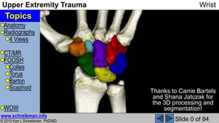
- 1. Anatomy Radiographs 4 Views CT/MR FOOSH Colles Torus Barton Scaphoid WOW © 2015 Ken L Schreibman, PhD/MD www.schreibman.info Upper Extremity Trauma Wrist Slide 0 of 84 Topics Thanks to Carrie Bartels and Shana Jatczak for the 3D processing and segmentation!
- 2. Anatomy Radiographs 4 Views CT/MR FOOSH Colles Torus Barton Scaphoid WOW © 2015 Ken L Schreibman, PhD/MD www.schreibman.info Upper Extremity Trauma Wrist Slide 1 of 84 3D Wrist CT Frontal view Ulna side view
- 3. Anatomy Radiographs 4 Views CT/MR FOOSH Colles Torus Barton Scaphoid WOW © 2015 Ken L Schreibman, PhD/MD www.schreibman.info Upper Extremity Trauma Wrist Slide 2 of 84 Ulna side view Radius: Frontal view Radial Styloid Arm:Radius rotates around ulna (radial head) Lister’s Tubercle (dorsal) Wrist: Radius is the foundation upon which the carpal bones reside Looking down on articular surface Lunate Fossa Scaphoid Fossa Anterior Normal anterior (volar) (palmar) tilt of distal radius Long axis of radius Perpen- dicular to long axis Normal 2-20° volar R R [L] “ray”
- 4. Anatomy Radiographs 4 Views CT/MR FOOSH Colles Torus Barton Scaphoid WOW © 2015 Ken L Schreibman, PhD/MD www.schreibman.info Upper Extremity Trauma Wrist Slide 3 of 84 Scaphoid: Frontal view Ulna side view aka “Navicular of hand” (confusing Navicular in foot) Scaphoid Fossa Waist Distal pole sticks out anteriorly Proximal Pole Scaphoid bridges the proximal and distal carpal rows Proximal Pole Distal PoleDistal Pole R R S S [Gr]“boat” Waist
- 5. Anatomy Radiographs 4 Views CT/MR FOOSH Colles Torus Barton Scaphoid WOW © 2015 Ken L Schreibman, PhD/MD www.schreibman.info Upper Extremity Trauma Wrist Slide 4 of 84 TFC Lunate: Frontal view Ulna side view Lunate Fossa R R S S L L Should have opening up Like a teacup holding tea Lunate sits ½ over radius (lunate fossa), ½ over Triangular Fibro Cartilage (TFC) Lunate is nearly surrounded by cartilage Lunate susceptible to AVN (Kienböck) One small artery anterior One small artery posterior [L]“moon” Scaphoid Fossa
- 6. Anatomy Radiographs 4 Views CT/MR FOOSH Colles Torus Barton Scaphoid WOW © 2015 Ken L Schreibman, PhD/MD www.schreibman.info Upper Extremity Trauma Wrist Slide 5 of 84 TFC Proximal Carpal Row:(S+L+Tq+P) Frontal view Ulna side viewR R S L L P Tq P Tq Triquetrum (Tq): [L] “three-cornered” Pisiform (P): [L] “pea” Radio- Carpal Joint SL Jt LT Jt PT Jt Pisiform stick out anterior Distal Pole
- 7. Anatomy Radiographs 4 Views CT/MR FOOSH Colles Torus Barton Scaphoid WOW © 2015 Ken L Schreibman, PhD/MD www.schreibman.info Upper Extremity Trauma Wrist Slide 6 of 84 Ulna major component of elbow, forearm Role at wrist is limited Doesn’t even normally touch carpal bones ½ Radius fxs have Ulnar styloid fxs** Often remain ununited Seldom require surgery (If DRUJ stable)TFC Ulna: Frontal view Ulna side viewR UU S L L P Tq P Tq Ulna Styloid [L] “arm”… related to “ell”, “cubit” * Unit of length equal to the forearm *www.etymonline.com DRUJ Forms the Distal Radio- Ulnar Joint Ulna Styloid **orthopedics.about.com [L]“elbow”
- 8. Anatomy Radiographs 4 Views CT/MR FOOSH Colles Torus Barton Scaphoid WOW © 2015 Ken L Schreibman, PhD/MD www.schreibman.info Upper Extremity Trauma Wrist Slide 7 of 84 TFC Frontal view R U S L P Tq P Tq C U Ulna side view L C Capitate: Head-shaped round proximal end sits inside open end of the lunate R Capitate Lunate Radius form a straight stack [L]“head”
- 9. Anatomy Radiographs 4 Views CT/MR FOOSH Colles Torus Barton Scaphoid WOW © 2015 Ken L Schreibman, PhD/MD www.schreibman.info Upper Extremity Trauma Wrist Slide 8 of 84 TFC Hamate: Frontal view Ulna side viewR UU S L L P Tq P Tq HC Hook-shaped process (H) sticks out anterior Pisiform Distal Pole Hook of Hamate sticks out anteriorH [L]“hook”
- 10. Anatomy Radiographs 4 Views CT/MR FOOSH Colles Torus Barton Scaphoid WOW © 2015 Ken L Schreibman, PhD/MD www.schreibman.info Upper Extremity Trauma Wrist Slide 9 of 84 TFC Metacarpals Frontal view Ulna side viewR UU S L L P Tq P Tq HHC Capitate articulates with Long finger MC Hamate articulates with Ring & Small finger
- 11. Anatomy Radiographs 4 Views CT/MR FOOSH Colles Torus Barton Scaphoid WOW © 2015 Ken L Schreibman, PhD/MD www.schreibman.info Upper Extremity Trauma Wrist Slide 10 of 84 TFC aka “Lesser Multangular” Frontal view Trapezoid: Ulna side viewR UU S L L P Tq P Tq HHC 2 parallel sides Trapezoid articulates with index finger MC Td [Gr] “table shaped”
- 12. Anatomy Radiographs 4 Views CT/MR FOOSH Colles Torus Barton Scaphoid WOW © 2015 Ken L Schreibman, PhD/MD www.schreibman.info Upper Extremity Trauma Wrist Slide 11 of 84 aka “Greater Multangular” TFC Trapezium: Frontal view Ulna side viewR UU S L L P Tq P Tq C HH Tm Tm no parallel sides TrapeziUM articulates with the ThUMb Td [Gr] “little table”
- 13. Anatomy Radiographs 4 Views CT/MR FOOSH Colles Torus Barton Scaphoid WOW © 2015 Ken L Schreibman, PhD/MD www.schreibman.info Upper Extremity Trauma Wrist Slide 12 of 84 Carpal Tunnel S PHTm Walls of the carpal tunnel are made of the carpal bones that stick out anteriorly
- 14. Anatomy Radiographs 4 Views CT/MR FOOSH Colles Torus Barton Scaphoid WOW © 2015 Ken L Schreibman, PhD/MD www.schreibman.info Upper Extremity Trauma Wrist Slide 13 of 84 Hand ≠ Wrist R,A 27yoM, fell off bike Hand PA Hand Lat Hand Obl All Negative
- 15. Anatomy Radiographs 4 Views CT/MR FOOSH Colles Torus Barton Scaphoid WOW © 2015 Ken L Schreibman, PhD/MD www.schreibman.info Upper Extremity Trauma Wrist Slide 14 of 84 Hand ≠ Wrist R,A 27yoM, fell off bike Hand ≠ Wrist Wrist PA Wrist Obl Wrist Lat Wrist Ul Dev Bennett Fracture! Still Negative…
- 16. Anatomy Radiographs 4 Views CT/MR FOOSH Colles Torus Barton Scaphoid WOW © 2015 Ken L Schreibman, PhD/MD www.schreibman.info Upper Extremity Trauma Wrist Slide 15 of 84 Hand ≠ Wrist G,M 44yoM PA Hand ? PA Wrist (next day) !
- 17. Anatomy Radiographs 4 Views CT/MR FOOSH Colles Torus Barton Scaphoid WOW © 2015 Ken L Schreibman, PhD/MD www.schreibman.info Upper Extremity Trauma Wrist Slide 16 of 84 Hand vs Wrist: X-ray Beam Hand radiographs: X-ray beam centered @ 3rd MC head G,M 44yoM Wrist radiographs: X-ray beam centered @ capitate
- 18. Anatomy Radiographs 4 Views CT/MR FOOSH Colles Torus Barton Scaphoid WOW © 2015 Ken L Schreibman, PhD/MD www.schreibman.info Upper Extremity Trauma Wrist Slide 17 of 84 Wrist: PA = Standard View Marty age 15 Elbow @ shoulder height Elbow @ 90° Low chair Raise cassette Shield X-rays X-ray beam PosteriorAnterior = “PA” X-ray beam centered on Capitate
- 19. Anatomy Radiographs 4 Views CT/MR FOOSH Colles Torus Barton Scaphoid WOW © 2015 Ken L Schreibman, PhD/MD www.schreibman.info Upper Extremity Trauma Wrist Slide 18 of 84 PA: Standard view Wrist: PA View Carpal Alignment Proximal Carpal Row Joint Alignment Radio-Carpal Joint Carpal-Metacarpal Jt Distal Radio-Ulnar Jt Ulnar Length Normally, Ulna same length as Radius DRUJ Ulna shorter than Radius R-C Jt C-MC D,H 21yoF
- 20. Anatomy Radiographs 4 Views CT/MR FOOSH Colles Torus Barton Scaphoid WOW © 2015 Ken L Schreibman, PhD/MD www.schreibman.info Upper Extremity Trauma Wrist Slide 19 of 84 Ulnar Variance Ulna shorter than Radius “Negative Ulnar Variance” Risk AVN Lunate (Kienböck) Ulna longer than Radius “Positive Ulnar Variance” Ulna can punch hole in TFC Ulna can impact upon Lunate “Ulna Abutment Syndrome” S,Z 18yoM Ulna is only slightly shorter than Radius AVN Lunate with collapse Radius shortening T,C 14yoM 2 y earlier, normal unfused growth plates Premature fusion radius, continued ulna growth UV Compared to normal side Treated with ulna shortening osteotomy
- 21. Anatomy Radiographs 4 Views CT/MR FOOSH Colles Torus Barton Scaphoid WOW © 2015 Ken L Schreibman, PhD/MD www.schreibman.info Upper Extremity Trauma Wrist Slide 20 of 84 Wrist: Lateral View R L C,S 48yoM CC RAnterior L Normal 2-20° volar
- 22. Anatomy Radiographs 4 Views CT/MR FOOSH Colles Torus Barton Scaphoid WOW © 2015 Ken L Schreibman, PhD/MD www.schreibman.info Upper Extremity Trauma Wrist Slide 21 of 84 Can see most carpal bones on Lateral C,S 48yoM C R L S S R L C P Hard to see Ulna as it overlaps Radius on a good lateral view U P Tq Can’t see Triquetrum on lateral view…
- 23. Anatomy Radiographs 4 Views CT/MR FOOSH Colles Torus Barton Scaphoid WOW © 2015 Ken L Schreibman, PhD/MD www.schreibman.info Upper Extremity Trauma Wrist Slide 22 of 84 Triquetral Fracture Classically presents as a tiny avulsion fracture dorsal to the mid-carpus There are no normal ossicles dorsal to the carpal bones If you see a small bone back there, it’s a fracture May be old, as these tiny fractures don’t always heal M,G 50yoM Fx
- 24. Anatomy Radiographs 4 Views CT/MR FOOSH Colles Torus Barton Scaphoid WOW © 2015 Ken L Schreibman, PhD/MD www.schreibman.info Upper Extremity Trauma Wrist Slide 23 of 84 Wrist: Standard 3 Views PA View Lateral View Thumb Down Thumb Up Oblique View Thumb Halfway Between
- 25. Anatomy Radiographs 4 Views CT/MR FOOSH Colles Torus Barton Scaphoid WOW © 2015 Ken L Schreibman, PhD/MD www.schreibman.info Upper Extremity Trauma Wrist Slide 24 of 84 Wrist: Oblique View Best view of: STT joint Thumb C-MC joint Common sites for OA Additional view of: Carpals (scaphoid) Metacarpals Radius (styloid) Sometimes a fracture is seen only on this view K,M 20yoF S Tm Td MC
- 26. Anatomy Radiographs 4 Views CT/MR FOOSH Colles Torus Barton Scaphoid WOW © 2015 Ken L Schreibman, PhD/MD www.schreibman.info Upper Extremity Trauma Wrist Slide 25 of 84 Scaphoid (Ulnar Deviation) View S,B 21yoF Patient holds wrist in ulnar deviation Yields an elongated view of the scaphoid. Helps when looking for fractures.
- 27. Anatomy Radiographs 4 Views CT/MR FOOSH Colles Torus Barton Scaphoid WOW © 2015 Ken L Schreibman, PhD/MD www.schreibman.info Upper Extremity Trauma Wrist Slide 26 of 84 4 View Series for Scaphoid Fracture K,T 32yoF Lateral View PA View Oblique View Scaphoid View Doesn’t show scaphoid well Dorsal swelling Negative Negative? Positive! scaphoid waist fx ?
- 28. Anatomy Radiographs 4 Views CT/MR FOOSH Colles Torus Barton Scaphoid WOW © 2015 Ken L Schreibman, PhD/MD www.schreibman.info Upper Extremity Trauma Wrist Slide 27 of 84 Wrist: CT Good for complex fractures Aid in surgical planning Good to assess fracture healing Even in the presence of metal E,A 18yoM PA view Scaphoid view CT:Coronal Acutrak® screw CT:Obl Sag Fx? Fx Fx! S L Tq HCTd CT:Obl Sag R Fx! S R Tm Healed!
- 29. Anatomy Radiographs 4 Views CT/MR FOOSH Colles Torus Barton Scaphoid WOW © 2015 Ken L Schreibman, PhD/MD www.schreibman.info Upper Extremity Trauma Wrist Slide 28 of 84 Wrist CT: Positioning We don’t scan patients with their wrist down at their side Excess radiation across torso X-ray scatter decreases res. We scan patients with their wrist over the head No excess radiation to body No x-ray scatter Mighty Mouse Position
- 30. Anatomy Radiographs 4 Views CT/MR FOOSH Colles Torus Barton Scaphoid WOW © 2015 Ken L Schreibman, PhD/MD www.schreibman.info Upper Extremity Trauma Wrist Slide 29 of 84 Wrist: CT NOT good for occult fractures Fractures non-displaced on radiographs… …are non-displaced on CT L,N 21yoF PA viewScaphoid view No fracture CT: Coronal MR: T1 Coronal Black fracture line MR: T2fs Coronal Acutrak® screw No fracture Marrow edema
- 31. Anatomy Radiographs 4 Views CT/MR FOOSH Colles Torus Barton Scaphoid WOW © 2015 Ken L Schreibman, PhD/MD www.schreibman.info Upper Extremity Trauma Wrist Slide 30 of 84 Wrist MR: Positioning Wrist coil We scan patients with their wrist over the head In a wrist coil Functions best in the center of the magnetic field
- 32. Anatomy Radiographs 4 Views CT/MR FOOSH Colles Torus Barton Scaphoid WOW © 2015 Ken L Schreibman, PhD/MD www.schreibman.info Upper Extremity Trauma Wrist Slide 31 of 84 Fall On Out-Stretched Hand (FOOSH) Most injuries to the wrist are due to one common mechanism Perhaps THE most common injury 1-in-6 ER fractures occur in the distal radius* Humans are a clumsy species We walk upright We’re top heavy When falling, we instinctively protect our head, by Extending our arm Striking the ground with our hand This mechanism of injury is perhaps UNIQUE to humans *orthopedics.about.com
- 33. Anatomy Radiographs 4 Views CT/MR FOOSH Colles Torus Barton Scaphoid WOW © 2015 Ken L Schreibman, PhD/MD www.schreibman.info Upper Extremity Trauma Wrist Slide 32 of 84 The most famous penguin on the Internet www.youtube.com www.youtube.com
- 34. Anatomy Radiographs 4 Views CT/MR FOOSH Colles Torus Barton Scaphoid WOW © 2015 Ken L Schreibman, PhD/MD www.schreibman.info Upper Extremity Trauma Wrist Slide 33 of 84Marty age 8½ HAND S U L N A R A D I U S Fall On Out-Stretched Hand (FOOSH) FOOSH Hyperextend Wrist Hyperextend Wrist
- 35. Anatomy Radiographs 4 Views CT/MR FOOSH Colles Torus Barton Scaphoid WOW © 2015 Ken L Schreibman, PhD/MD www.schreibman.info Upper Extremity Trauma Wrist Slide 34 of 84 Fall On Out-Stretched Hand (FOOSH) Hyperextension of wrist Hyperextensive forces on: Radius Colles fracture Torus fracture (children) Carpal bones Barton fracture Scaphoid fracture Lunate/perilunate dislocations S
- 36. Anatomy Radiographs 4 Views CT/MR FOOSH Colles Torus Barton Scaphoid WOW © 2015 Ken L Schreibman, PhD/MD www.schreibman.info Upper Extremity Trauma Wrist Slide 35 of 84 Transverse Fx distal radius Hyperextension forces cause: Dorsal angulation ± Dorsal displacement Colles Fracture S Fx R,C 92yoF O,M 20yoM DORSAL ANGULATION ALWAYS ABNORMAL! Lateral view Lateral view
- 37. Anatomy Radiographs 4 Views CT/MR FOOSH Colles Torus Barton Scaphoid WOW © 2015 Ken L Schreibman, PhD/MD www.schreibman.info Upper Extremity Trauma Wrist Slide 36 of 84 Dorsal Angulation is Bad To measure angle: Draw line along distal radius From front corner To back corner Draw line along shaft of radius Perpendicular to this Measure this angle Normal is VOLAR 2-20° Dorsal = Abnormal R,C 92yoF Lateral view 5° Dorsal 20° Dorsal 2 weeks later… The ligaments are not designed to support carpal bones on a dorsal sloped radius Lateral view
- 38. Anatomy Radiographs 4 Views CT/MR FOOSH Colles Torus Barton Scaphoid WOW © 2015 Ken L Schreibman, PhD/MD www.schreibman.info Upper Extremity Trauma Wrist Slide 37 of 84 Must reduce angle to heal right M,D 59yoF ER lateral view: Marked dorsal angulation Following reduction & casting in ER: Volar angulation 6 weeks later: Healing, normal volar angulation
- 39. Anatomy Radiographs 4 Views CT/MR FOOSH Colles Torus Barton Scaphoid WOW © 2015 Ken L Schreibman, PhD/MD www.schreibman.info Upper Extremity Trauma Wrist Slide 38 of 84 Colles fractures very common In children Fall a lot Torus fracture In women Osteopenia 2 women in my life… In the media… Secretary Judy Wife Lynn
- 40. Anatomy Radiographs 4 Views CT/MR FOOSH Colles Torus Barton Scaphoid WOW © 2015 Ken L Schreibman, PhD/MD www.schreibman.info Upper Extremity Trauma Wrist Slide 39 of 84 Colles vs Smith Fracture Anatomically impossible? Season 15, Episode 2 original air date 9/22/04
- 41. Anatomy Radiographs 4 Views CT/MR FOOSH Colles Torus Barton Scaphoid WOW © 2015 Ken L Schreibman, PhD/MD www.schreibman.info Upper Extremity Trauma Wrist Slide 40 of 84 S Colles: Hyper- extension DORSAL angulation Smith: Hyper- flexion VOLAR angulation Smith Fracture = Reverse Colles S,K 51yoF Lateral view: Too much volar angulation Reduction & cast: Normal volar angulation
- 42. Anatomy Radiographs 4 Views CT/MR FOOSH Colles Torus Barton Scaphoid WOW © 2015 Ken L Schreibman, PhD/MD www.schreibman.info Upper Extremity Trauma Wrist Slide 41 of 84 FOOSH Colles: Hyper- extension DORSAL angulation Mechanisms: Colles vs Smith
- 43. Anatomy Radiographs 4 Views CT/MR FOOSH Colles Torus Barton Scaphoid WOW © 2015 Ken L Schreibman, PhD/MD www.schreibman.info Upper Extremity Trauma Wrist Slide 42 of 84 Mechanisms: Colles vs Smith FOOSH Hyperextension Colles whether fall Forwards or Backwards
- 44. Anatomy Radiographs 4 Views CT/MR FOOSH Colles Torus Barton Scaphoid WOW © 2015 Ken L Schreibman, PhD/MD www.schreibman.info Upper Extremity Trauma Wrist Slide 43 of 84 Mechanisms: Colles vs Smith Fall onto Back of hand Hyper- flexion Smith Fx VOLAR angulation Colles: Hyper- extension DORSAL angulation Smith fracture is much less common than Colles
- 45. Anatomy Radiographs 4 Views CT/MR FOOSH Colles Torus Barton Scaphoid WOW © 2015 Ken L Schreibman, PhD/MD www.schreibman.info Upper Extremity Trauma Wrist Slide 44 of 84 Abraham Colles (1773-1843) “The injury to which I wish to direct the attention of surgeons, had not, as far as I know, been described by any author.” “I should consider this as by far the most common injury to which the wrist or carpal extremities of the radius and ulnar are exposed.” babel.hathitrust.org (81 years before Roentgen)
- 46. Anatomy Radiographs 4 Views CT/MR FOOSH Colles Torus Barton Scaphoid WOW © 2015 Ken L Schreibman, PhD/MD www.schreibman.info Upper Extremity Trauma Wrist Slide 45 of 84 Robert William Smith (1807-1873) google.com books.google.com (1847?) MDCCCL=1850 Page 162
- 47. Anatomy Radiographs 4 Views CT/MR FOOSH Colles Torus Barton Scaphoid WOW © 2015 Ken L Schreibman, PhD/MD www.schreibman.info Upper Extremity Trauma Wrist Slide 49 of 84 R A D I U S PowerPoint Model Adult Lateral Fractures in Children A R A D I U S PowerPoint Model Child Lateral Epiphysis physis (growth plate) Metaphysis Diaphysis K,V 2yoM Lateral PA view
- 48. Anatomy Radiographs 4 Views CT/MR FOOSH Colles Torus Barton Scaphoid WOW © 2015 Ken L Schreibman, PhD/MD www.schreibman.info Upper Extremity Trauma Wrist Slide 50 of 84 FOOSH Fractures in Children Adult bones: Brittle Snap under force Child bones: Soft Bend under force FOOSH Hyperextension distal radial metaphysis Buckling metaphysis- diaphysis junction Buckle Fracture “Torus Fracture” R A D I U S PowerPoint Model Child Lateral R A D I U S A G,A 5yoM Lateral
- 49. Anatomy Radiographs 4 Views CT/MR FOOSH Colles Torus Barton Scaphoid WOW © 2015 Ken L Schreibman, PhD/MD www.schreibman.info Upper Extremity Trauma Wrist Slide 51 of 84 Torus Fractures: Lateral View Cortex buckles IN FOOSH (Colles) Dorsal cortex Fall on back of wrist (Smith) Volar cortex Nature does not make angles… Nature makes smooth curves If you see cortex angulation in a child that should be smooth, it’s likely a torus fracture! R A D I U S S,A 5yoF Lateral Cortex of radius & ulna overlap A,C 6yoM Lateral
- 50. Anatomy Radiographs 4 Views CT/MR FOOSH Colles Torus Barton Scaphoid WOW © 2015 Ken L Schreibman, PhD/MD www.schreibman.info Upper Extremity Trauma Wrist Slide 52 of 84 Torus Fractures: PA View FOOSH Axial Load R A D I U S Axial Load R A D I U S Axial Load Cortex buckles OUTWARD PowerPoint Model Child PA View PA view H,T 8yoF
- 51. Anatomy Radiographs 4 Views CT/MR FOOSH Colles Torus Barton Scaphoid WOW © 2015 Ken L Schreibman, PhD/MD www.schreibman.info Upper Extremity Trauma Wrist Slide 53 of 84 Torus Fractures: Common… Run eyes along cortex Focus on metaphysis PA view Buckles outward Not sure? Compare to normal side Use other views! Subtle A,B 14yoF PA view Symptomatic side PA view Asymptomatic side
- 52. Anatomy Radiographs 4 Views CT/MR FOOSH Colles Torus Barton Scaphoid WOW © 2015 Ken L Schreibman, PhD/MD www.schreibman.info Upper Extremity Trauma Wrist Slide 54 of 84 Torus Fractures: Common… Run eyes along cortex Focus on metaphysis Lat view Buckles inward Not sure? Compare to normal side Use other views! Subtle PA view Symptomatic side Lat view Asympt. Lat view Sympt. A,B 14yoF
- 53. Anatomy Radiographs 4 Views CT/MR FOOSH Colles Torus Barton Scaphoid WOW © 2015 Ken L Schreibman, PhD/MD www.schreibman.info Upper Extremity Trauma Wrist Slide 55 of 84 “Torus” Capital S h a f t Base Plinth Torus RadioGraphics 2004; 24:p1025 [L]:“swelling,protuberance,bulge” [Architecture]: A large convex molding, semicircular in cross section, at base of a classical column. Wisconsin State Capitol
- 54. Anatomy Radiographs 4 Views CT/MR FOOSH Colles Torus Barton Scaphoid WOW © 2015 Ken L Schreibman, PhD/MD www.schreibman.info Upper Extremity Trauma Wrist Slide 56 of 84 Fall On Out-Stretched Hand (FOOSH) Hyperextension of wrist Hyperextensive forces on: Radius Colles fracture Torus fracture (children) Carpal bones (Proximal carpal row) Barton fracture S
- 55. Anatomy Radiographs 4 Views CT/MR FOOSH Colles Torus Barton Scaphoid WOW © 2015 Ken L Schreibman, PhD/MD www.schreibman.info Upper Extremity Trauma Wrist Slide 57 of 84 Barton Fracture Hyperextension of wrist Impaction of carpal bones on radius dorsal rim Fracture radius rim Intra-articular fracture Potentially more serious than Colles (extra-articular fracture) May require surgical fixation Surgeon may order CT for planning S
- 56. Anatomy Radiographs 4 Views CT/MR FOOSH Colles Torus Barton Scaphoid WOW © 2015 Ken L Schreibman, PhD/MD www.schreibman.info Upper Extremity Trauma Wrist Slide 58 of 84 Dorsal Barton Fracture Dorsal Barton Due to FOOSH is much more common than Volar Barton Due to blow to back of wrist (Just as Colles is much more common than Smith fracture) S S,G 37yoM Lateral view M,M 58yoF Lateral view
- 57. Anatomy Radiographs 4 Views CT/MR FOOSH Colles Torus Barton Scaphoid WOW © 2015 Ken L Schreibman, PhD/MD www.schreibman.info Upper Extremity Trauma Wrist Slide 59 of 84 Volar Barton Fracture S Lateral view A,D 43yoM CT: Sagittal Open Reduction Internal Fixation
- 58. Anatomy Radiographs 4 Views CT/MR FOOSH Colles Torus Barton Scaphoid WOW © 2015 Ken L Schreibman, PhD/MD www.schreibman.info Upper Extremity Trauma Wrist Slide 60 of 84 John Rhea Barton (1794-1871) www.kmle.co.krThe Medical Examiner Nov 7, 1838; 1, 23; p 365-8 It was said that Barton was ambidextrous and that once he had positioned himself for an operation, he did not move about. whonamedit.com
- 59. Anatomy Radiographs 4 Views CT/MR FOOSH Colles Torus Barton Scaphoid WOW © 2015 Ken L Schreibman, PhD/MD www.schreibman.info Upper Extremity Trauma Wrist Slide 61 of 84 Fall On Out-Stretched Hand (FOOSH) Hyperextension of wrist Hyperextensive forces on: Radius Colles fracture Torus fracture (children) Carpal bones (Proximal carpal row) Barton fracture (Distal carpal row) Scaphoid fracture S
- 60. Anatomy Radiographs 4 Views CT/MR FOOSH Colles Torus Barton Scaphoid WOW © 2015 Ken L Schreibman, PhD/MD www.schreibman.info Upper Extremity Trauma Wrist Slide 62 of 84 Scaphoid Fractures Scaphoid THE most common carpal bone to be fractured. 71% of all carpal fxs* Scaphoid bridges the carpal rows Traumatic shear forces between the rows … shearing fracture across the scaphoid *emedicine.com
- 61. Anatomy Radiographs 4 Views CT/MR FOOSH Colles Torus Barton Scaphoid WOW © 2015 Ken L Schreibman, PhD/MD www.schreibman.info Upper Extremity Trauma Wrist Slide 63 of 84 Scaphoid Fractures Locations S,A 24yoM PA View Wrist Scaphoid Waist 70% of scaphoid fractures occur at the waist www.gentili.net B,J 21yoM Scaphoid Proximal Pole 20% occur at scaphoid proximal pole Increased risk of non-union/AVN Ulnar Deviation View Wrist
- 62. Anatomy Radiographs 4 Views CT/MR FOOSH Colles Torus Barton Scaphoid WOW © 2015 Ken L Schreibman, PhD/MD www.schreibman.info Upper Extremity Trauma Wrist Slide 64 of 84 Scaphoid Fractures Locations PA View Wrist Scaphoid Distal Pole 10% occur at distal pole These are usually uneventful* PA View Wrist Scaphoid Tubercle Rare, usually uncomplicated. If nonunion, usually asympt.* *emedicine.com B,T 44yoMT,B 20yoM
- 63. Anatomy Radiographs 4 Views CT/MR FOOSH Colles Torus Barton Scaphoid WOW © 2015 Ken L Schreibman, PhD/MD www.schreibman.info Upper Extremity Trauma Wrist Slide 65 of 84 Scaphoid & Radius Fractures Same common mechanism (FOOSH) Distal Radius Fracture Scaphoid Fracture …BOTH! Watch out for “satisfaction of search” “Aha, I found the fracture… I’d done looking” Old Radiology Axiom: The hardest fracture to find is the 2nd fracture
- 64. Anatomy Radiographs 4 Views CT/MR FOOSH Colles Torus Barton Scaphoid WOW © 2015 Ken L Schreibman, PhD/MD www.schreibman.info Upper Extremity Trauma Wrist Slide 66 of 84 Scaphoid with Radius Fracture W,M 19yoF PA View Wrist Obl View Wrist PA View Wrist Colles Ulna Styloid Proximal Pole Plate fixates Colles fracture Screw fixates scaphoid fracture
- 65. Anatomy Radiographs 4 Views CT/MR FOOSH Colles Torus Barton Scaphoid WOW © 2015 Ken L Schreibman, PhD/MD www.schreibman.info Upper Extremity Trauma Wrist Slide 67 of 84 22yo M 03:00 Unbelted passenger High speed MVC T-boned by minivan Air bags deployed Took 20 minutes to extract from car Intubated Scaphoid doesn’t heal as well as other bones V,G 22yoM FB … Acetabular fracture PA View Hand 2 months later… Healing FB OK
- 66. Anatomy Radiographs 4 Views CT/MR FOOSH Colles Torus Barton Scaphoid WOW © 2015 Ken L Schreibman, PhD/MD www.schreibman.info Upper Extremity Trauma Wrist Slide 68 of 84 Scaphoid doesn’t heal as well as other bones after 4 months… FB OK CT: Coronal OK FBCT: Sagittal Oblique Non-union scaphoid waist V,G 22yoM
- 67. Anatomy Radiographs 4 Views CT/MR FOOSH Colles Torus Barton Scaphoid WOW © 2015 Ken L Schreibman, PhD/MD www.schreibman.info Upper Extremity Trauma Wrist Slide 69 of 84 Scaphoid has a tenuous blood supply Radial artery supplies: Distal Pole (DP) of Scaphoid (S) NotProximalPole (PP) The more proximal the fracture, the greater the risk of non-union. The more distracted the fracture, the greater the risk of non-union. PA Hand Obl Hand S DP PP Radial Artery S Heavy arterial calcification Pt w/ diabetes, renal failure L,T 60yoM
- 68. Anatomy Radiographs 4 Views CT/MR FOOSH Colles Torus Barton Scaphoid WOW © 2015 Ken L Schreibman, PhD/MD www.schreibman.info Upper Extremity Trauma Wrist Slide 70 of 84 Scaphoid Non-Union AVN Q,B 62yoF PA View Wrist CT: Coronal CT: Sagittal Oblique Non-union scaphoid waist fx Collapse & fragmentation PP = AVN
- 69. Anatomy Radiographs 4 Views CT/MR FOOSH Colles Torus Barton Scaphoid WOW © 2015 Ken L Schreibman, PhD/MD www.schreibman.info Upper Extremity Trauma Wrist Slide 71 of 84 Proximal Row Carpectomy Lateral View WristPA View Wrist Resection: Scaphoid, Lunate, Triquetrum Radius articulates with Capitate (distal row) Only treatment for fragmented scaphoid AVN R C Q,B 62yoF
- 70. Anatomy Radiographs 4 Views CT/MR FOOSH Colles Torus Barton Scaphoid WOW © 2015 Ken L Schreibman, PhD/MD www.schreibman.info Upper Extremity Trauma Wrist Slide 72 of 84 To avoid non-unionAVNPRC All scaphoid fxs require early treatment! Probably with a screw if displaced At least with a splint or cast if non-displaced But non-displaced fractures are hard to see because they are non-displaced So how do we know if a patient has a non-displaced scaphoid fracture? SNUFFBOX TENDERNESS = PRESUMED SCAPHOID FRACTURE
- 71. Anatomy Radiographs 4 Views CT/MR FOOSH Colles Torus Barton Scaphoid WOW © 2015 Ken L Schreibman, PhD/MD www.schreibman.info Upper Extremity Trauma Wrist Slide 73 of 84 Anatomical Snuffbox Extensor Pollicis Longus Tendon Extensor Pollicis Brevis Tendon
- 72. Anatomy Radiographs 4 Views CT/MR FOOSH Colles Torus Barton Scaphoid WOW © 2015 Ken L Schreibman, PhD/MD www.schreibman.info Upper Extremity Trauma Wrist Slide 74 of 84 Snuffbox Tenderness = PRESUMED SCAPHOID FRACTURE What if the radiographs are normal? Beautiful! Then it’s a non-displaced fracture Treat anyway with a cast/splint Make sure radiologist agrees they’re negative Have patient follow-up in 2 weeks Re-examine… if still tender… back into the splint Get repeat radiographs (out of the cast/splint) We’re taught occult fxs become visible after 1-2 weeks from bone resorption at fx margins… I’m not sure it’s true… If you really need to know… MRI (we don’t miss fractures on MRI)
- 73. Anatomy Radiographs 4 Views CT/MR FOOSH Colles Torus Barton Scaphoid WOW © 2015 Ken L Schreibman, PhD/MD www.schreibman.info Upper Extremity Trauma Wrist Slide 75 of 84 Resorbtion of Fracture Margins? M,D 55yoM, cutting tree branches, FOOSH 15ft Scaphoid View Oblique View No scaphoid fx No scaphoid fx No scaphoid fx… Radius fractures Importance of multiple views! PA View
- 74. Anatomy Radiographs 4 Views CT/MR FOOSH Colles Torus Barton Scaphoid WOW © 2015 Ken L Schreibman, PhD/MD www.schreibman.info Upper Extremity Trauma Wrist Slide 76 of 84 Resorbtion of Fracture Margins? M,D 55yoM, cutting tree branches, FOOSH 15ft PA View 8 days later Still snuffbox tenderness Still no scaphoid fx MRI: 19 days after injury Coronal T1 Coronal T2fs Bone marrow edema in Radius Black fx line Black fx line Bone marrow edema in Scaphoid Black fx line Occult scaphoid fracture! No resorbtion scaphoid fracture margins
- 75. Anatomy Radiographs 4 Views CT/MR FOOSH Colles Torus Barton Scaphoid WOW © 2015 Ken L Schreibman, PhD/MD www.schreibman.info Upper Extremity Trauma Wrist Slide 77 of 84 Resorbtion of Fracture Margins? M,D 55yoM, cutting tree branches, FOOSH 15ft Scaphoid View Oblique ViewPA View after 29 days… Still see lucent radius fractures Still no resorbtion scaphoid fracture margins Negative radiographs do not exclude a scaphoid fracture Snuffbox Tenderness = Presumed Scaphoid Fracture!
- 76. Anatomy Radiographs 4 Views CT/MR FOOSH Colles Torus Barton Scaphoid WOW © 2015 Ken L Schreibman, PhD/MD www.schreibman.info Upper Extremity Trauma Wrist Slide 78 of 84 Anatomical Snuffbox? snuffhouse.org www.snuffstore.co.uk schmalzlerfranzl.de
- 77. Anatomy Radiographs 4 Views CT/MR FOOSH Colles Torus Barton Scaphoid WOW © 2015 Ken L Schreibman, PhD/MD www.schreibman.info Upper Extremity Trauma Wrist Slide 79 of 84 Wrist: What to Order When (WOW) Radiographs: TraumaPain Arthritis (Hand radiographs) CT Surgical planning known fractures MR Occult fractures (scaphoid) Synovitis (w/Gd) (Usually includes MCPs ± IPs) …pain? RG 95% CT 2% MR 3% UW data 2005 & 2014
- 78. Anatomy Radiographs 4 Views CT/MR FOOSH Colles Torus Barton Scaphoid WOW © 2015 Ken L Schreibman, PhD/MD www.schreibman.info Upper Extremity Trauma Wrist Slide 80 of 84 Wrist: What to Order When (WOW) Wrist Radiographs (95%) 3-view wrist series PA (not AP) Lateral Oblique If snuffbox tenderness, add 4th view Scaphoid (ulnar deviation) If snuffbox tenderness+negative radiographs TREAT AS PRESUMED SCAPHOID FRACTURE Cast/splint, follow-up in 2 weeks If still has snuffbox tenderness, keep treating
- 79. Anatomy Radiographs 4 Views CT/MR FOOSH Colles Torus Barton Scaphoid WOW © 2015 Ken L Schreibman, PhD/MD www.schreibman.info Upper Extremity Trauma Wrist Slide 81 of 84 Wrist: What to Order When (WOW) Wrist CT Predominantly used for surgical planning of known radius/carpal bone fractures Ordered by Orthopedics from ER or clinic Assess healing of known scaphoid fracture With or without prior screw fixation Small screws cause virtually no CT artifacts We always reformat in 3 orthogonal planes For scaphoid, we add oblique sagittal We have a specialized protocol for DRUJ instability All protocols at: www.radiology.wisc.edu
- 80. Anatomy Radiographs 4 Views CT/MR FOOSH Colles Torus Barton Scaphoid WOW © 2015 Ken L Schreibman, PhD/MD www.schreibman.info Upper Extremity Trauma Wrist Slide 82 of 84 Wrist: What to Order When (WOW) Wrist MR Occult fractures (scaphoid) Persistent symptoms despite negative radiographs Synovitis (RA) Needs IV contrast Normal synovium does not enhance Vascularized pannus greatly enhances Ordering provider should specify area of concern Just intercarpal joints Also Metacarpal-phalangeal joints Also Interphalangeal joints Field of View = Resolution
- 81. Anatomy Radiographs 4 Views CT/MR FOOSH Colles Torus Barton Scaphoid WOW © 2015 Ken L Schreibman, PhD/MD www.schreibman.info Upper Extremity Trauma Wrist Slide 83 of 84 Wrist: What to Order When (WOW) Wrist Charges Wrist Radiographs 3 views = 4 views = $137 It costs nothing to add the scaphoid view to a 3 view series 1 view = 2 views = $128 Going from 2 views to 4 views adds only $9 (7%) Wrist CT (without contrast) = $1,460 Wrist MR (without contrast) = $2,921 (with contrast) = $3,377 UWMF charges 2012
- 82. Anatomy Radiographs 4 Views CT/MR FOOSH Colles Torus Barton Scaphoid WOW © 2015 Ken L Schreibman, PhD/MD www.schreibman.info Upper Extremity Trauma Wrist Slide 84 of 84 That’s all we have on wrists… Marty age 7