Infrared spectroscopy
•Download as PPTX, PDF•
1 like•150 views

Infrared Spectroscopy

Recommended
More Related Content
What's hot
What's hot (20)
Similar to Infrared spectroscopy
Similar to Infrared spectroscopy (20)
Recently uploaded
Recently uploaded (20)
Infrared spectroscopy
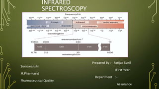
- 1. INFRARED SPECTROSCOPY Prepared By :- Parijat Sunil Suryawanshi (First Year M.Pharmacy) Department :- Pharmaceutical Quality Assurance
- 2. INTRODUCTI ON • Infrared Spectroscopy is one of the most important analytical techniques among the spectroscopic techniques. IR provides a valuable tool for probing the structure of organic molecules. • When infrared light is passed through a sample of an organic compound, some of the frequencies are absorbed, while other frequencies are transmitted through the sample without being absorbed. If we plot absorbance or transmittance against frequency, the result is an infrared spectrum • Infrared (IR) Spectroscopy uses a beam of infrared light to analyze the structure of organic compounds. IR analyzes the bonds present , produces broad absorptions which may frequently overlap. • IR spectroscopy gives sufficient information about structure of compound and also determine the functional group.
- 3. THEORY • IR spectra are commonly measured in units of wavenumbers (units of cm- 1). • Wave Number = 1/Wavelength (in Centimeter) • The infrared part of the electromagnetic spectrum covers the range from roughly 300 GHz to 400 THz (1 mm – 750 nm). It can be divided into three parts: Wavelength range • Far-infrared, from 1 mm – 10 μm • Mid-infrared, from 10–2.5 μm • Near-infrared, from 2,500–750 nm Region Wavelength range (µm) Wave number range (cm-1) Near 0.8-2.5 12500-4000 Middle 2.5-25 4000-400 Far 25-1000 400-10
- 4. IR ABSORPTION PROCESS • Molecules are excited to higher energy when they absorb IR radiation. • Absorption of IR radiation is Quantized Process – Molecules absorbs only selected frequencies of IR radiation. • IR radiation absorbtion corresponds to energy change on order of 8 to 40 KJ/mole • Radiation in this energy range corresponds to the stretching and bending vibrational frequencies of the bonds in most covalent molecules. • In the absorption process, those frequencies of infrared radiation which match the natural vibrational frequencies of the molecule, serves to increase the amplitude of the vibrational motions of the bonds in the molecule. • Only those bonds that have a dipole moment that changes as a function of time are capable of absorbing infrared radiation. To absorb light a molecule must have a bond within its structure that can exhibit what is referred to as a ‘dipole moment’ which means electrons in bond are not shared equally. • Symmetric bonds, such as those of H2 or Cl., do not absorb infrared radiation. A bond must present an electrical dipole that is chang ing at the same frequency as the incoming radiation in order for energy to be transferred. The changing electrical dipole of the bond can then couple with the sinusoidally changing electromagnetic field of the
- 5. MOLECULAR VIBRATIONS • Types :- 1) Stretching Vibration : that changes the Bond Length 2) Bending Vibration : that changes the Bond Angle 1) STRETCHING VIBRATION :- • Change in inter-atomic distance along bond axis • Types :- a) Symmetric Stretching :- Change in inter-atomic distance in equal manner. Two or more bonds vibrate in and out together. The atom of a molecule either move away or towards the central atom, but in the same direction.
- 6. b) Asymmetric Stretching :- One atom approach towards the central atom while other departs from it. It generally occurs at higher frequencies than that of symmetric stretching. 2) Bending Vibrations :- • Change in angle between two bond. Also called as Deformation Vibrations. • Types :-
- 7. i) Scissoring :- There will be vibrations of atoms in the plane of molecule towards each other like the movement of a scissor. ii) Rocking :- The atoms are vibrated in the plane of molecules towards one direction. The angle of the bond changes towards the same direction. b) Out-of-plane Bending :- i) Wagging :- The vibrations in which atoms vibrates near to the attached atom at either side away from the plane. ii) Twisting :- The Vibrations in which the atoms vibrate in opposite directions away the plane in twisted manner.
- 10. SAMPLE HANDLING 1) Sampling for Solids :- I. Mull technique : In this technique, the finely crushed sample is mixed with Nujol (mulling agent) in n a marble or agate mortar, with a pestle to make a thick paste. A thin film is applied onto the salt plates. This is then mounted in a path of IR beam and the spectrum is recorded. II. KBr disks : The solids are dissolved in suitable solvents to prepare a concentrated solution and it is used directly in the IR plates or else the sample is placed in the IR plates and a drop of solvent is placed on it. The standard method to prepare solid sample for FTIR spectrometer is to use KBr. About 2 mg of sample and 200 mg KBr are dried and ground. The particle size should be unified and less than two micrometers. Then, the mixture is squeezed to form transparent pellets which can be measured directly. III. Solid films : It can be deposited onto NaCI plates by allowing a solution in a volatile solvent to evaporate drop by drop on the surface of the flat. Polymers
- 11. 2) Sampling for Liquids : Liquid sample cells can be sandwiched using liquid sample cells of highly purified alkali halides, normally NaCl. Other salts such as KBr and CaF2 can also be used. Aqueous solvents cannot be used because they cannot dissolve alkali halides. Organic solvents like chloroform can be used. The sample thickness should be selected so that the transmittance lies between 15-20%. For most liquids, the sample cell thickness is 0.01-0.05 mm. Some salt plates are highly soluble in water, so the sample and washing reagents must be anhydrous 3) Sampling for Gases : The gas sample is introduced into a gas cell. The sample cell is made up of NaCl, KBr etc. and it is similar to the liquid sample cell. A sample cell with a long path length (5 – 10 cm) is needed because the gases show relatively weak absorbance.
- 12. INSTRUMENTATI ON There are two basic types of infrared spectrophotometer characterized by the manner in which the infrared frequencies are handled. 1) Dispersive Instrument :- In this type the infrared light is separated into its individual frequencies by dispersion, using a grating monochromator 2) Fourier Transform Infrared Instrument (FTIR):- In this type the infrared frequencies are allowed to interact to produce an interference pattern, and this pattern is then analyzed mathematically, using Fourier Transforms, to determine the individual frequencies and their intensities. Both types of instrument require, in addition, a suitable source of infrared light and a detector to measure light intensity.
- 13. A) INFRARED SOURCES • Common sources are rods of the following, electrically heate1d to near 1800°C 1) Nernst glower or Nernst filament :- (L :- 22mm ; D :- 1-2mm) • Cylinder made up of Sintered mixtures of the oxides of Zr, Th , Ce, Y, Er, etc . • At the end of cylinder platinum wire is attached and current is passed through the cylinder. • Temperature reach upto 2200K 2) Globar (silicon carbide) :- (L-50mm ; D-5mm) • Temperature reach upto 1500K 3) Incandescent wire Source :- • It is tightly wound coil of Nichrome wire , electrically heated upto 1100K , produces low intensity.
- 14. C) DETECTORS 1. Thermopile detectors :- These consist of several thermocouples connected in series so that their outputs are added together for greater sensitivity. A thermocouple works on the principle that if wires of two dissimilar metals (such as bismuth and antimony) are joined head to tail, then a difference in temperature between head and tail causes a current to flow in the wires 2. Thermal detectors :- Based on pyroelectric materials or on solid state semiconductor devices based on photovoltaic or photoconductive principles. 3. Photovoltaic detector :- This Is an extremely sensitive detectors, uses a diode-type device based on doped silicon or indium antimonide (InSb) or indium-gallium arsenide (InGaAs) 4. Photoconductive detectors :- It based on the photoconductive properties of mercury cadmium telluride (MCT). This is a homogeneous (undoped)
- 15. 5. Pyroelectric detectors :- Substances such as deuteriated triglycine sulphate (DTGS) or lithium tantalate (LiTaO3), which are used at room temperature. Crystals of these highly polar materials exhibit an electric polarization along certain axes. This detector is also used in the more expensive 'ratio recording' dispersive instruments.
- 16. MODE OF OPERATION - DISPERSIVE INSTRUMENT
- 18. SCHEMATIC DIAGRAM OF DISPERSIVE IR SPECTROMETER
- 19. FOURIER TRANSFORM INFRARED SPECTROMETER • Infrared light from a suitable source passes through a scanning Michelson interferometer, and Fourier Transformation gives a plot of intensity versus frequency. When a sample compound is placed in the beam (either before or after the interferometer), it absorbs particular frequencies, so that their intensities are reduced in the interferogram and the ensuing Fourier Transform is the infrared absorption spectrum of the sample. • The scan time for the moving mirror dictates the speed with which the infrared spectrum can be recorded; digitization of the data and calculation of the Fourier Transform take a few seconds more, but the information which constitutes the spectrum can be acquired in exceedingly short times, even in a few milliseconds.
- 20. INTERFEROMETER • The interferometer is a basically different component than a monochromator. The interferometer consists of two mirrors, an infrared light source, an infrared detector, and a beam splitter. The light passes through a beam splitter, which splits the light in two directions at right angles. One beam goes to a stationary mirror then back to the beam splitter. The other goes to a moving mirror. The motion of the mirror makes the total path length variable versus that taken by the stationary-mirror beam. When the two meet up again at the beam splitter, they recombine, but the difference in path lengths creates constructive and destructive interference thus an interferogram. and this subtracts specific wavelengths from the interferogram. • The detector reports variation in energy versus time for all wavelengths. The recombined beam passes through the sample. The sample absorbs all the different wavelengths characteristic of its spectrum, simultaneously. A laser beam is superimposed to provide a reference for
- 22. ADVANTAGES OF FTIR i. The entire energy from the source gets to the sample, thus signal- to-noise ratio is improved. ii. Resolution is limited by use of interferometer. iii. The accuracy of wave number is high. iv. Scan time is short. v. The resolution is extremely high. vi. Scan range is wide. vii.The interference from stray light is reduced.
- 23. READ OUT DEVICES • The read out devices is the computing system. • Now a days a sophisticated software is involved for readout.
- 24. APPLICATION • Infrared spectroscopy is widely used in industry as well as in research. It is a simple and reliable technique for measurement, quality control and dynamic measurement. It is also employed in forensic analysis in civil and criminal analysis Identification of functional group and structure elucidation. • In group frequency region, the peaks corresponding to different functional groups can be observed. According to corresponding peaks, functional group can be determined. • Each atom of the molecule is connected by bond and each bond requires different IR region so characteristic peaks are observed. This region of IR spectrum is called as finger print region of the molecule. It can be determined by characteristic peaks. Identification of substances R spectroscopy is used to establish whether a given sample of an
- 25. Studying the progress of the reaction • Progress of chemical reaction can be determined by examining the small portion of the reaction mixure withdrawn from time to time. The rate of disappearance of a characteristic absorption band of the reactant group and/or the rate of appearance of the characteristic absorption band of the product group due to formation of product is observed. Detection of impurities IR spectrum of the test sample to be determined is compared with the standard compound. If any additional peaks are observed in the IR spectrum, then it is due to impurities present in the compound. Quantitative analysis The quantity of the substance can be determined either in pure form or as a mixure of two or more compounds. In this, characteristic peak corresponding to the drug substance is chosen and log I0/It of peaks for standard and test sample is compared. This is called base line technique to determine the
- 26. CASE STUDY
- 30. REFERENCE 1. Donald L Pavia, Gary M Lampman, George S Kriz, “ Introduction to Spectroscopy”, Thomson Learning Academic Resource Center , Third edition, pg no.13-101 2. William Kemp, “ Organic Spectroscopy” Published by PAlGRAVE , 3rd edition, pg no. 19-38 3. https://www.pharmatutor.org/pharma-analysis/analytical- aspects-of-infra-red-spectroscopy-ir/application-ir- spectrophotometry/ 4. https://chem.libretexts.org/Bookshelves/Physical_and_Theoretical _Chemistry_Textbook_Maps/Supplemental_Modules_(Physical_and_ Theoretical_Chemistry)/Spectroscopy/Vibrational_Spectroscopy/Inf rared_Spectroscopy/How_an_FTIR_Spectrometer_Operates