turkey haemoragic enteritis.pdf
•
0 likes•45 views

HEMORRHAGIC ENTERITIS OF TURKEYS Hemorrhagic enteritis (HE) is a viral disease of young turkeys characterized by sudden onset, depression, bloody droppings, and variable but often high mortality. It is caused by a turkey adenovirus and transmitted through the fecal-oral route. Clinical signs include sudden deaths, bloody droppings, and an enlarged mottled spleen. At necropsy, the intestines are distended and dark purple with hemorrhagic content. The diagnosis is confirmed by finding intranuclear inclusions in reticuloendothelial cells of the spleen or intestine. Vaccines using avirulent HEV strains provide control of the

Recommended
More Related Content
Similar to turkey haemoragic enteritis.pdf
Similar to turkey haemoragic enteritis.pdf (20)
More from Mohamed Alashram
More from Mohamed Alashram (20)
Recently uploaded
Recently uploaded (20)
turkey haemoragic enteritis.pdf
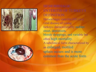
- 1. HEMORRHAGIC ENTERITIS OF TURKEYS DEFINITION Hemorrhagic enteritis (HE) is a viral disease of young turkeys characterized by sudden onset, depression, bloody droppings, and variable but often high mortality. A subclinical form characterized by an enlarged, mottled spleen occurs and is more common than the acute form
- 2. • OCCURRENCE • HE has a worldwide distribution and typically occurs in 6-12-week-old turkeys, but has been seen in poults as young as 2 weeks. • It is rare in turkeys less than 4 weeks • of age, presumably because of maternal antibody.
- 3. history HE in turkeys was first reported in 1937 In 1972 the disease was demonstrated to be caused by a viral infection. ETIOLOGY HE is caused by a turkey adenovirus, hemorrhagic enteritis virus.
- 4. EPIZOOTIOLOGY 1. The virus is very resistant to environmental factors and is shed in feces, hence the transmission route is fecal- oral. Infection frequently reoccurs on the same farm in successive flocks. 2. There is no evidence of egg transmission. 3. Infection of turkeys with HE virus results in a transient immunosuppression, often involving secondary colibacillosis.
- 5. CLINICAL SIGNS 1. Sudden deaths are often the first sign of HE in a flock. A concurrent drop in feed and water consumption may be noted. Droppings containing fresh blood or melena can be seen, especially around waterers.
- 6. Blood may be seen oozing from the vent of dead or moribund birds or may be adhered to feathers around the vent. Blood may be expelled from the vent if the abdomen is squeezed. Most birds with bloody feces die.
- 7. The disease usually runs its course in a flock in 10-14 days. Most mortality occurs over a 10 day period. Mortality averages 5-10% but may exceed 60%
- 8. Outbreaks of coli septicemia often follow clinical and subclinical infections with hemorrhagic enteritis virus 12 to14 days later. Coli septicemia may be the only indication of prior HE subclinical infection.
- 9. LESIONS Dead poults often appear pale due to intestinal blood loss but are well fleshed with feed in their crops. The skin and feathers around the vent can be stained with blood or blood stained feces.
- 10. duodenal loop, is distended, dark purple, and filled with hemorrhagic content The intestinal mucosa, especially of the duodenum is congested, and may be covered with a yellow layer of fibrin necrotic exudates.
- 11. the spleen is typically very enlarged and mottled (Fig. 2) and as the disease progresses, the spleen becomes smaller and pale.
- 12. Microscopically, early in the course of the disease, reticuloendothelial cells of the spleen contain numerous large intranuclear adenoviral inclusions and the condensed nuclear chromatin around the inclusions often resembles a signet ring
- 13. DIAGNOSIS 1. Typical history and gross lesions strongly suggest the diagnosis. Demonstration of intranuclear inclusions in reticuloendothelial cells in the spleen or intestine confirms the diagnosis unless the turkeys have received HE vaccine.
- 14. The disease can be reproduced in 6-week-old or older, susceptible poults by giving minced splenic tissue or its supinate intravenously, orally, or interlocally. Typical intestinal content also will reproduce the disease when given orally or cloacal.
- 15. it is possible to use the agar-gel diffusion test to demonstrate antigen in the spleen of an infected turkey or to demonstrate antibody in the convalescent sera of recovered birds.
- 16. HE must be differentiated from acute bacterial septicemia including coli septicemia, salmonellosis, fowl cholera and erysipelas. Gastrointestinal hemorrhage/ mucosal congestion may be associated with acute septicemic/viremic/bacteremia conditions.
- 17. CONTROL 1. Avirulent strains of HEV and related marble spleen disease (of pheasants) virus are used as vaccines. 2. Vaccines are prepared as crude splenic homogenates or are cell culture derived.
- 18. Intestinal coccidiosis should also be considered. HEV infection in growing turkeys results in immunosuppression predisposing birds to secondary infections such as Escherichia coli septicemia