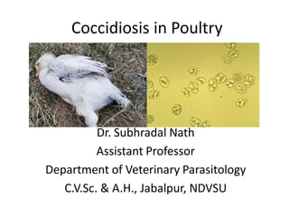
1. Coccidiosis in Poultry
Dr. Subhradal Nath
Assistant Professor
Department of Veterinary Parasitology
C.V.Sc. & A.H., Jabalpur, NDVSU
2. Introduction
• Poultry coccidiosis is one of the most important
disease
• Generates great economic losses due to mortality,
reduced body weight plus the expenses related to
preventive and therapeutic control
• Poultry coccidia are generally host specific, and the
different species parasitize specific parts of the
intestine.
• However, in game birds, including quail, the
coccidia may parasitize the entire intestinal tract.
Coccidia are distributed worldwide in poultry,
game birds reared in captivity, and wild birds
• Coccidial infection is self-limiting
3. Etiology
• Nine species of Eimeria have been described
that infect Gallus gallus var. domesticus at
various locations along the intestinal tract: E.
acervulina, E. brunetti, E. hagani, E. maxima,
E. mitis, E. mivati, E. necatrix, E. praecox, and
E. tenella.
• Except E. hagani and E. mivati the other 7
causes clinical coccidiosis
4. Life cycle
1. The life-cycle is short and starts with the
bird ingesting sporulated oocysts
2. The sporulated oocysts contain four sporocysts, each
containing two sporozoites and the mechanical and acidic
environmentin the gut result in the release of these sporocysts
and sporozoites into the gut.
3. The sporozoites invade the duodenal mucosa epithelial cells
before undergoing phases of growth and multiplication with
periodic release of merozoites into the gut.
4. Merozoites develop within the duodenal cells as gametes, in
the form of both macro- and microgametocytes
5. These develop into a zygote and then an oocyst which is shed in
the faeces
6. These oocysts require moist conditions to undergo sporulation,
a process that requires oxygen and takes about 24 hours, at
which point they become infective.
5.
6.
7.
8. Transmission
• Young chickens pick up the infection from
contaminated premises (soil, houses, utensils, etc.).
• These premises may have been contaminated
previously by other young infected birds or by adult
birds that have recovered from the condition.
• Wet areas around water fountains are a source of
infection. Oocysts remain viable in litter for many
months.
• In this way, they can contaminate a farm from year
to year. Oocysts are killed by freezing, extreme
dryness and high temperatures.
9. Prediposing Factors
• High stocking density
• Bad quality litter and lighting schedule
• Antinutritional factors (ANF's) in the feed,
• Brooder and grower management
• overcrowding
• Number of oocysts ingested by the bird and strain
of coccidia
• Environmental factors affecting the survival of the
oocysts. Viz Season
• Site of development within the host
• Nutritional status and age of the host
10. Pathogenesis
• The disease is seen in birds of 3-6 weeks old,
before they have acquired immunity.
• The most virulent strains will cause diarrhoea
and a sudden increase in flock mortality. Less
virulent strains will result in poor growth and
reduced feed efficiency
• Coccidiosis rarely occurs in layers and
breeders as they have generally acquired
immunity to this disease
11. • Pathogenicity depends on a number of factors
a. the number of host cells destroyed per infecting oocyst (which
depends upon the number of merozoite generations and the
number of merozoites per generation)
b. location of the parasite in the host tissues and within the host
cells.
c. The size of the infecting dose or doses, the degree of reinfection
d. degree of acquired or natural immunity of the host
The most common and pathogenic form of the disease is caecal
coccidiosis caused by E. tenella followed by intestinal coccidiosis
caused by E. necatrix
12. • E. necatrix and E. tenella
are the most pathogenic in
chickens, because
schizogony occurs in the
lamina propria and crypts
of Lieberkühn of the small
intestine and ceca,
respectively, and causes
extensive hemorrhage
• Most species develop in
epithelial cells liningthe villi.
Intestinal Crypts
13. Clinical Signs
1. Decreased feed and water consumption.
2. Decreased growth rate with high percentage of
visibly sickbirds.
3. Weight loss.
4. Severe diarrhea, bloody diarrhea.
5. Development of culls.
6. Decreased egg production.
7. High mortality.
14. 8. Mild infections (subclinical) may cause
depigmentation andpotentially lead to
secondary infection, particularly Clostridium
spp infection.
9. Survivors of severe infections recover in
10–14 days but never recover lost
performance
10. Lesions are present along the intestinal
tract and often have a distinctive location
and appearance that is useful in diagnosis.
15. Caecal coccidiosis
Characterized by:
1. Accumulation of blood in
the caeca.
2. Bloody droppings.
3. Caecal cores, which are
accumulations of clotted
blood,
tissue debris, and oocysts,
may be found in birds
surviving
the acute stage.
Coccidiosis site parasitized by E tenella
in poultry.
16. • Gross lesions of E tenella with frank
hemorrhaging into cecal
• pouches in a broiler chicken.
Gross lesions of E tenella with frank hemorrhaging into
caecal pouches in a broiler chicken.
17. E. tenella, a marked typhlitis is present and
haemorrhages are seen through the intestinal wall.
19. E. tenella, a later stage, the caecal content becomes thicker, mixed
with fibrinous exudate and acquires a cheese like appearance.
20. Intestinal coccidiosis
E. necatrix
• This species of
Eimeria is highly
pathogenic in
chickens and it is
often seen in birds
from 9 to 14
weeks of age
• Mortality, severe
weight losses, and
feces with blood
and mucus are
frequent findings.
Coccidiosis site parasitized by E necatrix in
poultry
21. • Small white spots, usually intermingled with
rounded, bright or dull-red spots of various
sizes, can be seen on the serosalsurface.
• This appearance is sometimes described as
“salt and pepper.”
• The white spots are diagnostic for E necatrix if
clumps of large schizonts can be
demonstrated microscopically.
22. • In severe cases
1. The intestinal wall is
thickened.
2. The infected area
dilated to 2–2.5 times the
normal diameter.
3. lumen filled with blood,
mucus, and fluid.
4. marked dehydration.
Although the damage is in the small intestine, the sexual
phase of the life cycle is completed in the caeca.
Oocysts of E necatrix are found only in the ceca.
Because of concurrent infections, oocysts of other
speciesmay be found in the area of major lesions,
misleading the diagnostician.
23. Rectal coccidiosis
• Caused by E brunetti
• E brunetti is found in the lower small intestine,
rectum, ceca and cloaca.
• In moderate infections, the mucosa is pale and
disrupted but lacking in discrete foci, and may
be thickened.
• In severe infections, coagulative necrosis and
sloughing of themucosa occurs throughout
most of the small intestine.
24. Coccidiosis site parasitized by E brunetti in poultry.
Gross lesions of E brunetti in small intestine of
a broiler chicken
25. E. maxima
• E maxima develops in the
small intestine, where it
causes:
• Dilatation and thickening of
the wall.
• Petechial hemorrhage.
• Reddish, orange, or pink
viscous mucous exudate
and fluid.
• midgut often has numerous
whitish pinpoint foci, and
the area may appear
engorged.
Coccidiosis site parasitized by E maxima in
poultry.
26. Speceies Lesion Clinical signs
E. Acervulina numerous whitish
transverse patches in
the upper half of the
small intestine
poor growth, an
increase in culls,
and slightly
increased mortality
E. Mitis pathogenic in the
lower small intestine
and lesions are
indistinct resemble
moderate infections
of E brunetti
Moderate infection
E. praecox infects the upper
small intestine, does
not cause distinct
lesions watery
intestinal contents
Less economic
importance
27. Species Site of
development
Pathogenicity Disease type
E. necatrix Jejunum,
ileum, caeca
+++++ Hemorrhagic
E. tenella Caeca +++++ Hemorrhagic
E. brunetti Caeca and
rectum
++++ Hemorrhagic
E. maxima Jejunum,
ileum
+++ Malabsorptive
E. mitis Ileum ++ Malabsorptive
E. acervulina Duodenum,
ileum
++ Malabsorptive
E. praecox Duodenum,
jejunum
+ Malabsorptive
28. Diagnosis
• The location in the host, appearance of
lesions, and the size of oocysts are used in
determining the species present.
• Coccidial infections are readily confirmed by
demonstration of oocysts in faeces or
intestinal scrapings; however, the number of
oocysts present has little relationship to the
extent of clinical disease.
29.
30. Prevention and treatment
• In poultry production several antimicrobials or
antiprotozoals have been used for decades to treat and
prevent coccidiosis. Depending on the type of poultry
production, the approaches for an effective control of
coccidiosis are different.
Methods of coccidiosis prevention or treatment:
• Coccidicides
• Coccidiostats
• vaccines
31. Coccidicides
• coccidiosis preventive program used usually aims
for eliminating Eimeria completely from the gut
by using coccidicides that kill the parasites.
• This results in optimal condition of the
gastrointestinal tract, improving
body weight, and reducing feed conversion
32. Coccidiostats
• In breeders and layers a different approach is usually
needed. Due to the relatively long life cycle of these birds,
development of protective immunity is desired. For this
purpose a minimal degree of exposure to Eimeria is
allowed.
• To achieve this objective, coccidiostats are used to arrest
the development of the parasites at different stages of
development allowing for a good balance between
intestinal damage and appropriate exposure for immunity
development.
• Of course, once the coccidiostats are withdrawn from the
diet, the infecting parasites may resume their life cycle
producing the clinical manifestations of the disease
33. Treatment
• sulfonamides are widely used
• sulfadimethoxine, sulfaquinoxaline,
sulfamethazine, but they should not be used in
layer hens.
• ionophores, which have an effect on membrane
function of the parasite and act as both
coccidiocides and coccidiostats ( monensin)
• quinolones, which have an effect on energy
metabolism of the parasite and act as both
coccidiocides and coccidiostats ( buquinolate)
• coccidiostatic thiamine analogs, which have an
effect on co-factor synthesis for the parasite
• The supplementation of vitamins A and K
promotes the recovery
34. Treatment Example Mechanism of Action
Ionophores Lasalocid, Monensin,
Narasinm Salinomycin, and
Semduramicin
Disruption of ion gradient
across the parasite cell
membrane
Chemicals Quinolone drugs (Decoquinate
and nequinatem buquinolate)
Pyridones (Meticlorpindol)
Inhibition of parasite
mitochondrial respiration
Sulphonamides Inhibition of the folic acid
pathway
Amprolium Diclazuril, Competitive inhibition of
thiamine uptake
Halofuginone, and Robenidine Mode of action unknown
Nicarbazin Inhibition of the
development of the first and
second generations of the
schizont stage of the
parasites
35. Antimicrobial resistance
• Eimeria parasites do develop drug resistance
due to regular use of drugs.
• The resistance is greatly enhanced if the same
family of antimicrobials is used for a long time
within a defined area.
• Selective pressure will favour the few parasites
within a population that are resistant, and
within few rearing cycles the initial parasites
would increase their population size to
numbers able to induce clinical disease in a
flock.
36. Shuttle programs
• A common practice to partially solve this problem is to use
anti-coccidial ‘shuttle’ programs that rotate through different
periods of the bird's life.
• This method has a good chance of eliminating the parasites
that demonstrated resistance to a single antimicrobial.
• A variation of the same principle consists on changing
coccidiostats between flocks.
• most suitable drug is used for starter, while another drug is
used for grower and finisher. Drug withdrawal period is the
most important consideration for drugs that will be used in
finisher feeds.;
• Examples of reasonable shuttles are:
• Coban:Stenorol:Clinacox (an ideal winter program)
• Coxistac:Avatec - ideal for summer program
• Coxistac:Stenorol - winter or summer program
• Coxistac:Clinacox - winter or summer shuttle
37. Rotation Programme
• Rotation: means that a conscious decision is made to change
the drug(s) used at a given time in the future i.e. every four
months, after two crops, go to a winter and summer
program etc. The alternative to a rotation program is a
continuous program where the same drug(s) are used
indefinitely, usually until a problem develops, or until a new
product is introduced on the market. Shuttle programs fit
into rotation programs
• An example of a rotation program (change every 4 months)
would be:
• 1st rotation (May-August) - ionophore i.e. Coxistac
• 2nd rotation (September-December) - non-ionophore i.e.
Clinacox
• 3rd rotation (January-April) - shuttle Coban: Stenorol
38. Vaccines
Passive or active immune responses induce immunity in
animals. This immunity can reduce the pathogenic effects of
coccidiosis such as less macroscopically visible lesions,
decreasing of oocyst production, and increasing
performance of birds.
• The first commercial live coccidiosis vaccine
was CocciVac® registered in the USA in 1952
• Currently, two types of vaccines are used with
the aim of controlling coccidiosis in a chemical
free way:
• A) Live Non-attenuated
• B) Live attenuated vaccines.
• The main risk of using live non-attenuated
vaccines (Coccivac, Advent, Immucox, and
Inovocox) is the live parasites that can
develop a severe reaction in birds.
• Many times their use is accompanied by
chemical treatments to control the inherent
pathogenicity of the parasites .
39. • On the contrary, the success of
live attenuated vaccines (Paracox
and HatchPak CocciIII) relies on
the low risk of disease occurring
because of the reduction in the
proliferation of parasites and
consequently a less damage in
birds’ tissue.
• Non-attenuated and attenuated
vaccines may have different
routes of administration (oral,
eyes drops, in ovo) in birds and
several Eimeria species as target.
40. Genetically engineered vaccines
• Genetically engineered Subunit vaccines consist
of purified antigenic determinants obtained from
Eimeria parasite.
• These vaccines are obtained from DNA
recombinant technology and may consist of
native antigens or recombinant proteins of
various stages (sporozoites, merozoites, and
gametes) of Eimeria.
• Distinct protective antigens used are
micronemes, rhoptries, refractile bodies,
merozoites, or gametocytes of Eimeria parasite
41. Coccidia of Turkey
• Generally there are seven species of coccidia
infecting turkeys :
• E. adenoides, E. dispersa, E gallopavonis, E
meleagrimitis, E. innocua, E meleagridis, and E
subrotunda .
• E. innocua, E. meleagridis, and E. subrotunda
are considered nonpathogenic.
• E. adenoides, E. dispersa, E. gallopavonis, and
E. meleagrimitis are pathogenic
42. • Oocysts sporulate within 1–2 days after
expulsion from the host; the prepatent period
is 4–6 days.
• E. adenoeides and E. gallopavonis infect the
lower ileum, ceca, and rectum. These species
often cause mortality.
• The developmental stages are found in the
epithelial cells of the villi and crypts. The
affected portion of the intestine may be
dilated and have a thickened wall.
• Thick, creamy material or caseous casts in the
gut or excreta may contain enormous
numbers of oocysts.
43. • E meleagrimitis chiefly infects the upper and mid
small intestine.
• The lamina propria or deeper tissues may be
parasitized, which may result in necrotic enteritis
• E. dispersa infects the upper small intestine and
causes a creamy, mucoid enteritis that involves the
entire intestine, including the ceca. Large numbers
of gametocytes and oocysts are associated with the
lesions.
• Common signs in infected flocks include reduced
feed consumption, rapid weight loss, droopiness,
ruffled feathers, and severe diarrhea. Wet
droppings with mucus are common.
• Clinical infections are seldom seen in poults >8 wk
old. Morbidity and mortality may be high
46. Coccidia of Duck
• A large number of specific
coccidia have been reported in
both wild and domestic ducks
• Presence of Eimeria,
Wenyonella, and Tyzzeria spp
has been confirmed
• T. perniciosa is a known
pathogen that balloons the
entire small intestine with
mucohemorrhagic or caseous
material
• Pathogenesis resembles E.
necatrix
47. Coccidia of Geese
The best known coccidial infection
of geese is that produced by E .
truncata, in which the kidneys are
enlarged and studded with poorly
circumscribed, yellowish white
streaks and spots.
The tubules are dilated with
masses of oocysts and urates.
Mortality may be high.
At least five other Eimeria spp
have been reported to parasitize
the intestine of geese, but these
are of lesser importance
48. Control by managemental practices
• Maintain vigilance and treat as soon as the first
symptoms are seen
• Keep different age groups separate
• Ensure that litter is dry but not dusty – avoid any
causes of wet litter
• Keep litter dry round watering points – do not allow
drinkers to overflow
• Ensure high standards of hygiene of personnel
• Ensure good hygiene of feeding and drinking
equipment Keep raising the level of drinkers as chicks
grow to reduce fouling
• Vaccinate if the risks of disease are high