80 ĐỀ THI THỬ TUYỂN SINH TIẾNG ANH VÀO 10 SỞ GD – ĐT THÀNH PHỐ HỒ CHÍ MINH NĂ...
Activity 2
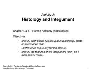
1. Activity 2:
Histology and Integument
Chapter 4 & 5 – Human Anatomy (4e) textbook
Objectives:
• Identify each tissue (26 tissues) in a histology photo
or microscope slide.
• Sketch each tissue in your lab manual.
• Identify the features of the integument (skin) on a
slide and/or model.
1
Compilation: Benjamin Sparks & Claudia Gonzales
Last Revision: Mohammad Tomaraei
2. Basic Tissue Types
• We’re going to look at four different tissue types:
1. Epithelium tissue
2. Connective tissue
3. Muscle tissue
4. Nervous tissue
2
3. Epithelium Tissues
• Epithelium is a tissue composed of cells that line the cavities and
surfaces of structures throughout the body.
• Epithelial cells have three general cell shapes: squamous,
cuboidal, and columnar.
• They are also found in varying number of cell layers, namely:
simple, stratified, and pseudostratified.
3
4. How to identify Epithelium?
1. Determine how many cell layers are there:
• If only one layer,
it’s simple.
• If more than one layer, it’s stratified.
2. Determine the shape of the epithelial cells (look at cells near the
apical surface):
• Squamous
• Cuboidal
• Columnar
4
7. Important Note!
• After you have determined the number of cell layers and their
general shape, make sure to write the word “epithelium” at the
end as well!
• For example, Simple Squamous Epithelium
7
8. Simple Squamous Epithelium
• Structure:
• Single layer of thin, flat, irregularly-shaped cells resembling floor tiles
• The single nucleus of each cell bulges at its center
• Function:
• Rapid diffusion, filtration, and some secretion in serous membranes
• Location:
• Air sacs in lungs (alveoli)
• Lining lumen of blood vessels (endothelium)
• Serous membranes of body cavities (mesothelium)
• Textbook References:
• Table: p. 86, table 4.3a
• Description: pp. 84-85
8
12. Keratinized Stratified Squamous Epithelium
• Structure:
• Multiple layers of cells
• Basal cells are typically cuboidal or columnar, while apical
(superficial) cells appear squamous
• More superficial cells are dead and filled with the protein keratin
• Function:
• Protection of the underlying tissue
• Location:
• Epidermis of the skin
• Textbook References:
• Table: p. 89 table 4.4a, b
• Description: pp. 87-88
12
16. Nonkeratinized Stratified Squamous Epithelium
• Structure:
• Multiple layers of cells
• Basal cells are typically cuboidal or polyhedral, while apical cells are
squamous
• Surface cells are alive (visible nuclei) and kept moist
• Function:
• Protection of the underlying tissue
• Location:
• Lining of oral cavity, part of pharynx, esophagus, vagina, and anus
• Textbook References:
• Table: p. 89 table 4.4a, b
• Description: pp. 87-88
16
20. Simple Cuboidal Epithelium
• Structure:
• Single layer of cells as tall as they are wide
• Contains spherical, centrally located nucleus
• Function:
• Absorption and secretion
• Location:
• Thyroid gland follicles
• Kidney tubules
• Ducts and secretory regions of most glands
• Textbook References:
• Table: p. 86 table 4.3b
• Description: p. 85
20
26. Stratified Cuboidal Epithelium
• Structure:
• Two or more layers of cells
• Cells at apical surface are cuboidal
• Function:
• Protection and secretion
• Location:
• Found in large ducts in most exocrine glands and in some parts of
the male urethra
• Textbook References:
• Table: p. 90 table 4.4
• Description: p. 88
26
29. Simple Columnar Epithelium (Ciliated and Non-Ciliated)
• Structure:
• Single layer of tall, narrow cells
• Oval shaped nucleus in the basal region of cells
• Function:
• Absorption and secretion (non-ciliated)
• Secretion of mucin and movement of mucus along apical surface of
epithelium by action of cilia (ciliated)
• Location:
• Lining of most of the digestive tract (non-ciliated)
• Lining of uterine tubes and larger bronchioles of respiratory tract
(ciliated)
• Textbook References:
• Table: p. 86 table 4.3c, d
• Description: pp. 85-86 29
35. Stratified Columnar Epithelium
• Structure:
• Two or more layers of cells
• Cells at the apical surface are columnar
• Function:
• Protection and secretion
• Location:
• Rare, found in large ducts of some exocrine glands and in some
regions of the male urethra
• Textbook References:
• Table: p. 90 table 4.4d
• Description: p. 88
35
40. Pseudostratified Columnar Epithelium
• Structure:
• Single layer of cells with varying heights that appear multi-layered
• All cells connect to the basement membrane but not all cells reach the
apical surface
• Function:
• Protection
• Ciliated form also involved with secretion of mucin and movement of
mucus across surface with ciliary action
• Location:
• Ciliated form lines most of the respiratory tract
• Non-ciliated form is rare and lines the epididymis and part of male
urethra
• Textbook References:
• Table: p. 91 table 4.5a
• Description: p. 88
40
46. Transitional Epithelium
• Structure:
• Epithelial appearance varies, depending on whether the tissue is
stretched or relaxed
• Shape of cells on the apical surface changes
• Function:
• Distention and relaxation to accommodate urine volume changes in
the bladder, ureters, and urethra
• Location:
• Lining of urinary bladder, ureters, and part of urethra
• Textbook References:
• Table: p. 91 table 4.5b
• Description: p. 88
46
50. Connective Tissues
• Connective tissue is the most diverse tissue. It is widespread
although not exposed to the outside of the body. Except for
cartilage, it is highly vascular. It has widely scattered cells with
lots of space between each cell.
• Functions: Protection (bone, fat), support (bone, cartilage),
binding together (tendons, ligaments), storage of fat (bone
marrow), disease fighting (blood), and production of certain blood
cells.
50
52. Blood (Fluid Connective Tissue)
• Structure:
• Contains erythrocytes, leukocytes, and platelets
• Soluble (dissolved) protein fibers and a watery ground substance
form a fluid extracellular matrix called plasma
• Function:
• Erythrocytes transport gases, leukocytes control immune response,
platelets help with blood clotting
• Plasma transports nutrients, wastes, and hormones throughout the
body and contains clotting elements to stop blood loss
• Location:
• Primarily found within blood vessels (arteries, veins, and capillaries),
and the heart
• Textbook References:
• Table: p. 108 table 4.13
• Description: p. 105 52
60. Reticular Connective Tissue (Loose Connective Tissue)
• Structure:
• Ground substance is a gel-like liquid
• Contains a scattered arrangement of reticular fibers and extracellular
matrix
• Function:
• Provides a supportive framework for spleen, lymph nodes, thymus,
and bone marrow
• Location:
• Forms stroma of lymph nodes, spleen, thymus, and bone marrow
• Textbook References:
• Table: p. 103 table 4.9c
• Description: p. 100
60
68. Dense Regular Connective Tissue (Dense Connective Tissue)
• Structure:
• Contains densely packed and parallel collagen fibers, fibroblast
nuclei, and scarce ground substance
• Function:
• Attaches muscle to bone and bone to bone
• Resists stress applied in one direction
• Location:
• Forms tendons
• Most ligaments
• Textbook References:
• Table: p. 104 table 4.10a
• Description: p. 101
68
69. Dense Regular Connective (Dense Connective Tissue)
69
Hint: notice the unidirectional pattern of this type of connective tissue and relate it to its one-directional support
70. Dense Regular Connective (Dense Connective Tissue)
70
Hint: notice the unidirectional pattern of this type of connective tissue and relate it to its one-directional support
71. Dense Regular Connective (Dense Connective Tissue)
71
Hint: notice the unidirectional pattern of this type of connective tissue and relate it to its one-directional support
72. Elastic Connective Tissue (Dense Connective Tissue)
• Structure:
• Contains parallel elastic fibers, fibroblast nuclei, and ground
substance
• Function:
• Allows stretching of some organs
• Location:
• Walls of elastic arteries
• Trachea
• Bronchial tubes
• True vocal cords
• Suspensory ligaments of penis
• Textbook References:
• Table: p. 105 table 4.10c
• Description: p. 101
72
73. Elastic Connective Tissue (Dense Connective Tissue)
73
Hint: note the wave-like, rubber-band structure of elastic fibers and relate it to their flexibility
75. Dense Irregular Connective Tissue (Dense Connective Tissue)
• Structure:
• Predominantly contains collagen fibers (bundled and randomly
arranged), fibroblasts, and ground substance (more than in dense
regular connective tissue)
• Function:
• Withstands stresses applied in all directions
• Durable
• Location:
• Dermis
• Periosteum covering bone
• Perichondrium covering cartilage
• Organ capsules
• Textbook References:
• Table: p. 104 table 4.10b
• Description: p. 101 75
76. Dense Irregular Connective Tissue (Dense Connective Tissue)
76
Hint: notice the multidirectional pattern of this type of connective tissue; you can remember dense irregular
connective tissue by its resemblance to steak
83. Compact Bone (Osseous Tissue)
83
osteocyte (in lacunae)
central canal
lamellae
(concentric)
canaliculi
Identify the structures at the arrows.
84. Hyaline Cartilage (Cartilage Tissue)
• Structure:
• Contains extracellular matrix, lacunae, chondrocytes, perichondrium (often visible)
• Function:
• Smooth surfaces for movement at joints
• Model for bone growth
• Supports soft tissue
• Location:
• Most of fetal skeleton
• Covers articular ends of long bones
• Costal cartilages
• Most of larynx, trachea, and nose
• Textbook References:
• Table: p. 106 table 4.11a
• Description: p. 103
84
97. Muscle Tissues
• Cells are long and narrow and are called fibers
• Functions through contraction in motion, posture, and heat
production
97
98. Types of Muscle Tissues
98
Type of Muscular
Tissue
Skeletal Muscle Smooth Muscle Cardiac Muscle
Shape of Fibers Elongated; blunt ends Elongated, Tapered
ends
Elongated; Blunt ends
Nucleus: # and Location Multinucleated,
peripheral
Uninucleated, central Uninucleated, central
Striated or
Non-striated
Striated Non-striated Striated
Branched or
Unbranched
Unbranched Unbranched Branched (Bifurcated
and Intercalated Discs)
Involuntary or Voluntary Voluntary Involuntary Involuntary
Location in Body Attached to bones Walls of hollow, internal
organs, and tubes
Only in wall of Heart
(Myocardium)
Speed of Contraction Fastest Slowest Intermediate
Ability to Remain
Contracted
Least Greatest Intermediate
99. Smooth Muscle
• Structure:
• Contains spindle-shaped muscle fibers that have a centrally located
nucleus
• Function:
• Involuntary movements and motion
• Moves materials through internal organs
• Location:
• Walls of hollow internal organs, such as vessels, airways, stomach,
bladder, and uterus
• Textbook References:
• Table: p. 111 table 4.14c
• Description: p. 109
99
102. Skeletal Muscle
• Structure:
• Contains long, cylindrical, unbranched muscle fibers with multiple
nuclei per fiber
• Striations are visible in each muscle fiber
• Function:
• Moves skeleton
• Responsible for voluntary body movements, locomotion, and heat
production
• Location:
• Attaches to bones or sometimes skin
• Textbook References:
• Table: p. 110 table 4.14a
• Description: p. 109
102
106. Cardiac Muscle
• Structure:
• Contains short and branched muscle fibers (also known as
cardiomyocytes) with one nucleus per cell
• Each muscle fiber exhibits striations
• Intercalated discs are located between cells
• Function:
• Involuntary contractions and relaxations pump blood in the heart
• Location:
• Heart wall (myocardium)
• Textbook References:
• Table: p. 110 table 4.14b
• Description: p. 109
106
118. Integumentary System – Hypodermis / Subcutaneous Layer
• Description:
• Not part of the integument proper
• Made of areolar connective tissue and adipose tissue
• Often called superficial fascia
118