
Recommended
More Related Content
What's hot
What's hot (20)
Similar to 1.EPITHELIAL TISSUE (1).pptx
Similar to 1.EPITHELIAL TISSUE (1).pptx (20)
Recently uploaded
Recently uploaded (20)
1.EPITHELIAL TISSUE (1).pptx
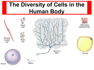
- 1. The Diversity of Cells in the Human Body
- 2. 2 ¶ Despite its complexity, the human body is composed of only four basic types of tissue: ● epithelial ● connective & supporting ● muscular & contractile ● nervous ¶ These tissues ● are formed by cells and molecules of the extracellular matrix ● exist not as isolated units but rather in association with one another and in variable proportions ● form different organs and systems of the body ¶ The main characteristics of these basic types of tissue are: Tissue Cells Extracellular Matrix Main Functions Nervous Intertwining elongated processes None Transmission of nervous impulses Epithelial Aggregated polyhedral cells Small amount Lining of surface surface or body body cavities, glandular secretion Muscle Elongated contractile cells Moderate amount Movement Connective Several types of fixed and wandering cells Abundant amount Support and protection
- 3. 3 Prepared by:-Dr. Alazar Etana May/2023
- 4. Epithelial Tissue • covers all free surfaces of the body (epi, on + thelium, surface). • consists of cells attached to one another to form an uninterrupted layer of cells that separates the underlying tissues from the outside world. • consists of closely apposed cells without (or very little) intervening intercellular substances. • is avascular, but all epithelia "grow" on an underlying layer of vascular connective tissue (CT). CT and epithelium are separated by a basement membrane • the body's epithelium - covers its obvious surfaces (such as the epidermis of the skin and the linings of respiratory, urinary, and digestive tracts) - extends into all of the complex invaginations which form lungs, kidneys, sweat glands, digestive glands, liver, etc. • provides the essential functions – protection - containment of body fluids - transport in and out across body surfaces (absorption and secretion). • Embryonically, most epithelial tissues are derived either from ectoderm (e.g., epidermis) or endoderm (e.g., epithelium of trachea and lung).
- 5. Ion transport Filtration Forms slippery surfaces
- 6. Special Characteristics of Epithelia • Cellularity – cells are in close contact with each other with little or no intercellular space between them. • Specialized contacts – may have junctions for both attachment and communication. • Polarity – epithelial tissues always have specific apical, lateral and basal surfaces. • Support by CT – at the basal surface, both the epithelial tissue and the CT contribute to the basement membrane. • Avascularity (generally) – nutrients must diffuse. • Innervation • Regeneration – epithelial tissues have a high capacity for regeneration ; plasticity. Epithelial tissue thus serves both as a protective barrier for the body and as an active interface with the environment ● Great diversity of function
- 7. Special Characteristics of Epithelia
- 8. • Epithelial cells exhibit distinct polarity. They have • an apical domain, • a lateral domain, and • a basal domain. • Specific biochemical characteristics are associated with each cell surface. • These characteristics and the geometric arrangements of the cells in the epithelium determine the functional polarity of all three cell domains. ●The free or apical domain is always directed toward the exterior surface or the lumen of an enclosed cavity or tube. ●The lateral domain communicates with adjacent cells and is characterized by specialized attachment areas. ●The basal domain rests on the basal lamina anchoring the cell to underlying CT.
- 9. Classification of epithelia 1. Surface epithelia (Covering epithelia) 2. Glandular epithelia 3. Special epithelia
- 11. I. First name of tissue indicates number of layers. • Simple –one layer of cells • Stratified –more than one layer of cells Classifications & Naming of Epithelia
- 12. Classification & Naming of Epithelia II. Last name of tissue describes shape of cells. – Squamous – cells wider than tall (plate or “scale” like) – Cuboidal – cells are as wide as tall, as in cubes – Columnar – cells are taller than they are wide, like columns
- 13. • Naming the epithelia includes both the layers (first) and the shape of the cells (second). – i.e. stratified cuboidal epithelium • The name may also include any accessory structures. – Goblet cells – Cilia – Keratin • Special epithelial tissues (don’t follow naming convention). – Psuedostratified – Transitional Classifications & Naming of Epithelia
- 14. Diagrams of simple epithelial tissue. A: Simple squamous epithelium. B: Simple cuboidal epithelium. C: Simple ciliated columnar epithelium. All are separated from the subjacent CT by a basement membrane. In C, note the terminal bars that correspond in light microscopy to the zonula occludens and the zonula adherens of the junctional complex.
- 15. Diagrams of stratified, transitional and pseudostratified epithelia. A: Stratified squamous epithelium. B: Transitional epithelium. C: Ciliated pseudostratified epithelium. The goblet cells secrete mucus, which forms a continuous mucous film over the ciliary layer.
- 17. Simple Squamous Epithelium • Description • single layer of flattened, scale- or plate-like cells with disc-shaped nuclei. (The nuclei are often flattened or ovoid, and they are located close to the centre of the cells). • Special types – Endothelium (inner covering) • slick lining of hollow organs: lines the internal surfaces of blood and lymph vessels – Mesothelium (middle covering) - lines the large internal body cavities • Lines peritoneal, pleural, and pericardial cavities • Covers visceral organs of those cavities
- 18. • Function – Passage of materials by passive diffusion and filtration. – Secretes lubricating substances. • Location – Renal corpuscles – Alveoli of lungs – Lining of heart, blood and lymphatic vessels – Lining of ventral body cavity …Simple Squamous Epithelium
- 19. Simple squamous lining the walls …Simple Squamous Epithelium
- 20. Simple Squamous Mesothelium Mesothelial cells
- 21. Simple Squamous – Endothelium (En) Simple Squamous – Endothelium (Ed) by TEM Simple Squamous (Mesothelium) by TEM
- 22. Capillary (endothelial cell) by TEM
- 23. Simple Cuboidal Epithelium • Description – single layer of cube-like cells with large, spherical central nuclei • Functions – secretion and absorption • Location – kidney tubules (PCT, DCT, small CDs) – secretory portions of small exocrine glands, surface of the ovary & the follicles of the thyroid follicles
- 24. Kidney Collecting Ducts: (cross section) Simple Cuboidal Epithelium (longitudinal section)
- 25. Simple cuboidal epithelium. Cells vary in their height but are roughly as tall as they are wide. Their greater thickness often includes cytoplasm rich in mitochondria providing energy for a high level of active transport of substances across the epithelium.
- 26. Simple Columnar Epithelium • Description – single layer of column-shaped (rectangular) cells with oval nuclei – (cells are taller than they are wide) • Some bear cilia at their apical surfaces • May contain goblet cells • Function – absorption – secretion of mucus, enzymes, and other substances – Ciliated type propel mucus in reproductive cells by ciliary action
- 27. • Location – Non-ciliated form • Lines digestive tract, • gallbladder, • ducts of some glands – Ciliated form • Lines small bronchi, uterine tubes, uterus …Simple Columnar Epithelium
- 29. Goblet Cell with PAS stain (red) Simple Columnar with Goblet Cells (Mu) by TEM (unicellular exocrine gland)
- 30. Absorbing cell: Single cell layer of tall columnar cells with basal ovoid nucleus. Although the cells appear retangular in this section, they are actually five- or six-sided when they are cross-sectioned. When underlying tissue folds or bends, these cells may have a pyramidal appearance. Nucleus: Ovoid situated in lower half of the columnar cell. The nuclei in tightly packed cells may appear elongated and staggered at different levels within the cell. (This is readily seen in pseudostratified ciliated columnar). Brush border: (also know as the straited border). Made up of fine, closely packed microvilli that vastly increase the surface area of the cell. Characteristic of absorptive surfaces. Adequate absorption of digestive products is dependent upon this cell surface specialization of absorbing columnar epithelial cells. Goblet cell: Unicellular mucous glands scattered among the tall columnar cells appear empty because mucin is extracted during tissue processing. These unicellular gland cells are a specialization of simple epithelium and serve a protective function for the principal epithelial cell type. Basement membrane: Delicate in appearance but a firm support for the columnar cells. Lamina propria: CT stroma. Reticular framework containing a variety of wandering cells as well as vascular and lymphatic channels. Cells commonly found in the lamina propria include lymphocytes, plasma cells, eosinophils, and mast cells.
- 32. Pseudostratified Columnar Epithelium • Description – All cells originate at basement membrane – Only tall cells reach the apical surface – May contain goblet cells and bear cilia – Nuclei lie at varying heights within cells •Gives false impression of stratification • Function – secretion of mucus – propulsion of mucus by cilia
- 33. • Locations – Non-ciliated type • Ducts of male reproductive tubes • Ducts of large glands – Ciliated variety • Lines trachea and most of upper respiratory tract …Pseudostratified Columnar Epithelium Cells appear to be in layers, but the basal ends of the cells are all in contact with the basement membrane, which is often very thick.
- 34. Pseudostratified Ciliated Epithelium with Goblet Cells Respiratory epithelium
- 35. Pseudostratified Columnar Epithelium (with microvilli) Actually a single layer of columnar tissue but the nuclei do not line up making the tissue layer appear stratified Pseudostratified columnar epithelium ●All cells of this type of epithelium are in contact with the basement membrane, but not all of them reach the surface of the epithelium. ● Nuclei of the epithelial cells are typically located in the widest part of the cell. ● Consequently, the nuclei of cells which do or do not reach the surface of the epithelium are often located at different heights within the epithelium and give the epithelium a stratified appearance. ● The epithelium will look stratified but it is not - hence its name "pseudostratified". ● Pseudostratified columnar epithelia are found in the excretory ducts of many glands.
- 36. ● Pseudostratified columnar epithelium of the trachea, formed by long and short cells. ● As some cells do not reach the surface of the epithelium their nuclei are present in different heights of the epithelial layer. ● Mucus-secreting cells, called goblet cells (blue arrow), intermingle with ciliated lining
- 38. Stratified Epithelia • Contain two or more layers of cells. – Many superficial layers of cells – squamous in shape. – Deeper layers of cells appear cuboidal or columnar. • Regenerate from below. • Major role is protection. • Thickest epithelial tissue. • Are named according to the shape of cells at apical layer. Description
- 39. • Specific types – Keratinized – contain the protective protein keratin. • Surface cells are dead and full of keratin. – Non-keratinized – forms moist lining of body openings. • Function – Protects underlying tissues in areas subject to abrasion. • Location – Keratinized – forms epidermis – Non-keratinized – forms lining of • esophagus • pharynx • mouth • anal canal, uterine cervix & vagina …Stratified Squamous Epithelium
- 40. Stratified squamous nonkeratinized (moist) epith. of the esophagus. The most superficial cells (arrow) have the form of very thin scales. PT stain. Medium magnification. Oesophagus, human - H&E The oesophagus is lined by a stratified squamous epithelium consisting of many cell layers. Basal cells often form a well defined layer at the border of the epithelium to the underlying CT. The underlying CT forms finger-like extensions towards the lumen of the oesophagus, which are called papillae. The border between epithelium and CT may appear quite irregular because of the papillae. This irregular border aids in anchoring of the epithelium to the CT. If these extensions are not cut exactly along their long axis, they may look like isolated small islands of CT and blood vessels within the epithelium.
- 41. Keratinized Stratified Squamous Epithelium Stratum germinativum (basale) Str. spinosum Str. granulosum Str. corneum Dermal ridge Melanocytes
- 42. 42 Stratified cuboidal & columnar epithelia are not common. Stratified cuboidal epithelium ●is thin, ● usually consists of only 2-3 layers of cuboidal cells. This type of epithelium is usually confined to the lining of the larger excretory ducts of exocrine glands ● the salivary glands ● ducts of sweat glands
- 43. STRATIFIED COLUMNAR EPITHELIUM Mucous gland duct tongue Stratified columnar epithelia are found in the excretory ducts of the mammary gland and the main excretory duct of the large salivary glands & Conjunctiva.
- 45. Transitional Epithelium • Description – Basal cells usually cuboidal or columnar – Superficial cells dome- shaped or squamous • Function – stretches and permits distension of urinary bladder • Location : Lines - renal calyces & pelvis – ureters, – urinary bladder and – part of urethra (prox)
- 46. Transitional epithelium The shape of the cells in the surface layer of a transitional epithelium varies with the degree of distension of the organs whose lumen is lined by this type of epithelium. [The term transitional epithelium does not imply that this epithelium is in actual transition from one type to another, but rather refers to the appearance of the cells, which change as the organs with which they are associated are stretched or relaxed]. ✹ In the 'relaxed' state of the epithelium, it seems to be formed by many cell layers. The most basal cells have a cuboidal or columnar shape. There are several layers of polyhedral cells, and, a layer of super-ficial cells, which have a convex, dome-shaped luminal surface. ✹ In the distended state of the epithelium only one or two layers of cuboidal cells are followed by a superficial layer of large, low cuboidal or squamous cells. In the distended state the epithelium will resemble a stratified squamous epithelium.
- 47. Transitional Epithelium When the tissue stretches the cells become thinner and appear to be cuboidal or squamous. (umbrella or dome cells U)
- 49. SPECIAL EPITHELIA A. Myoepithelium 1. Contractile, branched epithelial cells. 2. Located between glandular epithelial cells and basement membrane. 3. Contain contractile proteins like actin filaments; smooth muscle-like. 4. Facilitate glandular secretion. B. Neuroepithelium 1. Non-neuronal a. Receptor cells of taste buds b. Hair cells of inner ear 2. Neuronal a. Sensory cells of olfactory epithelium b. Rods and cones of retina C. Seminiferous epithelium 1. Surface epithelium of testis a. Spermatogenic (germ) cells b. Supporting (Sertoli) cells i). Sertoli cells: simple epithelium ii). Spermatogenic cells: stratified
- 50. Common types of covering epithelia in the human body. Number of cell layers Cell form Examples of distribution function Simple (one layer) squamous Lining of vessels (endothelium). Serous lining of cavities; pericardium, pleura, peritoneum (mesothelium). . Facilitates the movement of the viscera (mesothelium) . active transport by pinocytosis (mesothelium and endothelium). cuboidal Covering the ovary, thyroid. Covering secretion. columnar Lining of intestine, gallbladder. Protection, lubrication, absorption, secretion. Pseudostratified (layers of cells with nuclei at different levels; not all cells reach surface but all adhere to basal lamina) Lining of trachea, bronchi, nasal cavity. Protection, secretion; cilia mediated transport of particles trapped in mucus out of the air passages. Stratified (two or more layers) Squamous keratinized (dry) Epidermis Protection prevents water loss. Squamous nokeratinized (moist) Mouth, esophagus, larynx, vagina, and anal canal. Protection, secretion prevents water loss cuboidal Sweet glands, developing ovarian follicles. Protection secretion transitional Bladder, ureters, renal calyces. Protection distensibility. columnar Conjunctiva. Protection.
Editor's Notes
- 2015