Histology of Muscle Tissues
•Download as PPT, PDF•
22 likes•13,855 views

A complete lecture of the Histology of Muscle Tissues, taught at First Moscow State Medical University, Moscow, in the Histology department, for the first year English medium foreign medical students.

Recommended
More Related Content
What's hot
What's hot (20)
Viewers also liked
Viewers also liked (13)
Similar to Histology of Muscle Tissues
Similar to Histology of Muscle Tissues (20)
More from Dr. Julius Kwedhi
More from Dr. Julius Kwedhi (20)
Recently uploaded
Recently uploaded (20)
Histology of Muscle Tissues
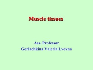
- 1. Muscle tissuesMuscle tissues Ass. Professor Goriachkina Valeria Lvovna
- 2. Muscle tissues are classified on the basis of their appearance. Two different types of muscle tissues are recognized: striated and smooth. Striated muscle tissue is subclassified on the basis of its location: skeletal muscle tissue and cardiac muscle tissue
- 4. Skeletal muscle tissue Each myofibril has characteristic banding patterns (dark and light bands). When observed under polarized light, the dark-staining bands are birefringent (anisotropic),while the light-staining ones are isotropic. Accordingly, the dark bands are called A bands ( A for anisotropic) and the light ones, I bands (I for isotropic).Owing to these alternations of dark and light bands, transverse striations in a muscle fiber can be seen with light microscope. The arrangement of the contractileThe arrangement of the contractile proteins within skeletal muscle fiberproteins within skeletal muscle fiber
- 5. Striated muscle tissue. Iron hematoxylinStriated muscle tissue. Iron hematoxylin
- 6. The structure of thick and thin filamentsThe structure of thick and thin filaments at the molecular levelat the molecular level
- 9. The structure of sarcomereThe structure of sarcomere
- 10. SarcotubularSarcotubular systemsystem is composed of agranular (smooth) sarcoplasmic reticulum (L-tubules) and T-tubules
- 11. Contraction cycle of skeletal muscleContraction cycle of skeletal muscle
- 12. Contraction cycle of skeletal muscleContraction cycle of skeletal muscle
- 14. Scanning electron micrograph of striated muscleScanning electron micrograph of striated muscle fibers (MF) endomysium (collagen fibers-KF)fibers (MF) endomysium (collagen fibers-KF) MFMF MFMF KFKF MFMF KFKF
- 15. Skeletal muscle fiber TypesSkeletal muscle fiber Types
- 16. Type of muscle fibresType of muscle fibres Aerobic (type I), anaerobic (type II) and intermediate fibres The activity of the specific mitochondrial enzyme succinate dehydrogenase ATP-ase activity
- 17. Histogenesis of Skeletal Muscle FibersHistogenesis of Skeletal Muscle Fibers 1. From the myotomes arise myoblasts (two cell populations). 2. a) On one side presumptive myoblasts differentiate into true myoblasts. b) On other, presumptive myoblasts remain undifferentiated and give rise to satellite cells. 3. a) True myoblasts range in rows, fuse together and form myotube. b) Satellite cells adhere to myotube. 4. Myotube gradually differentiates into skeletal muscle fiber.
- 19. General Features of Cardiac Muscle TissueGeneral Features of Cardiac Muscle Tissue 1. It is formed of striated muscle cells. 2. Each muscle cell contains one or two nuclei. They are located in the central part of the cell. 3. Each muscle cell contains parallel myofibrils (40% of the myofibrils in the cell), which are transversely striated. 4. Each cardiac muscle cell contains many (40% of the mitochondria in the cell). 5. Cardiac muscle cells are joined end - to - end by the intercalated discs (junctional complex of the two cell membranes of two adjacent cardiac muscle cells). 6. Cardiac muscle is involuntary striated muscle.
- 20. Cardiac muscle tissue 11 11 11 1 - intercalated disks
- 21. CardiacCardiac muscle cellsmuscle cells 33 11 22IDID 11 ID - Intercalated disk: 1 - desmosomes; 2 - fascia adherens; 3 - nexus.
- 22. General Features of Smooth Muscle TissueGeneral Features of Smooth Muscle Tissue 1. It is formed of smooth muscle cells. 2. Each smooth muscle cell contains one rod-shape nucleus. It is located in the center of the cell. 3. Each smooth muscle cell contains myofibrils. They lack any cross striations. 4. Smooth muscle tissue is called involuntary muscle, because it is not controlled by the will.
- 23. Smooth muscle tissueSmooth muscle tissue
- 25. Smooth muscle tissueSmooth muscle tissue
- 26. Smooth muscleSmooth muscle cellscells Molecular biology of concraction 1 - thin (actin) filaments; 2 - thick (myosin) filaments; 3 - dense bodies