Gram Staining Technique
•Download as PPTX, PDF•
9 likes•2,290 views

Gram staining is the most widely used staining in microbiology. Different steps and reagents used in this method are described here. Basic principle behind the procedure is explained in detail.

Recommended
More Related Content
What's hot
What's hot (20)
Similar to Gram Staining Technique
Similar to Gram Staining Technique (20)
More from Meera C R
More from Meera C R (13)
Recently uploaded
Recently uploaded (20)
Gram Staining Technique
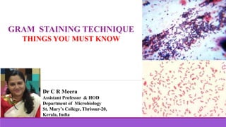
- 1. GRAM STAINING TECHNIQUE THINGS YOU MUST KNOW Dr C R Meera Assistant Professor & HOD Department of Microbiology St. Mary’s College, Thrissur-20, Kerala, India
- 2. Gram Staining Technique, Dr C R Meera, Assistant Professor & HOD, Dept. of Microbiology, St Mary’s College, Thrissur-20, Kerala. • Developed in 1880 by the Danish bacteriologist Christian Gram "I have therefore published the method, although I am aware that as yet it is very defective and imperfect; but it is hoped that also in the hands of other investigators it will turn out to be useful." Gram Staining Technique
- 3. • Most important and widely used differential staining in Microbiology • Bacteria can be differentiated into two major groups called Gram positive and Gram negative bacteria. • 24 hr. old cultures are usually used for gram staining Gram Staining Gram Staining Technique, Dr C R Meera, Assistant Professor & HOD, Dept. of Microbiology, St Mary’s College, Thrissur-20, Kerala.
- 4. • Why bacteria stain differently in Gram Staining? • The difference in the chemical and physical nature of the bacterial cell wall • G –ive cell wall is thin, complex, multi-layered, relatively high lipid contents and low peptidoglycan content. • G +ive cells have less lipid and thick peptidoglycan layer. Gram Staining Technique, Dr C R Meera, Assistant Professor & HOD, Dept. of Microbiology, St Mary’s College, Thrissur-20, Kerala.
- 5. • What is Gram Staining? • A type of differential staining • Four reagents & Four steps 1. Primary stain - Crystal Violet 2. Mordant-Gram’s Iodine 3. Decolorizing agent- 95% ethanol or ethanol-acetone 4. Counter stain or secondary stain –Safranin • G +ive cells- appear Violet in colour • G –ive cells- appear Red in colour Image courtesy: laboratoryinfo.com Gram Staining Technique, Dr C R Meera, Assistant Professor & HOD, Dept. of Microbiology, St Mary’s College, Thrissur-20, Kerala.
- 6. • The heat fixed smear treated with the primary stain called Crystal violet 30 sec • Crystal violet - A basic dye and function is to impart its colour to all cells • At this stage all the organisms appear violet in colour. • First step in Gram Staining Gram Staining Technique, Dr C R Meera, Assistant Professor & HOD, Dept. of Microbiology, St Mary’s College, Thrissur-20, Kerala.
- 7. • Second step in Gram Staining • Smears are treated with Gram’s Iodine • Gram’s Iodine - acts as the killing agent as well as the mordant • Mordant- a substance that increases the cells’ affinity for a particular stain • It binds with the primary stain and forms an insoluble crystal violet- iodine (CV-I) complex • All cells appear violet or purple at this stage. Gram Staining Technique, Dr C R Meera, Assistant Professor & HOD, Dept. of Microbiology, St Mary’s College, Thrissur-20, Kerala.
- 8. • Third step in Gram Staining • The smear is treated with the decolorizing agent, like 95% ethanol or ethanol-acetone solution • Add ethyl alcohol drop by drop, until no more colour flows from the smear • Excess decolourization will make G +ive organisms lose stain and give false results • G -ive bacteria lose the CV-I complex, whereas G +ive cells retain the same Gram Staining Technique, Dr C R Meera, Assistant Professor & HOD, Dept. of Microbiology, St Mary’s College, Thrissur-20, Kerala.
- 9. • Third step in Gram Staining (Conti..) • Decolorizing agents act as both lipid solvent and dehydrating agent • G-ive bacteria, the decolorizing agent dissolves the higher amount of lipids leading to the formation of large number pores in the cell wall • Dehydration and flattening of the cell wall proteins is taking place, but do not close the pores on the cell wall appreciably as numerous pores • Through these pores the CV-I complex escape and the cells become colourless • G +ive cell walls are thick and chemically simple, composed mainly of protein and cross-linked polypeptides • Lipid is dissolved and few pores are produced • Protein dehydration causes closure of cell wall pores, preventing the loss of CV-I complex Gram Staining Technique, Dr C R Meera, Assistant Professor & HOD, Dept. of Microbiology, St Mary’s College, Thrissur-20, Kerala.
- 10. • Also peptidoglycan content plays an important role in this step • G +ive bacteria peptidoglycan content is high which is cross linked well • Porosity is less to allow the escape of the CV-I Complex • G -ive bacteria, peptidoglycan content is less and are poorly cross-linked • Hence more porosity and CV- I complex can escape easily • Third step in Gram Staining (Conti..) Gram Staining Technique, Dr C R Meera, Assistant Professor & HOD, Dept. of Microbiology, St Mary’s College, Thrissur-20, Kerala.
- 11. • Smear is treated with the counter stain or secondary stain called Safranin for 30 sec • Counterstain - a basic dye having a different colour from that of the primary stain Crystal Violet • The G -ive organisms take up this red dye through the pores created by decolorizing agents and appear red in colour • The G +ive organisms which did not lose CV-I complex will not take up the secondary stain and remain violet in colour • Fourth step in Gram Staining Gram Staining Technique, Dr C R Meera, Assistant Professor & HOD, Dept. of Microbiology, St Mary’s College, Thrissur-20, Kerala.
- 12. 1. Gram Positive cells- walls retain Crystal Violet and appear deep violet in colour 2. Gram Negative cells- do not retain Crystal Violet and hence take up safranin 3. Gram non reactive organisms- do not stain or which stain poorly Atypical bacteria remain colourless to Gram staining procedure. Egs:Organisms under Chlamydiaceae and the Mycoplasmataceae (including mycoplasma) Rickettsiaceae which are actually G-ive, but too small to stain well by the procedure 4. Gram variable organisms - which stain unevenly during Gram staining Gram variable reaction - Very old cultures of Gram Positive bacteria Changes in the environment of the organism Slight changes in the staining technique Gram staining procedure divide the bacteria into 4 groups Gram Staining Technique, Dr C R Meera, Assistant Professor & HOD, Dept. of Microbiology, St Mary’s College, Thrissur-20, Kerala.