1. Case scenario: 1
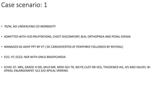
• 70/M, NO UNDERLYING CO-MORBIDITY
• ADMITTED WITH H/O PALPITATIONS, CHEST DISCOMFORT, BLN, ORTHOPNEA AND PEDAL EDEMA
• MANAGED AS ADHF PPT BY VT ( DC CARDIOVERTED AT PERIPHREE FOLLOWED BY REFERAL)
• ECG: VT, ECG2: NSR WITH SINUS BRADYCARDIA
• ECHO: EF: 48%, GRADE III DD, MILD MR, MOD-SEV TR, NO PE,CLOT OR VEG, THICKENED IAS, IVS AND VALVES. BI-
ATRIAL ENLARGEMENT. GLS S/O APICAL SPARING
2. •CAG: N
•INVESTIGATIONS: N HEMOGRAM, CREAT: 2.18, ALP: 125, NT- PRO BNP: 470.45
•MYELOMA PROFILE: NEGATIVE
•FAT PAD BIOPSY : POSITIVE FOR AMYLOID ON CONGO RED STAIN
•CMR: REVERSE NULLING PATTERN OF MYOCARDIUM AND BLOOD POOL, SUBENDOCARDIAL LGE, EDV : 126 ML, EF 68%
, BAE, LV SEPTUM : 13 MM
3.
4.
5.
6.
7.
8.
9. Case scenario II
• 42/M, INITIALLY PRESENTED TO SMHS WITH HISTORY OF CHEST PAIN (
ATYPICAL), POSITIVE TROPONINS, ECG : LOW VOLTAGE COMPLEXES IN LIMB
LEASDS.
• LABELLED NSTEMI, CAG: NON-OBSTRUCTIVE CAD
• PRESENTED TO GE WITH H/O DIARHHEA, WEIGHT LOSS, ABDOMINAL
SWELLING.
• INVESTIGATIONS REVEALED TRANSAMINITIS. ASCITIC FLUID ANALYSIS REVEALED
LYMPHOCYTIC EXUDATIVE PICTURE, NORMAL ADA AND ALB: 18. CRP: 18.8, Ig
ATTG: <0.1, STOOL CALPROTECTIN: NEGATIVE.
• CT ENTEROGRAPHY: F/O MALABORPTION SYNDROME
• USG: CONGESTED IVC WITH MILD PERICARDIAL EFFUSION
22. INTRODUCTION
DEFINITIONS AND CLASSIFICATION
CLINICAL FEATURES AND DIAGNOSIS OF AMYLOID HEART DISEASE
OUTCOMES AND PROGNOSIS
LABORATORY EXAMINATION AND BIOMARKERS
TREATMENT
SUMMARY AND FUTURE DIRECTIONS
23. INTRODUCTION
• Systemic process resulting in extracellular deposition of abnormal insoluble fibrils
• Derived from a variety of serum proteins
• Can affect one or more organs
• Results in organ dysfunction due to tissue infiltration
• considered a rare disease, recent data suggest that cardiac amyloidosis is
underappreciated as a cause of common cardiac diseases or syndromes
Falk RH, Circulation 2005, 2011
24. • light microscope: eosinophilic amorphous substance in hematoxylin–
eosin stained sections.
• polarized light: binds Congo red dye and when stained produces
apple green birefringence, which is used as ‘‘gold’’ standard in
diagnosis
26. • The affinity to Congo red dye is caused by special b-plated sheet confirmation of
amyloid, as could be seen by X-ray crystallography.
• Ultrastructurally, randomly oriented fibrils with a diameter of 7.5–10 nm can be
shown by electron microscopy
• Thioflavin T is another molecule which binds amyloid fibrils but is less frequently
used than Congo red.
27.
28. INTRODUCTION
DEFINITIONS AND CLASSIFICATION
CLINICAL FEATURES AND DIAGNOSIS OF AMYLOID HEART DISEASE
OUTCOMES AND PROGNOSIS
LABORATORY EXAMINATION AND BIOMARKERS
TREATMENT
SUMMARY AND FUTURE DIRECTIONS
29. Types of Amyloidosis and heart involvement
• Based on the spectrum of involved organs
• Systemic amyloidosis - different organs and tissues
• Localized forms - deposits present in only one particular tissue or
organ.
• The heart is frequently the predominant organ affected
• Amyloid deposition - all anatomical distributions, including the atria,
ventricles, and perivascular space as well as valves and conduction
system.
• Amyloid cardiomyopathy – prognosis.
30. Types of cardiac amyloidosis
• > 30 proteins are known to be capable of aggregating as amyloid in vivo
• 9 amyloidogenic proteins accumulate in the myocardium to cause significant
cardiac disease
• >98% of currently diagnosed cardiac amyloidosis results from fibrils composed of
monoclonal immunoglobulin light chains (AL) or transthyretin (ATTR), either in its
hereditary (ATTRv) or acquired (ATTRwt)
31.
32. Light chain amyloidosis ( AL)
• Light chain (AL) amyloidosis is the most commonly diagnosed form of
amyloid disease in developed countries.
• slight predominance of men over women
• usually diagnosed age of 55–60 years
• Associated with
• B cell lymphoproliferative disorders (Myeloma)
• Malignant lymphomas
• Macroglobulinemia
33. • Monoclonal plasma cell population produces abnormal monoclonal light
chains or more frequently their fragments which misfold to form amyloid.
• In vast majority of cases (69%), there is a systemic organ involvement in AL
amyloidosis.
• Kidneys (74%)
• Liver in 27%
• Peripheral nerves in 22%
• Autonomic nerves in 18%
35. Transthyretin (TTR) Cardiac Amyloidosis
There are 2 forms of disease:
1. Wild type amyloidosis or senile systemic amyloidosis (ATTRwt)
2. Familial (Hereditary) amyloid cardiomyopathy (hATTR or ATTRm)
Ruberg FL, Berk JL, Circulation 2012
36. Mutant TTR
monomers
misfold
Formation of
insoluble amyloid
fibrils and amyloid
deposition in the
nerves, heart, and
other tissues
Mutant TTR
monomers
aggregate
TTR gene mutation
destabilizes the
tetramer →
dissociation
Identical TTR
monomers are
synthesized in the
liver and form
tetramers
TTR tetramers
ATTR Amyloidosis
1. Hawkins et al. Ann Med 2015;47:625–38; 2. Hanna. Curr Heart Fail Rep 2014;11:50–7; 3. Damy et al. J Cardiovasc Transl Res 2015;8:117–27
41. Familial Amyloidosis (Hereditary amyloidosis)
• Autosomal dominant diseases with variable penetrance
• Mutation in transthyretin (mTTR) –
• methionine for valine at position 30,
• isoleucine for valine at position 122 (4 % African americans; severe
cardiomyopathy with no neuropathy)
• Transthyretin - synthesized in liver with small portion in choroid plexus.
• middle or old age
• male to female ratio of 50:50
• associated with cardiac and/or nervous system involvement
• carpal tunnel syndrome may be an early sign of the disease.
42. Familial Amyloidosis (Hereditary amyloidosis)
• Neuropathy - progressive sensorimotor and/or autonomic neuropathy
and its clinical aspects predominate in majority of patients.
• Although cardiomyopathy is usually severe even before heart failure
symptoms develop, it may not be diagnosed if the leading clinical
manifestation is neuropathy and echocardiography is not performed.
• Definite diagnosis - Endomyocardial biopsy.
43. Familial Amyloidosis (Hereditary amyloidosis)
• Rare effects of familial non-transthyretin amyloidoses are caused by
mutations in genes coding for fibrinogen, gelsolin, lysozyme and
apolipoproteins A1 and A2.
• Fibrinogen and apolipoprotein mutations lead predominantly to
amyloid renal disease.
• Gelsolin mutations, which are endemic in Finland and occur
sporadically worldwide, are almost exclusively associated with cardiac
conduction system disorders
44. Senile Systemic Amyloidosis
• Wild type TTR/prealbumin (wTTR) represents the precursor protein of
this type of amyloidosis.
• Exclusively men older than 65 years.
• Deposition of wTTR occurs predominantly in the heart
45. Senile Systemic Amyloidosis
• Systemic disease with deposits being found in the gastrointestinal tract,
liver, spleen, bone marrow, tongue and endocrine glands, other clinically
significant manifestations than cardiac amyloidosis and carpal tunnel
syndrome are very rare.
• Presenting features almost always symptoms of congestive heart failure
and final diagnosis is usually based on positive endomyocardial biopsy.
• According to autopsy studies, 22–36% of individuals older than 80 years
will have demonstrable amyloid deposits in the heart, but in the amount
not sufficient to cause apparent myocardial dysfunction
46. Isolated atrial amyloidosis (IAA)
• Atrial natriuretic peptide is a precursor protein for amyloid formation
and deposition, which occurs only in atria.
• This disease is the representative of a true localized form of
amyloidosis not affecting any other organ
• Almost always diagnosed only by autopsy as performing
endomyocardial biopsy from the thin atrial wall is associated with
unacceptably high risk of wall perforation.
47. Isolated atrial amyloidosis (IAA)
• Usually a disease of elderly women
• The prevalence of IAA increases with age and reaching 95% in subjects 81–
90 years of age according to one autopsy study.
• In spite of its high prevalence IAA does not represent clinically significant
type of amyloidosis, not-being usually responsible for eventual heart
failure.
• However, some studies suggest its potential role in the development of
atrial conduction defect and atrial fibrillation in older patients
48. Secondary systemic (AA) amyloidosis
• Infrequent complication of chronic inflammatory states like
rheumatoid arthritis, inflammatory bowel diseases, familial
Mediterranean fever or chronic infective conditions such as
tuberculosis.
• The amyloid fibrils are made of acute phase reactant protein, serum
amyloid A (SAA), which is synthetized by liver.
49. Secondary systemic (AA) amyloidosis
• Renal disease represents the major clinical feature of AA amyloidosis.
• Although myocardial deposits may be often found in histology,
clinically apparent cardiac involvement is very rare, occurring in about
2% of the affected subjects
50. Definition of cardiac amyloidosis. Diagnostic criteria
• diagnosed when amyloid fibrils are found within cardiac tissue.
• invasive and non-invasive diagnostic criteria have been proposed.
• Invasive diagnostic criteria apply to all forms of cardiac amyloidosis whereas non-
invasive criteria are accepted only for ATTR
51.
52. INVASIVE CRITERIA
confirmed when an endomyocardial biopsy demonstrates amyloid deposits after Congo red
staining irrespective of the degree of left ventricular (LV) wall thickness.
Identification of amyloid should be followed by classification of the amyloid fibril protein.
the gold standard for defining the type of amyloid remains mass spectrometry,
immunohistochemistry or immunoelectron microscopy
53. also confirmed if amyloid deposits within an extracardiac biopsy are accompanied either by
characteristic features of cardiac amyloidosis by echocardiography
not yet externally validated
a score ≥8 points in the presence of LV wall thickness ≥12 mm in combination with amyloid
deposits in an extra-cardiac biopsy could also be considered diagnostic of cardiac amyloidosis
54.
55. Non-invasive diagnostic criteria
• Cardiac ATTR amyloidosis can be diagnosed in the absence of histology
• Typical echocardiographic/ CMR findings when 99mTc-pyrophosphate (PYP),
99mTc-3, 3-diphosphono-1,2-propanodicarboxylic acid (DPD) or 99mTc-
hydroxymethylene diphosphonate (HMDP) scintigraphy shows grade 2 or 3
myocardial uptake of radiotracer
• Exclusion of Clonal dyscariasis using serum free light chain (FLC) assay, serum
(SPIE) and urine (UPIE) protein electrophoresis with immunofixation
• In the absence of a detectable monoclonal protein and/or an abnormal serum
FLC ratio, the specificity of grade 2 or 3 bone scintigraphy for cardiac ATTR when
the disease is suspected has been proposed to be almost 100%
56. Once cardiac ATTR amyloidosis is
confirmed, genetic counselling and
testing should be performed to assess
for the presence of TTR mutations in
order to differentiate between ATTRwt
and ATTRv
57.
58. Essential concepts
Although nine types of cardiac amyloidosis are known, AL and ATTR currently account for the
vast majority of cardiac amyloidosis.
Both invasive and non-invasive diagnostic criteria are accepted to diagnose cardiac amyloidosis.
While invasive diagnostic criteria apply to all forms of cardiac amyloidosis, non-invasive criteria
are accepted only for ATTR.
59. INTRODUCTION
DEFINITIONS AND CLASSIFICATION
CLINICAL FEATURES AND DIAGNOSIS OF AMYLOID HEART DISEASE
OUTCOMES AND PROGNOSIS
LABORATORY EXAMINATION AND BIOMARKERS
TREATMENT
SUMMARY AND FUTURE DIRECTIONS
60. Clinical presentation and diagnosis of amyloid heart disease
• Cardiac involvement - very ominous prognosis in almost all types of
amyloidosis
• Early diagnosis of amyloid heart disease - crucial with respect to
subsequent management of affected individuals including the choice of
therapeutic strategy as well as its efficiency, especially in AL amyloidosis.
• With an earlier diagnosis and less cardiac involvement, more aggressive
treatment can be employed resulting in better long-term outcome for the
patient
61. Amyloidosis is a Multisystem Disease
CNS MANIFESTATIONS
• Progressive dementia
• Headache
• Ataxia
• Seizures
• Spastic paresis
• Stroke-like episodes
AUTONOMIC NEUROPATHY
• Orthostatic hypotension
• Recurrent urinary tract
infection (due to urinary retention)
• Sexual dysfunction
• Sweating abnormalities
CV MANIFESTATIONS
• Conduction blocks
• Cardiomyopathy/cardiac hypertrophy
• Systolic and/or diastolic dysfunction
• Atrial and/or ventricular arrhythmias
• Mild valvular regurgitation
• Shortness of breath
• Fluid retention (edema, ascites)
PERIPHERAL SENSORY MOTOR NEUROPATHY
• Neuropathic pain
• Altered sensation (i.e., change
in sensitivity to pain and temperature)
• Numbness and paresthesia
• Muscle weakness
• Impaired balance
• Difficulty walking
RENAL
• Proteinuria
• Renal failure
CARPAL TUNNEL SYNDROME
OCULAR MANIFESTATIONS
• Vitreous opacification
• Glaucoma
• Abnormal conjunctival vessels
• Papillary abnormalities
GI MANIFESTATIONS
• Nausea and vomiting
• Changes in GI motility
(i.e., diarrhea, constipation, gastroparesis,
early satiety)
• Unintentional weight loss
Conceição et al. J Peripher Nerv Syst 2016;21:5–9
63. Diagnosis of cardiac amyloidosis
• two critical phases: (i) suspicious phase, and (ii) definite diagnosis phase.
• The latter phase also includes appropriate typing of the amyloid, which is critical
to guide specific treatment.
• When to suspect cardiac amyloidosis?
typically appears within a constellation of extracardiac signs and symptoms
these signs and symptoms are termed ‘red flags’
64.
65. Red flag for Cardiac Amyloidosis
Shah KB et al., Circ HF 2016
68. Diagnostic algorithm
• large majority of cases of cardiac amyloidosis are AL and ATTR,
• we propose a diagnostic algorithm focusing on identifying these subtypes by the initial use of 99mTc-
PYP, DPD or HMDP scintigraphy coupled to assessment for monoclonal proteins by SPIE, UPIE and
quantification of serum FLC
• The results of these tests could lead to four scenarios
1. Scintigraphy does not show cardiac uptake and assessments for monoclonal proteins are negative
2. Scintigraphy shows cardiac uptake and assessments for monoclonal proteins are negative.
3. Scintigraphy does not show cardiac uptake and at least one of the monoclonal protein tests is
abnormal
4. Scintigraphy shows cardiac uptake and at least one of the monoclonal protein tests is abnormal
69. INTRODUCTION
DEFINITIONS AND CLASSIFICATION
CLINICAL FEATURES AND DIAGNOSIS OF AMYLOID HEART DISEASE
OUTCOMES AND PROGNOSIS
LABORATORY EXAMINATION AND BIOMARKERS
TREATMENT
SUMMARY AND FUTURE DIRECTIONS
70. Outcome and prognosis
• focus has moved to multiparametric biomarker-based prognostic scores, and biomarker-based
staging systems have been developed for AL and ATTR cardiac amyloidosis
• provide an initial prognostic stratification
• prognostic impact of any change of the scores during follow-up has not yet been validated
71.
72.
73. no studies have yet addressed the optimal follow-up scheme in patients with cardiac amyloidosis,
a common scheme consists of 6-month visits with electrocardiogram (ECG) and complete blood tests (including NT-proBNP and
troponin) and yearly echocardiogram and 24 h Holter ECG.
assessment of penetrance in allele carriers is generally recommended to start ∼10 years prior to the age of disease onset in
affected members of the family (or other individuals with the same mutation), or as soon as symptoms compatible with
amyloidosis develop
74. INTRODUCTION
DEFINITIONS AND CLASSIFICATION
CLINICAL FEATURES AND DIAGNOSIS OF AMYLOID HEART DISEASE
OUTCOMES AND PROGNOSIS
LABORATORY EXAMINATION AND BIOMARKERS
TREATMENT
SUMMARY AND FUTURE DIRECTIONS
75. Laboratory examination and biomarkers
• Standard blood tests represent an essential part of examinations in suspected
amyloidosis and shall include the assessment of urea and creatinine levels, liver
enzymes, glucose, thyroid function tests, CRP, full blood count and blood clotting tests
together with the analysis of serum and urine for the presence of an abnormal
monoclonal immunoglobulin.
• Unfortunately, standard serum protein electrophoresis does not detect a monoclonal
band in many cases of AL amyloidosis as the amount of paraprotein is small.
• Immunofixation is more sensitive than plain electrophoresis and should always be
performed.
• in about 20% of affected subjects the circulating paraprotein will not be detected with
immunofixation
76. • serum immunoglobulin free light chain (FLC) assay has become most useful laboratory
method not only for establishing the diagnosis, but also for prognostic and follow-up of
AL amyloidosis.
• The quantitative analysis of k and l FLCs levels is declared to be 10 times more sensitive
than immunofixation electrophoresis for the detection of circulating paraprotein.
• Normal FLCs values basically disprove the diagnosis of AL amyloidosis.
• Importantly, the ratio of k to l should always be assessed.
• With renal impairment, serum levels of both k and l FLCs are increased and their ratio
remains unchanged.
77. • On the contrary, monoclonal production of paraprotein k or l significantly alters FLC
ratio.
• Compared to multiple myeloma, the predominance of l over k chains is more
common in AL amyloidosis leading to l to k ratio approximately 3:1.
• When examining monoclonal protein one must be aware that abnormal FLC assay
is not specific for AL amyloidosis.
• Monoclonal FLCs are found in about 50% of patients with monoclonal gammopathy
of unknown significance (MGUS), which is present in 5–10% of elderly individuals.
• Nevertheless, the FLC ratio is generally much less abnormal in MGUS than in AL
amyloidosis.
• Of course, all patients with multiple myeloma express monoclonal FLCs.
78. • Cardiac biomarkers represented by troponins and N-terminal of BNP (NT-proBNP)
are elevated in patients with amyloid heart disease.
• In AL amyloidosis, NT-proBNP levels are often elevated disproportionately to the
severity of symptoms of congestive heart failure.
• elevation of NT-proBNP levels is not only a result of heart failure but may also
reflect hormone production by myocytes that are compressed by extracellular
amyloid deposits.
79. • Troponins and NT-proBNP or BNP: important prognostic information in AL
amyloidosis
• used for staging the severity of organ involvement in AL amyloidosis and in
connection with this for stratification of patients regarding their suitability and
risk for high dose chemotherapy and autologous stem cell transplantation
(ASCT).
• useful for monitoring of disease progression and response to therapy.
80. Lab diagnosis
• SPEP/UPEP
• Immunofixation
• Serum FLC assay – quantifies k and lambda free light chains
• Lambda : kappa 3:1 (Multiple myeloma 2:3)
• Bone marrow biopsy – presence of Multiple myeloma
• Plasma cell number and degree of clonality, quantity of light chain
and light chain isotype – Prognosis.
81. Extracardiac and endomyocardial biopsy
• diagnosis requires histologic demonstration of amyloid deposits and subtyping of
amyloid.
• For safety reasons, extracardiac biopsy shall be the first step.
• specimen taken either non-specifically from rectal mucosa or abdominal fat
presumably involved organ or tissue like kidney, liver or peripheral nerve.
• The positivity of extracardiac biopsy together with the presence of typical
echocardiographic or MRI findings, especially in otherwise unexplained thickening
of LV walls, makes the diagnosis of amyloid heart disease almost certain and
cardiac biopsy is not necessary
82. • applies almost exclusively for the diagnosis of AL amyloidosis because of its common
systemic nature and in the case of rarely performed peripheral nerve biopsy also for
familial mTTR amyloidosis.
• If extracardiac biopsy is negative and the results of other examinations are suggestive
for amyloid cardiomyopathy, then endomyocardial biopsy must be performed.
• The presence of amyloid deposits in any biopsy specimen is firstly confirmed or
excluded by Congo red dye and in the case of positivity with apple-green birefringence
when placed under polarized light.
• other techniques : modified Sirius red staining and electron microscopy examination is
usually performed
83. • necessary to determine the type of amyloid as this determines the treatment.
• classification of amyloid based on the immunohistolabeling techniques with a
panel of antibodies against known amyloidogenic proteins (usually initial and
secondary panel).
• Immunoperoxidase staining used with formalin-fixed paraffin embedded tissue
and immunofluorescence ( preferred) staining typically performed on fresh frozen
tissue are used.
• For AL amyloidosis bone marrow biopsy to exclude the coexistence of multiple
myeloma.
85. Scintigraphy
• Radiolabelled serum amyloid P component (SAP) scintigraphy is a method
capable of evaluating the whole-body-amyloid burden.
• Serum amyloid P component is a plasma glycoprotein of the pentraxin
family which is specifically concentrated in amyloid deposits of all types.
• If 123 I-labelled P component is supplied intravenously it distributes
between the circulating and the amyloid-bound SAP pools in proportion to
their size and can be imaged and quantified on a gamma camera.
86. • provides information on the distribution as well as the extent of amyloid deposits
throughout the body.
• regression of amyloid infiltration may be documented by SAP scintigraphy when the
supply of the culprit amyloid precursor protein is significantly reduced.
• SAP scintigraphy is not able to image in the moving heart due to blood pool uptake.
• Another disadvantage : possible infectious risk as SAP is obtained from blood donors.
• used only in few centers, mainly in the Great Britain.
• 99m-Tc-aprotinin has been shown to be fairly specific for cardiac amyloidosis, the
experience with its use is limited
90. • Perugini et al. demonstrated, that 99mTc- 3,3-diphosphono-1,2-
propanodicarboxylic acid, a bone scanning agent, is significantly taken up by the
myocardium infiltrated by transthyretin amyloid.
• dicarboxypropane diphosphonate (DPD) scintigraphy serves a as a very elegant
noninvasive test for the presence of transthyretin cardiac amyloidosis and help to
differentiate it from AL amyloidosis, in which myocardial uptake of DPD is
minimal
91. Electrocardiogram
• In 46–71%, typical picture of low voltage of QRS complexes defined as QRS voltage amplitude
<0.5 mV in all limb leads or < 1.0 mV in all precordial leads.
• This contrasts with marked increase in LV wall thickness seen by imaging techniques, which is,
however, caused by extracellular amyloid deposition and not a result of true myocyte
hypertrophy as in hypertensive heart disease or hypertrophic cardiomyopathy.
• the combination of increased LV wall thickness on the echocardiogram with low ECG voltage is
highly suggestive for infiltrative process, especially AL or familial amyloidosis.
92. • Pseudo-infarct pattern may be present in half of affected subjects, most
often in anterior precordial leads.
• Both low ECG voltage and pseudo-infarct pattern are expressed in about
25% cases.
• sinus rhythm is present in majority of patients, although atrial fibrillation
and conduction system disease may occur.
93. • patients with senile systemic amyloidosis may have normal ECG voltage, often
with left anterior hemi-block pattern, and atrial fibrillation and flutter are
common.
• conduction disorders requiring an implantation of permament pacemaker are
frequent in these individuals.
• In patients with AL cardiac amyloidosis, signal-averaged ECG is often abnormal
and heart variability on 24-h ECG monitoring is reduced, both of which may be
related to the risk of sudden death
96. Right heart catheterization
• invasive cardiologic examination is currently performed only when
endomyocardial biopsy is indicated or in rare unclear cases, typically in order to
differentiate constrictive pericarditis as the other cause of congestive heart
failure.
• In typical advanced stages of amyloid cardiomyopathy, left and right heart
catheterization confirms the presence of restrictive hemodynamics.
97. • This is characterized by rapid and sustained elevation of diastolic ventricular
pressures (so called dip-and-plateau configuration or ‘‘square root sign’’) with
equalization of ventricular and atrial pressures at end-diastole.
• Similar pattern is also seen in constrictive pericarditis.
• However, the difference between left and right ventricular end-diastolic
pressures is less than 5 mmHg, right ventricular systolic pressure is less than
50mmHg and the ratio of right ventricular end-diastolic to systolic pressure is
more than 1/3 all favors constriction.
98. • The most important hemodynamic sign, that differentiates restriction
from constriction, is concordant behavior of systolic ventricular
pressures during respiration in restriction, while in constriction
respiratory discordance of left and right ventricular pressure curves is
seen in systole due to increased ventricular dependence.
99. Echocardiography
• Echocardiography provides comprehensive morphological and functional assessment of
the heart.
• ‘‘classical’’ features of amyloid cardiomyopathy are present only in advanced phases of
the diseases.
• wide spectrum of echocardiographic findings
• none is itself specific and thus they should be interpreted in the context of the clinical
picture and other investigations.
• Echocardiography cannot confirm diagnosis in isolation and also is not able to
distinguish between various types of amyloidosis
100. • Amyloid infiltration typically leads to the thickening of ventricular
walls .
• Usually there is a concentric pattern of increased wall thickness but
sometimes, at early stages, only interventricular septum may be
thickened.
• Because LV cavity is not dilated, the term ‘‘concentric hypertrophy’’ is
incorrectly used as the pathological process is amyloid deposition, not
myocyte hypertrophy.
101.
102.
103.
104. • Increased echogenicity of thickened ventricular myocardium, also
referred to as ‘‘granular’’ or ‘‘sparkling’’ appearance, has been
reported in several studies.
• can occur in other causes of LV hypertrophy and its specificity is
probably not sufficiently high.
• seen only on standard echocardiographic imaging, because scanning
with tissue harmonic frequencies imparts increased echogenicity of
myocardium in general
105. • thickened valves and interatrial septum may be noted in some patients,
especially in more advanced disease.
• The thickening of valve leaflets does not lead to hemodynamically significant
regurgitant lesions.
• some case reports in the literature on the occurrence of systolic anterior motion
of anterior mitral leaflet in amyloid cardiomyopathy, resulting in dynamic LV
outflow obstruction; however, this phenomenon is definitely exceptional in this
disease.
106. • Pericardial effusion is found in 40–60% of patients.
• It is usually small and reflects high right atrial pressure although
pericardial infiltration by amyloid may also play a role.
107.
108. • The dilatation of atria caused by high ventricular filling pressures is
often present, but it is not specific for amyloid cardiomyopathy.
• thrombi may form within the atria due to their standstill.
• High LV filling pressures lead to significant postcapillary pulmonary
hypertension and this may result in right ventricular cavity
enlargement.
109. • Diastolic dysfunction is the hallmark of amyloid heart disease.
• Traditionally, restrictive pattern of left ventricular filling indicating
increased ventricular stiffness with high filling pressures is regarded
as pathognomonic for amyloid cardiomyopathy (Fig. 4).
• severe impairment of diastolic function is present in advanced stages
of myocardial infiltration.
110.
111. • Mild to moderate LV diastolic dysfunction may be detected earlier in the
course of the disease.
• absence of restrictive filling pattern type as such should not lead to exclude
the diagnosis of amyloidosis if other echocardiographic and clinical
features and investigations, for example low voltage ECG, are present.
• according to the current European classification of cardiomyopathies,
amyloid cardiomyopathy may be regarded either as hypertrophic or
restrictive cardiomyopathy based on the severity of diastolic filling
impairment
112. • Global LV systolic function as assessed by ejection fraction is usually
normal until the more severe stages of the disease are present.
• However, longitudinal contractile dysfunction that can be evaluated
either by mitral annular displacement or velocity or by deformation
imaging is present early in the course of amyloid cardiomyopathy.
• Importantly, decreased longitudinal contractile function of LV walls is
disproportionately severe compared to other types of LV hypertrophy
with preserved ejection fraction
130. Echo - Summary
• Echocardiography can provide many features suggesting amyloid
heart disease though none of them are absolutely specific.
• The presence of several echocardiographic findings increases the
likelihood of the diagnosis, especially the combination of markedly
increased walls of nondilated LV with restrictive filling pattern, biatrial
enlargement, thickened valves and pericardial effusion.
• most useful approach is to combine echocardiographic findings with
results of other investigations like ECG and CMR.
131. Magnetic resonance imaging
• In the last few years, CMR has emerged as very useful imaging
modality in the diagnosis of cardiac amyloidosis.
• Compared to echocardiography, CMR more accurately measures the
thickness and volumes of both ventricles.
• It is also able to assess biatrial dilatation, interatrial septal thickness,
the presence of pericardial effusion and, by using phase velocity
mapping, to evaluate LV filling.
132.
133.
134.
135.
136. • major and unique advantage of CMR is the possibility of tissue characterization
by late gadolinium enhancement (LGE).
• First study interested in the use of LGE in patients with cardiac amyloidosis has
suggested the global subendocardial LGE pattern to be pathognomic pattern for
amyloid heart disease
• Nevertheless, other authors have shown that several LGE patterns, localized or
diffuse, and subendocardial or transmural may be present in patients with
amyloid cardiomyopathy.
137.
138.
139.
140.
141.
142. • The histological basis for LGE is interstitial expansion from amyloid infiltration as
demonstrated by Maceira et al.
• The prevalence of LGE in patients with cardiac amyloidosis has been reported
from 69% to 97%.
• global subendocardial to transmural LGE pattern is most common, being present
in 80–85% of affected individuals, and represent unique CMR feature that may
noninvasively ‘‘phenotype’’cardiac amyloidosis.
143. • Myocardial and blood pool kinetics of gadolinium uptake are also unusual in
patients with cardiac amyloidosis with similar myocardial and blood T1 relaxation
values as a result of high myocardial uptake and fast blood pool washout.
• Therefore, blood pool has atypically dark appearance in LGE images and the
selection of the appropriate inversion time may be difficult.
• If suboptimal ‘‘nulling’’ of the hypertrophied myocardium occurs, amyloid
infiltration shall be suspected.
144.
145.
146.
147.
148.
149.
150.
151. • Thickening of the LV wall has of course poor specificity for amyloidosis
as it also occurs in other conditions, such as hypertensive heart
disease, hypertrophic cardiomyopathy and other infiltrative
cardiomyopathies.
• The contemporary presence of RV wall thickening and, more
importantly, the absence of high ECG voltages favors the diagnosis of
infiltrative disorder, of which amyloidosis is the most common
152.
153.
154. INTRODUCTION
DEFINITIONS AND CLASSIFICATION
CLINICAL FEATURES AND DIAGNOSIS OF AMYLOID HEART DISEASE
OUTCOMES AND PROGNOSIS
LABORATORY EXAMINATION AND BIOMARKERS
TREATMENT
SUMMARY AND FUTURE DIRECTIONS
155. General Principles for Treatment of Cardiac
Amyloidosis
• Treatment depends on the identification of the precursor protein
• Diuretics/sodium restriction
• Surveillance for and treatment of atrial/ventricular arrhythmias
• Monitoring for conduction disease
• Avoidance of negative inotropic/chronotropic drugs (beta blockers,
calcium channel blockers)
• Avoidance of vasodilator therapy
156.
157. TREATMENT
Congestive heart failure and other cardiac therapy
• Restricted salt intake and loop diuretics together with aldosterone antagonists like
spironolactone or eplerenone are the mainstay of heart failure therapy.
• In patients with severe congestion and/or nephrotic syndrome, high doses of
diuretics are usually necessary.
• may lead to under-filling of small and stiff LV with further reduction of already
compromised cardiac output and consequently to hypotension, vertigo, syncope as
well as prerenal worsening of renal function.
158. • Hypotension is common especially in AL cardiac amyloidosis.
• This is due to low cardiac output, autonomic neuropathy or to impaired vascular
tone caused by amyloid infiltration.
• Ortostatic hypotension accompanied by peripheral edema may respond to thigh-
high support stockings.
• Midodrine, an a-agonist, can be effective if autonomic neuropathy is present.
• On the other hand, fludrocortizone is not recommended because of its sodium
retaining effect that worsens congestion.
159. • ACE I and ARBS poorly tolerated as they can lead to profound hypotension namely in AL
amyloidosis because vascular tone is disproportionately dependent on angiotensin receptors
due to impaired sympathetic nervous system function in this condition .
• Betablockers: not a standard part of heart failure therapy in cardiac amyloidosis worsen
hypotension and decrease myocardial contractility due to their negative inotropic effect.
• One possible exception for the use of cautiously dosage betablockers represents rate control of
atrial fibrillation.
160. • Recently, an interesting study has been published demonstrating a positive
effect of carvedilol treatment on mTTR deposition in a familial amyloidotic
polyneuropathy mouse model.
• Digoxin and some calcium channel blockers bind to amyloid fibrils thus
increasing susceptibility to digoxin toxicity and to negative inotropic effects
of calcium blockers.
• Therefore, these drugs are contraindicated in cardiac amyloidosis, again
with possible exception for low dose and carefully monitored digoxin
therapy in atrial fibrillation with rapid ventricular response.
161. • Amiodarone: well tolerated when used for rate-control therapy in patients with atrial
fibrillation.
• no solid data exist whether its use in treatment of VT is of any significance in amyloid
heart disease.
• Rhythm-control of atrial fibrillation either with amiodarone or with catheter ablation
techniques is ineffective due to abnormal atrial electrical properties.
• Anticoagulation therapy using warfarin is absolutely indicated in patients with atrial
fibrillation and flutter or after cardioembolic episode.
• anticoagulation shall be administered also in the setting of sinus rhythm, when
echocardiographic signs of minimal or absent atrial mechanical activity are present as
the prevalence of atrial thrombi and the risk of embolic event are considerably high.
162. • The presence of advanced atrioventricular block necessitates the implantation of permanent
pacemaker.
• Although not an indication supported by current guidelines, strong consideration should be given to
biventricular pacing as conventional right ventricular pacing may lead to LV dyssynchrony and thus
further decrease the stroke volume.
• Most cases of sudden cardiac death in amyloidosis are due to electromechanical dissociation and thus
implantation of cardioverter-defibrillator is less successful in prevention of fatal arrhythmic events.
• The study from Mayo Clinic clearly showed that despite high rate of appropriate shocks in patients
with cardiac amyloidosis, mainly AL, this therapy did not lead to survival benefit.
164. Specific treatment of systemic amyloidoses
AL amyloidosis
• Current therapies are targeted to eradicate the pathologic plasma
cells and to eliminate misfolded free light chains.
• The efficacy of a treatment is assessed by hematologic response,
which reflects reduction in the burden of plasma cell disease, and by
organ response representing improvement in the organ function.
• The consensus criteria for hematologic and organ response were
recently updated at the 12th International Symposium on Amyloidosis
and are summarized in Table 2.
165. AL Cardiac Amyloidosis Treatment
Chemotherapy is the cornerstone therapy
Hanna M, Cleveland Clinic Journal of Medicine, 2017
166.
167. AL amyloidosis
• The extent of cardiac involvement is the major determinant of prognosis in
amyloidosis.
• Echocardiographic features like LV wall thickness, severity of diastolic
dysfunction and reduced systolic function are associated with a poor
outcome.
• The prognostic value of new promising parameters derived from
echocardiographic deformation imaging as well as the presence and extent
of late gadolinium enhancement must be confirmed in larger trials.
168. AL amyloidosis
• Cardiac biomarkers, troponins I or T and BNP or NT-proBNP: the most important
predictors of outcome in patients with AL amyloidosis.
• quantitative measure of myocardial damage and natriuretic peptides reflect
cardiomyocyte stress.
• a staging system has been developed.
• The patients are classified to be at stage III if both biomarkers are elevated with
mean survival of 3.5 months, stage II when one of the biomarkers is abnormal
(survival 10.5 months) and stage I characterized by normal levels of troponins or
NT-proBNP (survival 26 months).
• The staging system based on the evaluation of cardiac biomarkers together with
the assessment of FLC levels and markers of plasma cell burden is important in
clinical management with respect to optimization of therapy, monitoring its
success and minimizing its toxicity
169. AL amyloidosis
• Treatment of AL amyloidosis has been largely based on experiences gained with
the therapy of multiple myeloma.
• high-dose melphalan followed by autologous stem cell transplantation
(HDM/SCT) represent a standard front-line therapy for patients with AL
amyloidosis who are suitable candidates for this aggressive treatment.
• Treatment-related mortality in the 90s reached 40% in some centers and
advanced age together with cardiac involvement has been recognized to identify
the most fragile population.
170. AL amyloidosis
• stem cell preparation requires administration of high-dose of granulocyte-colony
stimulating factor, patients with cardiac involvement and autonomic dysfunction
are particularly susceptible to hypotension and fluid shifts induced by this factor.
• patients with cardiac amyloidosis may experience life-threatening arrhythmias
during stem cell infusion presumably related to dimethyl sulfoxide preservative
toxicity.
• The above mentioned cardiac staging system represented a significant
improvement in careful selection of patients for HDM/SCT.
• The data from experienced centers show that hematologic response may be
achieved in more than three quarters of patients including 39% with complete
response and with treatment related mortality about 10%.
171. AL amyloidosis
• Complete hematologic response has been consistently documented
as the strongest predictor of outcome.
• The median survival of patients achieving complete hematologic
response may be more than 10 years compared to a few dozen
months in those with no hematologic response.
• In patients with no or partial response to HDM/SCT, regimens
combining dexamethasone with either thalidomide or bortezomib
have been successfully tested, especially with the latter one
172. Current indication criteria for HDM/SCT
• Age < 65 years
• < 2 organs involved,
• NT-proBNP and troponin I levels < 35 ng/l and <0.1 ug/l,
• LV ejection fraction > 45%,
• Creatinine clearance > 50 ml/min,
• Diffusion lung capacity for carbon monoxide > 50%
• Systolic blood pressure > 90 mmHg.
• That is why only about 25% of patients with Al amyloidosis are
suitable candidates for this effective therapy.
173. • In individuals not eligible for HDM/SCT, melphalan combined with high-dose dexamethasone
(MDex) became a standard of care.
• Studies evaluating this therapeutic regimen in patients ineligible for HDM/SCT demonstrated
that hematologic response was achieved in two thirds of patients and complete hematologic
response in about 30%.
• Similar to HDM/ SCT, the survival was much longer for patients who responded to the therapy.
• Lower response rates and outcome was again documented in subjects with advanced cardiac
involvement, with median survival rate of 10.5 months.
• Current trials are testing the efficacy of MDex combined with the third agent like thalidomide,
lenalidomide or bortezomib.
• Patients with severe cardiac disease may benefit from palliative melphalan therapy.
174. • novel agents have been recently introduced in the treatment of AL
amyloidosis.
• Thalidomide and lenalidomide have been combined with
dexamethasone and with dexamethasone plus melphalan or
cyclophosphamide.
• Hematologic responses have ranged generally between 40% and 50%;
however, considerable treatment toxicity has been documented
including most worrying myelosupression induced by lenalidomide.
175. • Bortezomib, a reversible proteasome inhibitor, has been successfully tested
either as a single agent or in combination with dexamethasone or mephalan
and dexamethasone (BMDex).
• The therapeutic regimens using bortezomib seem to be very promising.
• High rates of hematologic responses as well as organ improvement were
achieved rapidly in conducted trials and overall treatment was safe with mild
to moderate neurotoxicity being the most common adverse effect.
• After being tested in randomized trials, the BMDex regimen has the potential
to become a standard of care for newly diagnosed patients with AL
amyloidosis.
176. Familial amyloidoses
• Congestive heart failure is usually easier to control in patients with
familial amyloidosis as compared to AL amyloidosis.
• Low doses of ACE-inhibitors and b-blockers are better tolerated in
these patients if autonomic neuropathy is not present.
• To date, the only specific therapy for TTR, fibrinogen and
apolipoprotein hereditary amyloidoses is organ transplantation.
177. Familial amyloidoses
• In TTR-associated familial amyloid neuropathy, orthotopic liver
transplantation represents causal and highly effective treatment.
• However, in many patients with non-Val30Met TTR an accelerated
progression of cardiac amyloidosis was observed after liver transplantation
• This is probably due to continued deposition of wTTR in the heart.
• Therefore, combined heart and liver transplantation shall be considered for
patients with familial TTR amyloidosis involving the heart
178. Familial amyloidoses
• New drugs on the basis of small molecule ligands that are able to
stabilize tetrameric structure of TTR are investigated.
• Diflunisal is a non-steroidal anti-inflammatory agent which reduces
tetramer dissociation and subsequent misfolding and amyloid
formation; however, it may worsen renal function and lead to fluid
retention.
• Another drug with similar effect is tafamidis, with which successful
clinical trial that demonstrated the slowing of progression of
neuropathy in ATTR amyloidosis has been recently conducted.
• Currently, multicenter trial exploring the efficacy of tafamidis in
cardiac mTTR amyloidosis is under way.
179. Senile systemic amyloidosis
• Treatment is symptomatic with diuretics being the mainstay.
• Carefully monitored therapy often leads to significant improvement
with long-term freedom from recurrence of congestion.
• Similar to familial TTR amyloidosis, the patients with senile
amyloidosis usually tolerate ACE-inhibitors and low doses of b-
blockers.
• Atrial fibrillation is common and electrical or pharmacological
cardioversion shall be strongly considered as atrial arrhythmia may
lead to substantial worsening of heart failure
180. Senile systemic amyloidosis
• Amiodarone is generally used to maintain the sinus rhythm.
• Anticoagulation is mandatory if atrial fibrillation or flutter persists as the
thromboembolic risk is very high.
• The implantation of permanent pacemaker is common because of
considerable occurrence of advanced conduction system disorder with
high-degree AV blocks.
• The treatment of senile systemic amyloidosis with tafamidis is also being
tested in currently conducted clinical trials.
181. Secondary amyloidosis
• The treatment is primarily orientated to control the underlying
chronic inflammatory disorder.
• Immunomodulating agents like TNFa-inhibitors or IL-1-inhibitors are
increasingly used; however, financial aspects limit greater use of this
therapeutic approach.
• Recently, eprodisate, a molecule that binds to glycosaminoglycan-
binding sites on SAA and inhibits fibril polymerization, has been
successfully tested in randomized clinical trial which has
demonstrated slowing down the progression of renal failure in
patients with AA amyloidosis.
183. Mutant TTR
monomers
misfold
Formation of
insoluble amyloid
fibrils and amyloid
deposition in the
nerves, heart, and
other tissues
Mutant TTR
monomers
aggregate
TTR gene mutation
destabilizes the
tetramer →
dissociation
Identical TTR
monomers are
synthesized in the
liver and form
tetramers
TTR tetramers
Treatment Strategies in TTR Disease
1. Hawkins et al. Ann Med 2015;47:625–38; 2. Hanna. Curr Heart Fail Rep 2014;11:50–7; 3. Damy et al. J Cardiovasc Transl Res 2015;8:117–27
197. INTRODUCTION
DEFINITIONS AND CLASSIFICATION
CLINICAL FEATURES AND DIAGNOSIS OF AMYLOID HEART DISEASE
OUTCOMES AND PROGNOSIS
LABORATORY EXAMINATION AND BIOMARKERS
TREATMENT
SUMMARY AND FUTURE DIRECTIONS
198. Conclusion
• Cardiac amyloidosis may or may not be associated with involvement of other
organs and represents a major negative prognostic factor in all types of
amyloidosis.
• The presence of various typical features derived from imaging techniques and
ECG shall always rise suspicion for the disease and lead to stepwise diagnostic
process that translates into the confirmation of the amyloid heart disease and
assessment of other organ involvement together with precise histological
verification of the type of infiltrating amyloid.
• Early diagnosis is critical especially in most common AL amyloidosis, because
patients with advanced heart disease are not suitable candidates for modern and
effective hematologic treatment regimens including intensive chemotherapy and
autologous stem cell transplantation.
199. Conclusion
• In patients with advanced amyloid cardiomyopathy, diuretics with
aldosterone antagonists remain the mainstay of the therapy.
• Heart transplantation may be offered only to a minority of patients with
isolated cardiac involvement.
• In familial transthyretin amyloidosis, combined heart and liver
transplantation represent a therapeutic method of choice.
• Novel pharmacological agents like tafamidis are being tested in clinical
trials in treatment of transthyretin-related amyloidosis.