
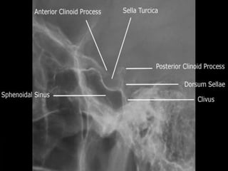
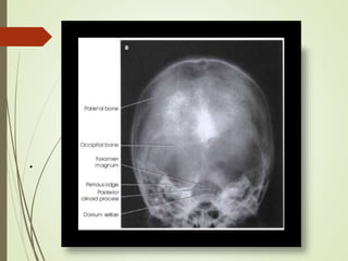
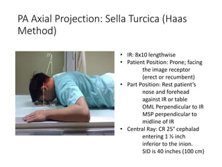
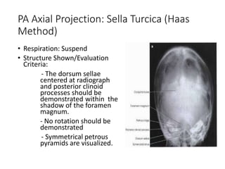

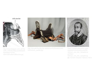
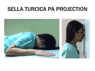
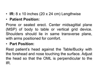
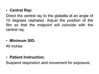
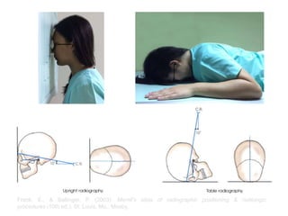
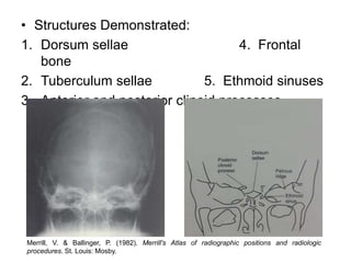
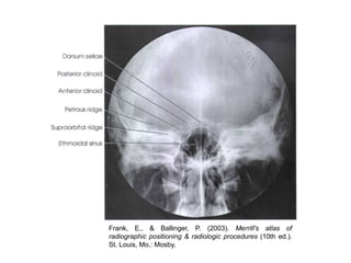
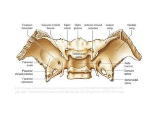
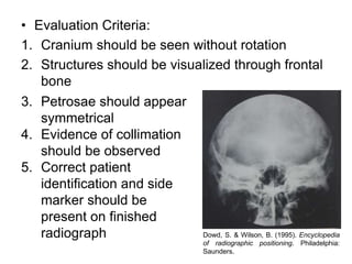


The document provides information about performing a PA projection radiograph of the sella turcica. It states that the patient should be positioned prone with their forehead and nose resting against the image receptor. The central ray should be directed at the glabella at a 10 degree angle cephalad. Structures that should be demonstrated include the dorsum sellae, tuberculum sellae, anterior and posterior clinoid processes, and frontal bone. Evaluation criteria include the cranium being seen without rotation and symmetrical petrous bones.


![Radiography of skull [Autosaved].pptxriuyowioehgg](https://cdn.slidesharecdn.com/ss_thumbnails/radiographyofskullautosaved-251211014507-1d75cfe3-thumbnail.jpg?width=640&height=640&fit=bounds)


