

Recommended
More Related Content
Similar to ffa.pptx
Similar to ffa.pptx (20)
Recently uploaded
Recently uploaded (20)
ffa.pptx
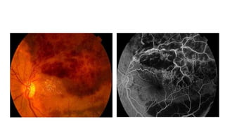
- 2. • Left superior branch retinal vein occlusion. • The fluorescein angiography on the left shows areas of hypofluorescence and hyperfluorescence. • Hypofluorescence - retina haemorrhages and the hard exudates block the choroidal fluorescein causing dark patches • pre-retinal haemorrhages, in addition to blocking the choroidal fluorescein also cover the retina vasculature • Hyperfluorescence - damaged retina veins have exposed collagen which are stained by fluorescein. Leakage around the damaged vein occurs due to damage to the endothelium walls • microaneurysms appear as multiple bright spots.
- 4. • This is the right funuds of a patient with dry form of age-related macular degeneration. The macula shows areas of retinal pigment epithelium (RPE) atrophy. • The fluorescein angiography shows hyperfluorescence in the macula due to RPE window defect allowing choroidal fluorescein to show through brightly.
- 6. • Fluorescein angiography of a diabetic patient with peripheral neovascularization. Hypofluroescence - dot and block haemorrhages Hyperfluorescence - new blood vessels with leakage - microaneurysms