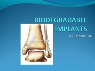
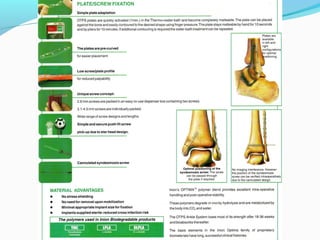
DR.IMRAN JAN
Biodegradable implants are derived by transforming
compounds that are present in nature to structural
plastics.

Organic molecules are polymerized to form strong
fibers and solid compounds.

When these polymers are implanted in patients,
they degrade and are eliminated from the body in a
period of time.
history
The first biodegradable material was made from
animal intestines and used as a suture material by
Galen in 175 BC.
 The first usage of these materials in
orthopaedic surgery was in 1984 by Rokkanen for the
treatment of internal fixation of ankle fractures .
materials
Widely used biodegradable materials include
polyglycolic acid (PGA),
 poly-L- lactic acid (PLLA),
poly-DL-lactic acid (PDLLA), PGA/trimethylene
carbonate compolymers (PGA/TMC),
poly-p-dioxanone (PDS) and
poly-beta-hydroxybutyric acid (PBHBA).
Biodegradable materials need to be hydrolytically
labile and sturdy, at least for a period of time.
To meet these requirements, they are produced by the
“self-reinforcing” (SR)technique .
The biodegradation process takes from one to 6
months starting around 6 onth post op.
 This period depends on contact with body
fluids,temperature, motion, molecular weight, crystal
form and geometry of material, and the tissue that is
implanted.
biodegradation
 begins with the polymer chains being broken into
smaller fragments by hydrolysis--- molecular weight
of the implant decreases --- the mechanical strength
decreases allowing subsequent mechanical
fragmentation and absorption of the implant to begin.
Actual mass loss of the implant occurs through the
release of soluble degradation products,
--phagocytosis by macrophages and histiocytes,
intracellular degradation and finally, metabolic
elimination through the citric acid (Krebs) cycle to
carbon dioxide and water, which are expelled from
the body via respiration and urine.
Clinical applications
 The mechanical properties of the materials permit
them to be used with metaphyseal and peri-articular
fractures where the loading is relatively low.
For treating small-bone fractures such as ankle
fractures .
Not suitable for comminuted and unstable fractures.
Clinical applications
Another suitable anatomic area for application is the
elbow joint, for fixing fractures of the radial head,
olecranon, capitellum and distal humerus.
Comminuted fractures --- not good candidates .
Also #s of the distal radial styloid, patella, glenoid
fossa and acetabulum; osteochondral fractures in the
knee, tibial plateau, phalanx, calcaneus and talus; and
also hallux valgus surgery.
Screws or rods may also be used for epiphyseal #s.
Clinical applications
Other uses--- as interference screws in knee ligament
surgery;
 for the promotion of osteogenesis in bone defects;
for the slow release of antibiotics and
growth factors; as an antiadhesive
membrane for preventing adhesions in flexor tendon
surgery;
as a matrix for cells in cartilage, bone or connective
tissue engineering.
Maintenance of alignment and fixation of bone fractures,
osteotomies, arthrodeses or bone grafts (OTPSTM Pin)
• Maintenance of reduction and fixation of cancellous bone
fractures, osteotomies or arthrodesis of the upper extremity,
ankle and foot (OTPSTM Ankle)
• Bone graft & fragment containment, cement restriction in
total joint arthroplaties, protective barrier for bone graft harvest
sites (OTPSTM Mesh)
• Fixation of non-comminuted diaphyseal fractures of the
metacarpal, proximal phalangeal middle phalangeal and
osteotomies (OTPSTM Hand/Mini)
advantages
 main advantage of biodegradable implants---
secondary operation for removal not necessary, in
contrast to metallic implants, which need to removed
because of osteopenia, corrosion and irritation of
adjacent tissues.
As biodegradable implants degrade, they lose
strength and this puts loading of bone--- prevent
bone resorption.
Will not interfere wth radiological imaging.
disadvantages
Disadvantage ---expensive, having less strength than
metals, tissue reactions, causing osteolysis around the
implant and sterile draining sinuses.
most cases, the symptoms of the tissue reaction are
subclinical and pass unnoticed, but in some patients a
clinically manifest inflammatory foreign-body
reaction ensues.
 synovitis when implant is put intra articularly
Danger of adverse tissue reaction more if the rate of
implant degradation produces more debris particles
than the tissue is able to tolerate. greatest risk when
the gross geometry of the implant is rapidly lost.
(Böstman and Pihlajamäki 2000, Middleton and
Tipton 2000)
 typically manifest in the latter stages of implant
degradation i.e. 6-12 months post operatively. In
children, the tissue reactions appear to be quite rare
(2.1 % of cases with polyglycolide implants) and
always mild in character (Rokkanen et al.
rate of bacterial infections not related to the implant
material, same after implantation of metal and
biodegradable materials. (Sinisaari 2004)
 In studies concerned with polylactide implants, the
adverse tissue reaction rate has usually been lower
than with polyglycolide
All implants cause tissue response but this varies
radically
Phases of Degradation
 Phases of Degradation of Amorphous Biodegradable
Implants and Tissue Reactions According to Pistner et
al.47
1. healing phase: Unchanged implant, development
of a fibrous capsule with a high amount of fibroblasts
2. Latency phase : Unchanged implant, fibrous
capsule gets thinner with less cells and more fibers or
direct implant contact to bone
3. Protracted resorptive phase :Mainly central
degradation of the implant, development of cracks,
mild to moderate cellular response with invasion of
macrophages and foreign-body giant cells
4. Progressive resorptive phase : Progressive
disintegration of the implant with a severe tissue
response (macrophages, foreign-body giant cells)
5. Recovery phase :No polymer remnants detectable,
development of scar tissue or osseous replacement of
the former implant site
Table 2. Classification of Osteolysis (O)
According to Hoffmann et al. and Weiler et
al.46,69
Osteolysis Radiological Findings
O-0 None
O-1 Mild

O - 2 Moderate
O-3 Severe
O-4 Disturbed healing
 No osteolytic changes visible
 Mild Osteolytic changes at the
implant site (osteolysis 1 mm or
larger than implant diameter)
 Moderate Cystic-like extended
osteolysis (osteolysis 3 mm or
larger than implant diameter
 Severe Confluence of osteolysis
into a resorption cavity (if more
than 1 implant is used)
 Disturbed healing Fracture
displacement, fragment
sequestration, or healing failure
of soft tissue due to osteolysis .
Osseous replacement
During or following implant degradation, osseous replacement
may follow 3 different patterns:
1. There is osseous ingrowth while the implant is degrading .
This phenomenon is most desirable but has rarely been found.
it has only been reported to occur during the degradation of
PLLA-co-PDLLA (70:30) or self-reinforced PLLA/PDLLA
composite rods.50,51
2. There is osseous ingrowth in the center of the former implant
site after the implant is degraded .46
3. There is an osseous scaring of the former implant site with a
slow marginal ingrowth of new bone.This kind of replacement
has been found in cases after an osteolytic lesion has occurred
and may progress over several months or years.46
Classification and Treatment of Extra-articular Soft-
Tissue Reactions (EA) According to Hoffmann et al.69
Extra-articular Soft-Tissue
Reactions
Symptoms/Findings/Treatment
 EA-0 None
 EA-1 Mild
 EA-2 Moderate
 EA-3 Severe
 EA-4 Bacterial
superinfection
 No or subclinical reaction
 Local, mild soft-tissue induration; no
treatment
 Fluctuant swelling, fluid
accumulation (ultrasound), local
warmth, reddening, swelling, pain;
single or repetitive puncture
necessary
 Spontaneous discharge of sinus,
primary sterile, secondary possible
bacterial contamination; debridement
and open wound treatment.
 Deep soft-tissue/bone infection
following EA-2 or EA-3; extensive and
repetitive debridement
Fixation of
longitudinal
vertical
meniscus
lesions
located in the
vascularized
area of the
meniscus
INTERFERENCE SCREW
Dr.Cyril G Cherian
A prosthesis is an artificial limb, which is fitted to an
individual who has lost a limb.
A pylon is the member which provides the
connection between the residual limb (leg stump) and
the prosthetic foot.
A method of immediate post op temporary rigid
dressing.
Postoperative management of below-knee
amputation varies widely.
Early rehabilitation oriented immediate post op
management.
 Pressure bandages do not always remain secure, tend
to apply uneven pressure and may increase stump
edema by a tourniquet effect.
Burgess and Romano (1968) recommended the use of
rigid plaster dressings immediately after operation.
Pylon prosthesis
Definite advantages, but there are concerns that it is
technically demanding.
Impractical in hospitals lacking an experienced
orthotist.
Air-splints, Unna paste dressings and removable rigid
dressings recommended, but
most of these also require skilled application, and are
expensive;
some do not allow either full weight-bearing or early
ambulation.
Removable rigid dressings (Wu et al 1979 ; Mueller
1982) shorten rehabilitation while allowing frequent
wound inspection
but they do not permit early weight bearing.
inflatable splints (Sher 1974) ---- this airbag apparatus
holds no advantage over conventional dressings.
The plaster-pylon technique --- advantages of rigid
dressings and early weight bearing.
Plaster technique.
At the completion of the operation,
gauze dressing is covered with a single layer of cotton
roll and a stockinette bandage.
The stump of the limb is lifted from the table using
the free end of the stockinette sleeve.
This is a simple but important step ----prevents
tension, shear stress and any direct pressure over the
transected tibia.
A rigid plaster dressing is then applied, extending to
upper mid thigh.
After operation the patient is allowed up in a chair,
walker or with crutches depending on physical
capability.
If no complications the original plaster is removed
one week later for wound inspection.
change of dressing done,
 A new well-moulded cast is then applied and a
copper-tube pylon is added.
patient is then allowed to
take partial or full weight as is
tolerated, and is encouraged
to continue to walk
throughout his hospital stay.
The pylon was applied on
average seven days after
operation (range 4 to 42).
The cast-pylon was changed
weekly until the stump was
ready for fitting with a
temporary prosthesis.
estimation of healing time ---- definition of Wu et a!
(1979) who recorded the interval between amputation
and the ordering of a temporary prosthesis.
Rehabilitation time is then defined as that between
amputation and final discharge, walking with a
temporary prosthesis.
For the pylon group, average time for healing was
40.4 days and for rehabilitation was 108.4 days.
For the soft bandage group, average healing time was
98.4 days and average rehabilitation time 200. 1 days.
Removable rigid dressings (Wu et al 1979 ; Mueller
1982) are reported to shorten rehabilitation while
allowing frequent wound inspection, but they do not
permit early ambulation.
 inflatable splints (Sher 1974) ---- airbag apparatus
holds no advantage over conventional dressings.
advantages
Decreases post op edema
Promotes maturation of scar and stump.
Help to produce a non tender stump
Decreases phantom limb sensation
Early adaptation of amputation
 The full length of the cast prevents knee flexion
contracture.
Stump healing and rehabilitation time decreas
advantages
The plaster-pylon technique advantages of an
immediate fit prosthesis, an inflatable splint device or
a removable rigid dressing,
 added benefit that it does not require the services of
a prosthetist for application.
 Early postoperative ambulation with full weight-
bearing is possible in most cases at one week.
In addition, the fitting of a pylon, even if the patient
cannot yet stand--- important in maintaining morale
and encouraging efforts to walk
advantages
The pylon allows easier transfer of patients from bed
to chair .
Facilitates nursing care, particularly of older patients.
Amputees can learn to balance and begin to walk long
before an artificial leg is available.
 Early out-patient management frees expensive
hospital beds.
The materials used for plaster-pylon application are
cheap, readily available in any hospital, and can be
used by paramedical staff without specialised
training.
Biodegradable implants

Biodegradable implants

  • 1.
  • 2.
    Biodegradable implants arederived by transforming compounds that are present in nature to structural plastics.  Organic molecules are polymerized to form strong fibers and solid compounds.  When these polymers are implanted in patients, they degrade and are eliminated from the body in a period of time.
  • 3.
    history The first biodegradablematerial was made from animal intestines and used as a suture material by Galen in 175 BC.  The first usage of these materials in orthopaedic surgery was in 1984 by Rokkanen for the treatment of internal fixation of ankle fractures .
  • 4.
    materials Widely used biodegradablematerials include polyglycolic acid (PGA),  poly-L- lactic acid (PLLA), poly-DL-lactic acid (PDLLA), PGA/trimethylene carbonate compolymers (PGA/TMC), poly-p-dioxanone (PDS) and poly-beta-hydroxybutyric acid (PBHBA).
  • 5.
    Biodegradable materials needto be hydrolytically labile and sturdy, at least for a period of time. To meet these requirements, they are produced by the “self-reinforcing” (SR)technique . The biodegradation process takes from one to 6 months starting around 6 onth post op.  This period depends on contact with body fluids,temperature, motion, molecular weight, crystal form and geometry of material, and the tissue that is implanted.
  • 6.
    biodegradation  begins withthe polymer chains being broken into smaller fragments by hydrolysis--- molecular weight of the implant decreases --- the mechanical strength decreases allowing subsequent mechanical fragmentation and absorption of the implant to begin. Actual mass loss of the implant occurs through the release of soluble degradation products, --phagocytosis by macrophages and histiocytes, intracellular degradation and finally, metabolic elimination through the citric acid (Krebs) cycle to carbon dioxide and water, which are expelled from the body via respiration and urine.
  • 7.
    Clinical applications  Themechanical properties of the materials permit them to be used with metaphyseal and peri-articular fractures where the loading is relatively low. For treating small-bone fractures such as ankle fractures . Not suitable for comminuted and unstable fractures.
  • 8.
    Clinical applications Another suitableanatomic area for application is the elbow joint, for fixing fractures of the radial head, olecranon, capitellum and distal humerus. Comminuted fractures --- not good candidates . Also #s of the distal radial styloid, patella, glenoid fossa and acetabulum; osteochondral fractures in the knee, tibial plateau, phalanx, calcaneus and talus; and also hallux valgus surgery. Screws or rods may also be used for epiphyseal #s.
  • 9.
    Clinical applications Other uses---as interference screws in knee ligament surgery;  for the promotion of osteogenesis in bone defects; for the slow release of antibiotics and growth factors; as an antiadhesive membrane for preventing adhesions in flexor tendon surgery; as a matrix for cells in cartilage, bone or connective tissue engineering.
  • 10.
    Maintenance of alignmentand fixation of bone fractures, osteotomies, arthrodeses or bone grafts (OTPSTM Pin) • Maintenance of reduction and fixation of cancellous bone fractures, osteotomies or arthrodesis of the upper extremity, ankle and foot (OTPSTM Ankle) • Bone graft & fragment containment, cement restriction in total joint arthroplaties, protective barrier for bone graft harvest sites (OTPSTM Mesh) • Fixation of non-comminuted diaphyseal fractures of the metacarpal, proximal phalangeal middle phalangeal and osteotomies (OTPSTM Hand/Mini)
  • 11.
    advantages  main advantageof biodegradable implants--- secondary operation for removal not necessary, in contrast to metallic implants, which need to removed because of osteopenia, corrosion and irritation of adjacent tissues. As biodegradable implants degrade, they lose strength and this puts loading of bone--- prevent bone resorption. Will not interfere wth radiological imaging.
  • 12.
    disadvantages Disadvantage ---expensive, havingless strength than metals, tissue reactions, causing osteolysis around the implant and sterile draining sinuses. most cases, the symptoms of the tissue reaction are subclinical and pass unnoticed, but in some patients a clinically manifest inflammatory foreign-body reaction ensues.  synovitis when implant is put intra articularly
  • 13.
    Danger of adversetissue reaction more if the rate of implant degradation produces more debris particles than the tissue is able to tolerate. greatest risk when the gross geometry of the implant is rapidly lost. (Böstman and Pihlajamäki 2000, Middleton and Tipton 2000)  typically manifest in the latter stages of implant degradation i.e. 6-12 months post operatively. In children, the tissue reactions appear to be quite rare (2.1 % of cases with polyglycolide implants) and always mild in character (Rokkanen et al.
  • 14.
    rate of bacterialinfections not related to the implant material, same after implantation of metal and biodegradable materials. (Sinisaari 2004)  In studies concerned with polylactide implants, the adverse tissue reaction rate has usually been lower than with polyglycolide All implants cause tissue response but this varies radically
  • 15.
    Phases of Degradation Phases of Degradation of Amorphous Biodegradable Implants and Tissue Reactions According to Pistner et al.47 1. healing phase: Unchanged implant, development of a fibrous capsule with a high amount of fibroblasts 2. Latency phase : Unchanged implant, fibrous capsule gets thinner with less cells and more fibers or direct implant contact to bone
  • 16.
    3. Protracted resorptivephase :Mainly central degradation of the implant, development of cracks, mild to moderate cellular response with invasion of macrophages and foreign-body giant cells 4. Progressive resorptive phase : Progressive disintegration of the implant with a severe tissue response (macrophages, foreign-body giant cells) 5. Recovery phase :No polymer remnants detectable, development of scar tissue or osseous replacement of the former implant site
  • 17.
    Table 2. Classificationof Osteolysis (O) According to Hoffmann et al. and Weiler et al.46,69 Osteolysis Radiological Findings O-0 None O-1 Mild  O - 2 Moderate O-3 Severe O-4 Disturbed healing  No osteolytic changes visible  Mild Osteolytic changes at the implant site (osteolysis 1 mm or larger than implant diameter)  Moderate Cystic-like extended osteolysis (osteolysis 3 mm or larger than implant diameter  Severe Confluence of osteolysis into a resorption cavity (if more than 1 implant is used)  Disturbed healing Fracture displacement, fragment sequestration, or healing failure of soft tissue due to osteolysis .
  • 18.
    Osseous replacement During orfollowing implant degradation, osseous replacement may follow 3 different patterns: 1. There is osseous ingrowth while the implant is degrading . This phenomenon is most desirable but has rarely been found. it has only been reported to occur during the degradation of PLLA-co-PDLLA (70:30) or self-reinforced PLLA/PDLLA composite rods.50,51 2. There is osseous ingrowth in the center of the former implant site after the implant is degraded .46 3. There is an osseous scaring of the former implant site with a slow marginal ingrowth of new bone.This kind of replacement has been found in cases after an osteolytic lesion has occurred and may progress over several months or years.46
  • 19.
    Classification and Treatmentof Extra-articular Soft- Tissue Reactions (EA) According to Hoffmann et al.69 Extra-articular Soft-Tissue Reactions Symptoms/Findings/Treatment  EA-0 None  EA-1 Mild  EA-2 Moderate  EA-3 Severe  EA-4 Bacterial superinfection  No or subclinical reaction  Local, mild soft-tissue induration; no treatment  Fluctuant swelling, fluid accumulation (ultrasound), local warmth, reddening, swelling, pain; single or repetitive puncture necessary  Spontaneous discharge of sinus, primary sterile, secondary possible bacterial contamination; debridement and open wound treatment.  Deep soft-tissue/bone infection following EA-2 or EA-3; extensive and repetitive debridement
  • 20.
    Fixation of longitudinal vertical meniscus lesions located inthe vascularized area of the meniscus INTERFERENCE SCREW
  • 28.
  • 29.
    A prosthesis isan artificial limb, which is fitted to an individual who has lost a limb. A pylon is the member which provides the connection between the residual limb (leg stump) and the prosthetic foot. A method of immediate post op temporary rigid dressing.
  • 30.
    Postoperative management ofbelow-knee amputation varies widely. Early rehabilitation oriented immediate post op management.  Pressure bandages do not always remain secure, tend to apply uneven pressure and may increase stump edema by a tourniquet effect. Burgess and Romano (1968) recommended the use of rigid plaster dressings immediately after operation.
  • 31.
    Pylon prosthesis Definite advantages,but there are concerns that it is technically demanding. Impractical in hospitals lacking an experienced orthotist. Air-splints, Unna paste dressings and removable rigid dressings recommended, but most of these also require skilled application, and are expensive; some do not allow either full weight-bearing or early ambulation.
  • 32.
    Removable rigid dressings(Wu et al 1979 ; Mueller 1982) shorten rehabilitation while allowing frequent wound inspection but they do not permit early weight bearing. inflatable splints (Sher 1974) ---- this airbag apparatus holds no advantage over conventional dressings. The plaster-pylon technique --- advantages of rigid dressings and early weight bearing.
  • 33.
    Plaster technique. At thecompletion of the operation, gauze dressing is covered with a single layer of cotton roll and a stockinette bandage. The stump of the limb is lifted from the table using the free end of the stockinette sleeve.
  • 35.
    This is asimple but important step ----prevents tension, shear stress and any direct pressure over the transected tibia. A rigid plaster dressing is then applied, extending to upper mid thigh. After operation the patient is allowed up in a chair, walker or with crutches depending on physical capability.
  • 36.
    If no complicationsthe original plaster is removed one week later for wound inspection. change of dressing done,  A new well-moulded cast is then applied and a copper-tube pylon is added.
  • 37.
    patient is thenallowed to take partial or full weight as is tolerated, and is encouraged to continue to walk throughout his hospital stay.
  • 38.
    The pylon wasapplied on average seven days after operation (range 4 to 42). The cast-pylon was changed weekly until the stump was ready for fitting with a temporary prosthesis.
  • 39.
    estimation of healingtime ---- definition of Wu et a! (1979) who recorded the interval between amputation and the ordering of a temporary prosthesis. Rehabilitation time is then defined as that between amputation and final discharge, walking with a temporary prosthesis. For the pylon group, average time for healing was 40.4 days and for rehabilitation was 108.4 days. For the soft bandage group, average healing time was 98.4 days and average rehabilitation time 200. 1 days.
  • 40.
    Removable rigid dressings(Wu et al 1979 ; Mueller 1982) are reported to shorten rehabilitation while allowing frequent wound inspection, but they do not permit early ambulation.  inflatable splints (Sher 1974) ---- airbag apparatus holds no advantage over conventional dressings.
  • 41.
    advantages Decreases post opedema Promotes maturation of scar and stump. Help to produce a non tender stump Decreases phantom limb sensation Early adaptation of amputation  The full length of the cast prevents knee flexion contracture. Stump healing and rehabilitation time decreas
  • 42.
    advantages The plaster-pylon techniqueadvantages of an immediate fit prosthesis, an inflatable splint device or a removable rigid dressing,  added benefit that it does not require the services of a prosthetist for application.  Early postoperative ambulation with full weight- bearing is possible in most cases at one week. In addition, the fitting of a pylon, even if the patient cannot yet stand--- important in maintaining morale and encouraging efforts to walk
  • 43.
    advantages The pylon allowseasier transfer of patients from bed to chair . Facilitates nursing care, particularly of older patients. Amputees can learn to balance and begin to walk long before an artificial leg is available.  Early out-patient management frees expensive hospital beds. The materials used for plaster-pylon application are cheap, readily available in any hospital, and can be used by paramedical staff without specialised training.