Urine concentration
•Download as PPTX, PDF•
42 likes•28,362 views

Urine concentration

Recommended
More Related Content
What's hot
What's hot (20)
Viewers also liked
Viewers also liked (20)
Similar to Urine concentration
Similar to Urine concentration (20)
More from DrChintansinh Parmar
More from DrChintansinh Parmar (20)
Recently uploaded
Recently uploaded (20)
Urine concentration
- 1. Urine Concentration - Dr. Chintan
- 2. Concentrated Urine The ability of the kidney to form a urine that is more concentrated than plasma is essential for survival of mammals that live on land, including humans. Water is continuously lost from the body through various routes, including the lungs by evaporation into the expired air, the gastrointestinal tract by way of the feces, the skin through evaporation and perspiration, and the kidneys through the excretion of urine. Fluid intake is required to match this loss, but the ability of the kidney to form a small volume of concentrated urine minimizes the intake of fluid required to maintain homeostasis, a function that is especially important when water is in short supply.
- 3. Concentrated Urine When there is a water deficit in the body, the kidney forms a concentrated urine by continuing to excrete solutes while increasing water reabsorption and decreasing the volume of urine formed. The human kidney can produce a maximal urine concentration of 1200 to 1400 mOsm/L, four to five times the osmolarity of plasma. Some desert animals, such as the Australian hopping mouse, can concentrate urine to as high as 10,000 mOsm/L. This allows the mouse to survive in the desert without drinking water
- 4. Obligatory Urine Volume The maximal concentrating ability of the kidney dictates how much urine volume must be excreted each day to rid the body of waste products of metabolism and ions that are ingested. minimal volume of urine that must be excreted, called the obligatory urine volume, - 0.5 L/Day (1) a high level of ADH, which increases the permeability of the distal tubules and collecting ducts to water, thereby allowing these tubular segments to avidly reabsorb water, and (2) a high osmolarity of the renal medullary interstitial fluid, which provides the osmotic gradient necessary for water reabsorption to occur in the presence of high levels of ADH.
- 5. Concentrated Urine The renal medullary interstitium surrounding the collecting ducts normally is very hyperosmotic, so that when ADH levels are high, water moves through the tubular membrane by osmosis into the renal interstitium; from there it is carried away by the vasa recta back into the blood. The countercurrent mechanism depends on the special anatomical arrangement of the loops of Henle and the vasa recta. In the human, about 25 per cent of the nephrons are juxtamedullary nephrons, with loops of Henle and vasa recta that go deeply into the medulla before returning to the cortex.
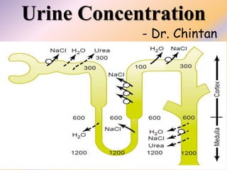
- 6. Countercurrent Mechanism The osmolarity of interstitial fluid in almost all parts of the body is about 300 mOsm/L, which is similar to the plasma osmolarity. The osmolarity of the interstitial fluid in the medulla of the kidney is much higher, increasing progressively to about 1200 to 1400 mOsm/L in the pelvic tip of the medulla. This means that the renal medullary interstitium has accumulated solutes in great excess of water. Once the high solute concentration in the medulla is achieved, it is maintained by a balanced inflow and outflow of solutes and water in the medulla.
- 7. Countercurrent Mechanism 1. Active transport of sodium ions and co-transport of potassium, chloride, and other ions out of the thick portion of the ascending limb of the loop of Henle into the medullary interstitium 2. Active transport of ions from the collecting ducts into the medullary interstitium 3. Facilitated diffusion of large amounts of urea from the inner medullary collecting ducts into the medullary interstitium 4. Diffusion of only small amounts of water from the medullary tubules into the medullary interstitium
- 9. Countercurrent Mechanism Because the thick ascending limb is almost impermeable to water, the solutes pumped out are not followed by osmotic flow of water into the interstitium. Thus, the active transport of sodium and other ions out of the thick ascending loop adds solutes in excess of water to the renal medullary interstitium. There is some passive reabsorption of sodium chloride from the thin ascending limb of Henle’s loop, which is also impermeable to water, adding further to the high solute concentration of the renal medullary interstitium.
- 10. Countercurrent Mechanism The descending limb of Henle’s loop, in contrast to the ascending limb, is very permeable to water, and the tubular fluid osmolarity quickly becomes equal to the renal medullary osmolarity. Therefore, water diffuses out of the descending limb of Henle’s loop into the interstitium, and the tubular fluid osmolarity gradually rises
- 11. Countercurrent Mechanism First, the loop of Henle is filled with fluid with a concentration of 300 mOsm/L, the same as that leaving the proximal tubule
- 12. Countercurrent Mechanism Next, the active pump of the thick ascending limb on the loop of Henle is turned on, reducing the concentration inside the tubule raising the interstitial concentration; this pump establishes a 200- mOsm/L concentration gradient between the tubular fluid and the interstitial fluid
- 13. Countercurrent Mechanism Step 3 is that the tubular fluid in the descending limb of the loop of Henle and the interstitial fluid quickly reach osmotic equilibrium because of osmosis of water out of the descending limb. The interstitial osmolarity is maintained at 400 mOsm/L because of continued transport of ions out of the thick ascending loop of Henle.
- 14. Countercurrent Mechanism Step 4 is additional flow of fluid into the loop of Henle from the proximal tubule, which causes the hyperosmotic fluid previously formed in the descending limb to flow into the ascending limb
- 15. Countercurrent Mechanism Once the fluid is in the ascending limb, additional ions are pumped into the interstitium, with water remaining behind, until a 200-mOsm/L osmotic gradient is established, with the interstitial fluid osmolarity rising to 500 mOsm/L
- 16. Countercurrent MechanismThen, once again, the fluid in the descending limb reaches equilibrium with the hyperosmotic medullary interstitial fluid as the hyperosmotic tubular fluid from the descending limb of the loop of Henle flows into the ascending limb, Still more solute is continuously pumped out of the tubules and deposited into the medullary interstitium
- 17. Countercurrent MechanismThese steps are repeated over and over, with the net effect of adding more and more solute to the medulla in excess of water; this process gradually traps solutes in the medulla and multiplies the concentration gradient established by the active pumping of ions out of the thick ascending loop of Henle, eventually raising the interstitial fluid osmolarity to 1200 to 1400 mOsm/L
- 19. Countercurrent Mechanism the repetitive reabsorption of NaCl by the thick ascending loop of Henle and continued inflow of new NaCl from the proximal tubule into the loop of Henle is called the countercurrent multiplier. NaCl reabsorbed from the ascending loop of Henle keeps adding to the newly arrived NaCl, thus “multiplying” its concentration in the medullary interstitium.
- 20. Role of Distal Tubule and Collecting Ducts When the tubular fluid leaves the loop of Henle and flows into the DCT in the renal cortex, the fluid is dilute, with an osmolarity of only about 100 mOsm/L The early distal tubule further dilutes the tubular fluid because this segment, like the ascending loop of Henle, actively transports NaCl out of the tubule but is relatively impermeable to water. As fluid flows into the cortical collecting tubule, the amount of water reabsorbed is critically dependent on the plasma concentration of ADH
- 21. Role of Distal Tubule and Collecting Ducts In the absence of ADH, this segment is almost impermeable to water and fails to reabsorb water but continues to reabsorb solutes and further dilutes the urine. When there is a high concentration of ADH, the cortical collecting tubule becomes highly permeable to water, so that large amounts of water are now reabsorbed from the tubule into the cortex interstitium, where it is swept away by the rapidly flowing peritubular capillaries. These large amounts of water are reabsorbed into the cortex, rather than into the renal medulla, helps to preserve the high medullary interstitial fluid osmolarity.
- 22. Role of Distal Tubule and Collecting Ducts As the tubular fluid flows along the medullary collecting ducts, there is further water reabsorption from the tubular fluid into the interstitium, but the total amount of water is relatively small compared with that added to the cortex interstitium. The reabsorbed water is quickly carried away by the vasa recta into the venous blood. When high levels of ADH are present, the collecting ducts become permeable to water, so that the fluid at the end of the collecting ducts has essentially the same osmolarity as the interstitial fluid of the renal medulla — about 1200 mOsm/L Thus, by reabsorbing as much water as possible, the kidneys form a highly concentrated urine.
- 24. Urea Contributes When there is water deficit and blood concentrations of ADH are high, large amounts of urea are passively reabsorbed from the inner medullary collecting ducts into the interstitium. As water flows up the ascending loop of Henle and into the distal and cortical collecting tubules, little urea is reabsorbed because these segments are impermeable to urea. In the presence of ADH, water is reabsorbed rapidly from the cortical collecting tubule and the urea concentration increases rapidly because urea is not very permeant in this part of the tubule.
- 25. Urea Contributes Then, as the tubular fluid flows into the inner medullary collecting ducts, still more water reabsorption takes place, causing an even higher concentration of urea in the fluid. This high concentration of urea in the tubular fluid of the inner medullary collecting duct causes urea to diffuse out of the tubule into the renal interstitium. This diffusion is greatly facilitated by specific urea transporters - UT-AI - activated by ADH, increasing transport of urea out of the inner medullary collecting duct
- 26. Urea Contributes people who ingest a high-protein diet, yielding large amounts of urea as a nitrogenous “waste” product, can concentrate their urine much better than people whose protein intake and urea production are low. Malnutrition is associated with a low urea concentration in the medullary interstitium and considerable impairment of urine concentrating ability.
- 29. Countercurrent Exchange 1. The medullary blood flow is low, accounting for less than 5 per cent of the total renal blood flow. This sluggish blood flow is sufficient to supply the metabolic needs of the tissues & helps to minimize solute loss from the medullary interstitium. 2. The vasa recta serve as countercurrent exchangers, minimizing washout of solutes from the medullary interstitium.
- 30. Countercurrent Exchange - As blood descends into the medulla, it becomes progressively more concentrated, partly by solute entry from the interstitium and partly by loss of water into the interstitium. - By the time the blood reaches the tips of the vasa recta, it has a concentration of about 1200 mOsm/L, the same as that of the medullary interstitium. - As blood ascends back toward the cortex, it becomes progressively less concentrated as solutes diffuse back out into the medullary interstitium and as water moves into the vasa recta.