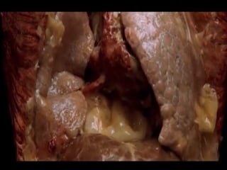
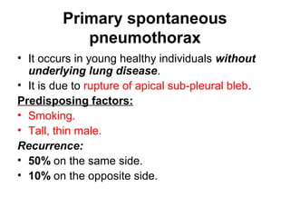
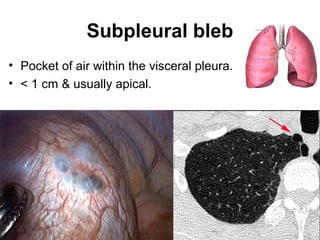
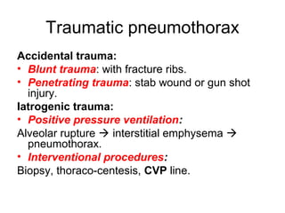
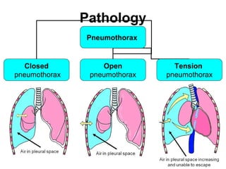
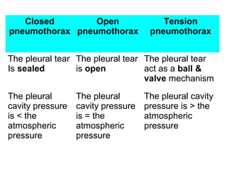
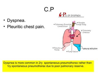
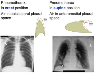
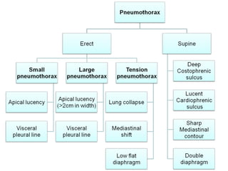
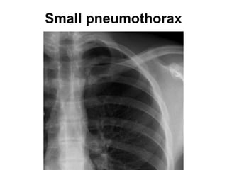
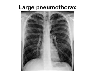
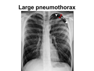
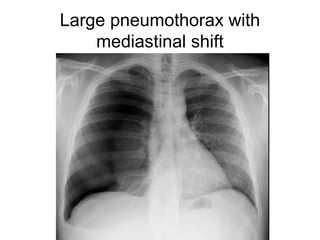
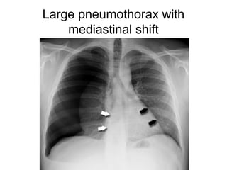
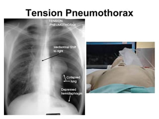
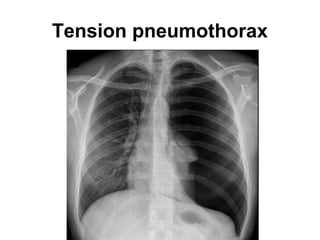
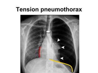
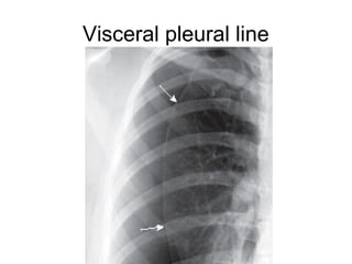
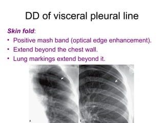
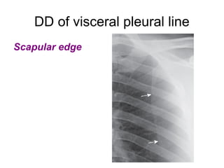
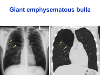
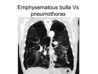
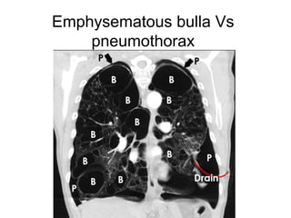
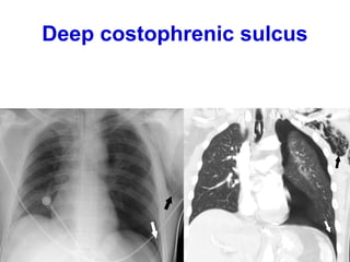
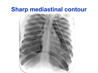
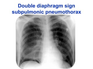
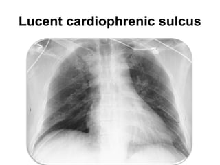
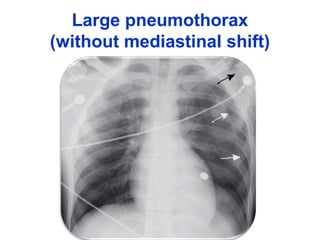
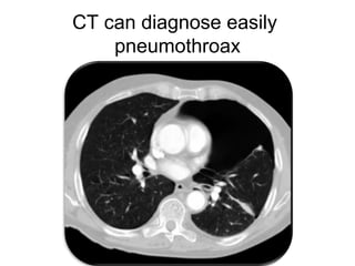
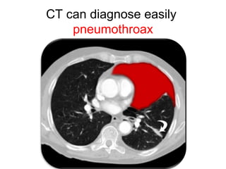
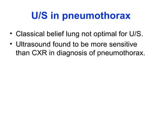
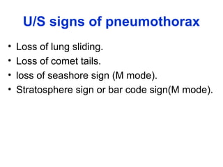
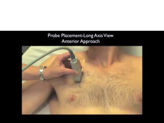
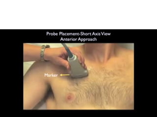
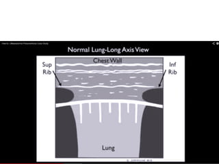
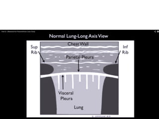
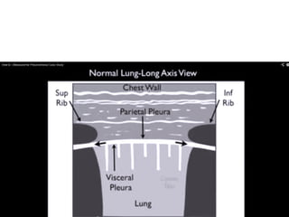
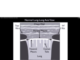
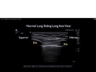
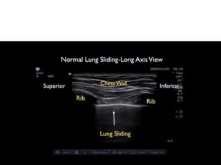
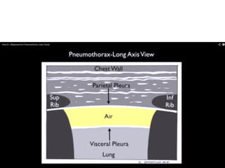
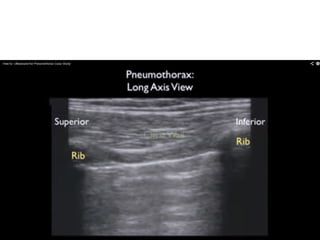
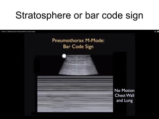
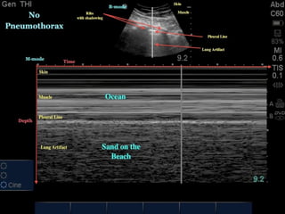
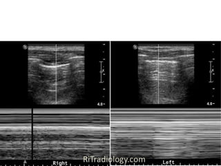
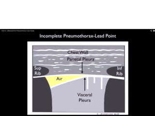
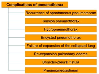
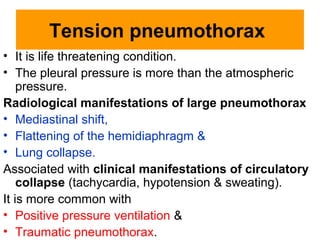
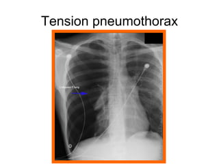
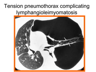
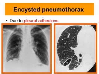
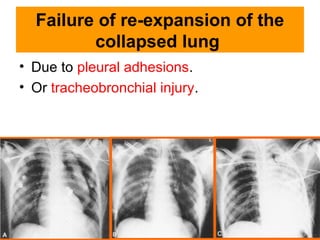
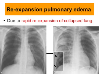
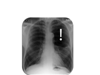
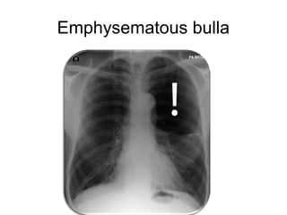
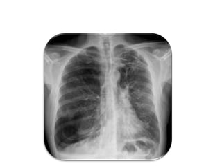
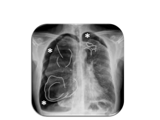
Pneumothorax
Dr. Hytham Nafady
Etiology
Primary spontaneous
pneumothorax
• It occurs in young healthy individuals without
underlying lung disease.
• It is due to rupture of apical sub-pleural bleb.
Predisposing factors:
• Smoking.
• Tall, thin male.
Recurrence:
• 50% on the same side.
• 10% on the opposite side.
Subpleural bleb
• Pocket of air within the visceral pleura.
• < 1 cm & usually apical.
Secondary spontaneous
pneumothorax:
Due to underlying lung disease.
• Cavitary lesion.
• Cystic lung disease.
• Emphysematous bullae.
• Pneumatocele.
Traumatic pneumothorax
Accidental trauma:
• Blunt trauma: with fracture ribs.
• Penetrating trauma: stab wound or gun shot
injury.
Iatrogenic trauma:
• Positive pressure ventilation:
Alveolar rupture  interstitial emphysema 
pneumothorax.
• Interventional procedures:
Biopsy, thoraco-centesis, CVP line.
Pathology
Pneumothorax
Closed
pneumothorax
Open
pneumothorax
Tension
pneumothorax
Closed
pneumothorax
Open
pneumothorax
Tension
pneumothorax
The pleural tear
Is sealed
The pleural tear
is open
The pleural tear
act as a ball &
valve mechanism
The pleural
cavity pressure
is < the
atmospheric
pressure
The pleural
cavity pressure
is = the
atmospheric
pressure
The pleural cavity
pressure is > the
atmospheric
pressure
C.P
Dyspnea is more common in 2ry spontaneous pneumothorax rather than
1ry spontaneous pneumothorax due to poor pulmonary reserve.
• Dyspnea.
• Pleuritic chest pain.
Radiological manifestations
Pneumothorax
in erect position
Pneumothorax
in supine position
Air in apicolateral pleural
space
Air in anteromedial pleural
space.
Small pneumothorax
Small pneumothorax
Large pneumothorax
Large pneumothorax
Large pneumothorax with
mediastinal shift
Large pneumothorax with
mediastinal shift
Tension Pneumothorax
Tension pneumothorax
Visceral pleural line
DD of visceral pleural line
Skin fold:
• Positive mash band (optical edge enhancement).
• Extend beyond the chest wall.
• Lung markings extend beyond it.
DD of visceral pleural line
Scapular edge
DD of apical radiolucency
Emphysematous bulla:
• Rounded (while pneumothorax is
crescentic & tapers toward the lung base).
• Double wall sign on CT is consistent with
ruptured bulla causing pneumothorax.
Giant emphysematous bulla
Emphysematous bulla Vs
pneumothorax
Emphysematous bulla Vs
pneumothorax
Signs of pneumothorax in
supine position
Deep costophrenic sulcus
Sharp mediastinal contour
Double diaphragm sign
subpulmonic pneumothorax
Lucent cardiophrenic sulcus
Large pneumothorax
(without mediastinal shift)
CT can diagnose easily
pneumothroax
CT can diagnose easily
pneumothroax
U/S in pneumothorax
• Classical belief lung not optimal for U/S.
• Ultrasound found to be more sensitive
than CXR in diagnosis of pneumothorax.
U/S signs of pneumothorax
• Loss of lung sliding.
• Loss of comet tails.
• loss of seashore sign (M mode).
• Stratosphere sign or bar code sign(M mode).
Stratosphere or bar code sign
Stratosphere
Bar code
Seashore
Recurrence of spontaneous
pneumothorax
• 50% on the same side.
• 15% on the contralateral side.
More common in
• 2ry spontaneous pneumothorax.
Tension pneumothorax
• It is life threatening condition.
• The pleural pressure is more than the atmospheric
pressure.
Radiological manifestations of large pneumothorax
• Mediastinal shift,
• Flattening of the hemidiaphragm &
• Lung collapse.
Associated with clinical manifestations of circulatory
collapse (tachycardia, hypotension & sweating).
It is more common with
• Positive pressure ventilation &
• Traumatic pneumothorax.
Tension pneumothorax
Tension pneumothorax complicating
lymphangioleimyomatosis
Hydropneumothorax
• Due to rupture of pleural adhesions.
• Bronchopleural fistula.
Encysted pneumothorax
• Due to pleural adhesions.
Failure of re-expansion of the
collapsed lung
• Due to pleural adhesions.
• Or tracheobronchial injury.
Re-expansion pulmonary edema
• Due to rapid re-expansion of collapsed lung.
Quizzes
Emphysematous bulla
Emphysema & bilateral pneumothorax
Pneumothorax

Pneumothorax