Downloaded 1,055 times

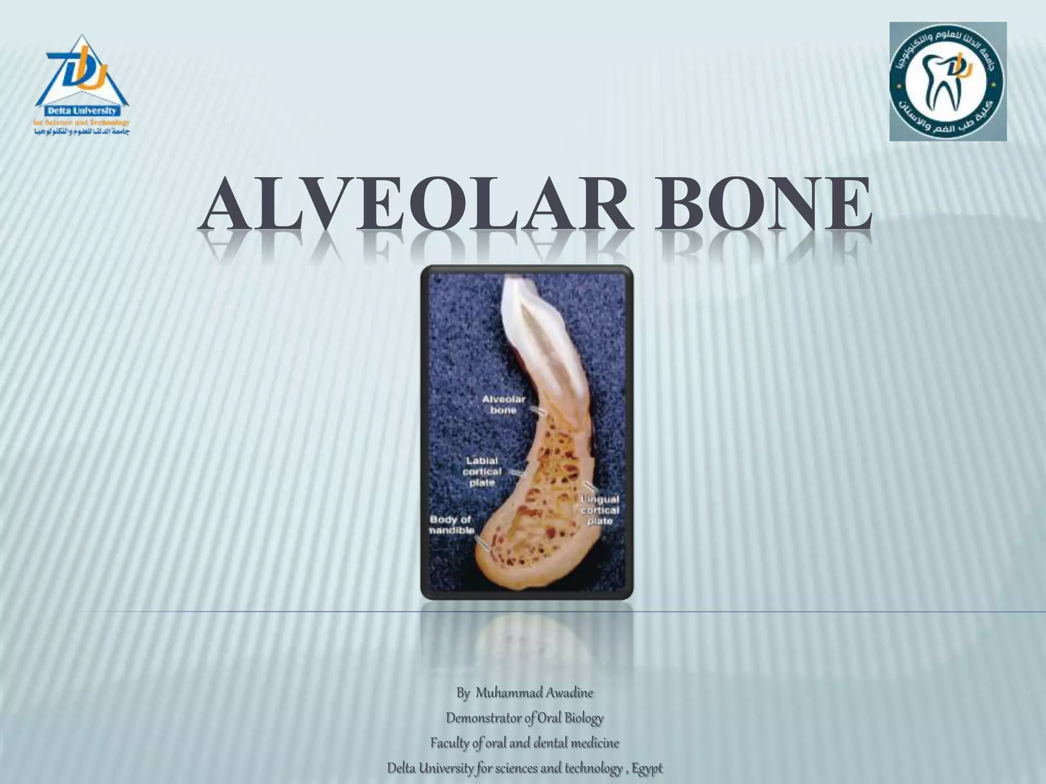
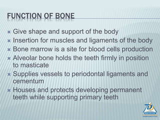
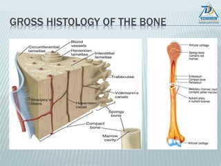
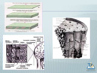
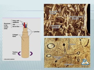
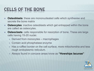
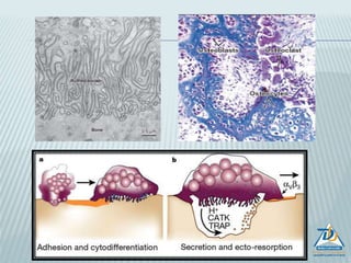
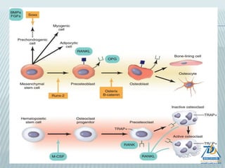
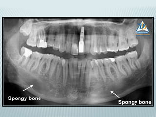
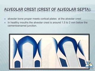
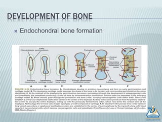
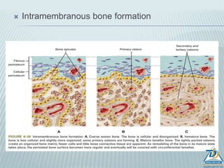

The document discusses alveolar bone, which forms the primary support structure for teeth. It defines alveolar bone and discusses its classification, composition, function, histology, cells, development, remodeling, and age-related changes. Alveolar bone holds teeth firmly in position, supplies vessels to periodontal ligaments and cementum, and houses developing permanent teeth. It is a specialized part of the maxilla and mandible composed of lamellar and bundle bone that surrounds tooth roots and provides attachment for periodontal ligament fibers. Alveolar bone is constantly remodeled through formation and resorption to adapt to functional forces.


