TABLE OF CONTENTS
Definition.
Development.
Composition and Structure.
Residual Ridge resorption.
Alveolar Bone from a Removable denture point
of view.
Alveolar Bone from a Fixed Prosthodontic point
of view.
Alveolar Bone from Implantology point of view.
Pathological Fate of Alveolar Bone.
References.
3.
DEFINITION
That part ofthe mandible and the maxilla in
which the teeth are located is referred to as the
ALVEOLAR PROCESS.
The alveoli that support the teeth are found
within the alveolar process, and the bone lining the
alveoli is called the ALVEOLAR BONE PROPER or
BUNDLE BONE.
4.
THE FACE OFA SIX-WEEK-OLD EMBRYO
The two mandibular processes (A) fuse in the midline to
form the tissues of the lower jaw. The mandibular and
maxillary (B) processes meet at the angles of the mouth,
thus defining its outline.
From the corners of the mouth, the
maxillary processes grow inwards
beneath the lateral nasal processes (C)
towards the medial nasal processes (D)
of the upper lip. Between the merging
maxillary and the lateral nasal
processes lie the naso-optic furrows (E).
5.
THE APPEARANCE OFTHE DEVELOPING
JAWS OF A HUMAN FOETUS
(14 weeks intra-uterine)
A:Body of mandible
B:Ramus of the
mandible
C:Secondary condylar
cartilage
D:Secondary coronoid
cartilage
E:Frontal bone
F:Parietal bone
G:Occipital bone
H:Squamous portion of
temporal bone
I:Maxilla.
6.
CONTRIBUTIONS TO THEADULT FACE
FROM THE EMBRYONIC FACIAL
PROCESSES.
A: Maxillary process.
B: Mandibular process.
C: Medial nasal process.
D: Lateral nasal process.
7.
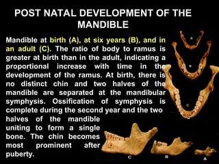
POST NATAL DEVELOPMENTOF THE
POST NATAL DEVELOPMENT OF THE
MANDIBLE
MANDIBLE
Mandible at birth (A), at six years (B), and in
an adult (C). The ratio of body to ramus is
greater at birth than in the adult, indicating a
proportional increase with time in the
development of the ramus. At birth, there is
no distinct chin and two halves of the
mandible are separated at the mandibular
symphysis. Ossification of symphysis is
complete during the second year and the two
halves of the mandible
uniting to form a single
bone. The chin becomes
most prominent after
puberty.
ORGANIC PART –33% - 35%
Collagen – 88% - 90% (Type – I)
Non collagen – 10% - 11%.
Glycoproteins – 6% - 9% (Mono, Di, Poly
and Oligosaccharides).
Proteoglycanes – 0.8% (sulfated and Non
sulfated)
Sialoproteins – 0.35%
Lipids – 0.4%
10.
INORGANIC PART –65% - 67%
Calcium & Phosphates – 95%
(Hydroxyapatite Crystals – Ca10(Po4)6 (OH)2)
Magnesium
Trace elements – Nickel, Iron, Fluoride,
Cadmium, Magnesium, Zinc and
Molybdenum.
11.
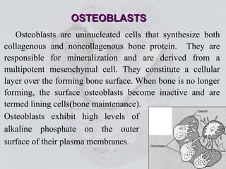
Osteoblasts are uninucleatedcells that synthesize both
collagenous and noncollagenous bone protein. They are
responsible for mineralization and are derived from a
multipotent mesenchymal cell. They constitute a cellular
layer over the forming bone surface. When bone is no longer
forming, the surface osteoblasts become inactive and are
termed lining cells(bone maintenance).
Osteoblasts exhibit high levels of
alkaline phosphate on the outer
surface of their plasma membranes.
OSTEOBLASTS
OSTEOBLASTS
12.
Other enzymes thatparticipate in there activity are
ATPase and pyrophosphates. Osteoblasts secrete, in
addition to type I and type V collagen and small amounts
of several noncollagenous proteins, a variety of cytokines.
Osteoblasts under the stimulation of interleukin 6
also produce their own hydrolytic enzymes that aid in
destroying or modifying the unmineralized matrix. Thus
freeing the osteoblast from its own secreted matrix.
The hormones most important in
bone metabolism are parathyroid
hormone. 1,25 dihydroxyvitamin
D, calcitonin, estrogen, and the
glucocorticoids,which have a
influence on osteoblasts.
13.
As osteoblasts secretebone matrix, some of them
become entrapped in lacunae and are then called osteocytes.
The number of osteoblasts that become osteocytes varies
depending on the rapidity of bone formation. The more
rapid the formation, a more osteocytes are present per unit
volume.
OSTEOCYTE
As a general rule,
embryonic bone and repair bone
have more osteocytes than does
lamellar bone.
14.
Osteocytes gradually losemost of their matrix
synthesizing machinery and become reduced in size. The
space in the matrix occupied by an osteocyte is called the
osteocytic lacuna. Narrow extensions of these or
canaliculi, that form radiating osteocytic processes
maintain contact with adjacent osteocytes and osteoblasts
the endosteum, periosteum, and Haversian canals.
Failure of any part of this inter connecting system result in
hyper mineralization (sclerosis) and death of the bone.
15.
OSTEOCLAST
Compared to allother bone cells and their
precursors, the multinucleated osteoclast is a much larger
cell. Because of their size, it can be identified under the
light microscopy, generally seen in a cluster rather than
singly. The osteoclast is characterized by acid phosphatase
within its cytoplasmic vesicles and vacuoles, which
distinguishes it from other giant cells and macrophages.
Typically osteoclasts are found
against the bone surface occupying
shallow, hollowed out depressions,
called Howship’s lacunae.
16.
Isolates a microenvironment between them and the bone
surface. The cell organelles consist of many nuclei, each
surrounded by multiple Golgi complexes, an array of
mitochondria and free polysomes, a rough endoplasmic
reticulum, many coated transport vesicles, and numerous
vacuolar structures. Osteoclast are also rich in lysosomal
enzymes.
Adjacent to the tissue
surface, their cell membrane is
thrown into a myriad of deep
folds that form a brush border.
This clear or “sealing” zone
attached the cells to the
mineralized surface.
17.
TThus the sequenceof resorptive events is considered to
be
11. Attachment of osteoclasts to the mineralized surface of
bone.
C2. Creation of a sealed acidic environment through action
of the proton pump, which demineralizes bone and
exposes the organic matrix.
D3. Degradation of this exposed organic matrix to its
constituent amino acids by the action of released
enzymes.
4. Uptake of mineral ions and
amino acids by the cell.
18.
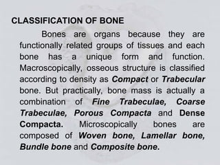
CLASSIFICATION OF BONE
Bonesare organs because they are
functionally related groups of tissues and each
bone has a unique form and function.
Macroscopically, osseous structure is classified
according to density as Compact or Trabecular
bone. But practically, bone mass is actually a
combination of Fine Trabeculae, Coarse
Trabeculae, Porous Compacta and Dense
Compacta. Microscopically bones are
composed of Woven bone, Lamellar bone,
Bundle bone and Composite bone.
19.
Woven bone
• Highlycellular.
• Formed rapidly (30-50 µm/ day or more) in
response to growth or injury.
• Low mineral content.
• Random fiber orientation and minimal
strength.
• Stabilize unloaded Endosseous implants
during initial healing.
20.
Lamellar bone
• Principleload bearing tissue of adult
skeleton.
• Predominant component of mature cortical
and trabecular bone.
• Formed relatively slowly (<1 µm/ day).
• Densely mineralized and highly organized
matrix.
21.
Bundle bone
• Characteristicof ligament and tendon
attachments along bone-forming
surfaces.
• Sharpey’s fibers from adjacent
connective tissue insert directly into
bone.
• Bundle bone is formed adjacent to the
periodontal ligament of natural teeth.
22.
Composite bone
• Highquality lamellar bone deposited
on a woven bone matrix.
• Got adequate strength for load
bearing.
• Important in achieving stabilization of
an implant during the rigid integration
process.
23.
Alveolar Bone formsthe bony sockets of the
jaw bones in which the roots of the natural teeth are
suspended by the attachment of the periodontal
ligament fibers (“Gomphosis” - Greek “Bolting
together). Some alveolar bone is formed during tooth
development, but the majority of alveolar bone
the majority of alveolar bone
formation occurs during tooth eruption.
formation occurs during tooth eruption.
24.
The presence ofalveolar bone in the jaw
bones is totally dependent on the roots of the
natural teeth; without the teeth the alveolar bone
need not exist.
25.
ERUPTION OF TEETHAND
ALVEOLAR BONE
The teeth move within the jaws throughout the life
and these movements include:
1. Pre-eruptive Movement
2. Eruptive (“Break Out”) Movement
a) Pre-functional Eruption
b) Functional Eruption
# Active eruption
# Passive eruption
26.
THEORIES OF ERUPTION
1.Bone remodeling.
2. Growth of the root.
3. Hydrostatic pressure.
4. Traction via the periodontal ligament.
27.
There is nosharp line or suture which
differentiates alveolar bone from the
surrounding basal bone of either the mandible
or the maxilla. There is no morphologic or
biochemical distinction between the two types
of bone.
28.
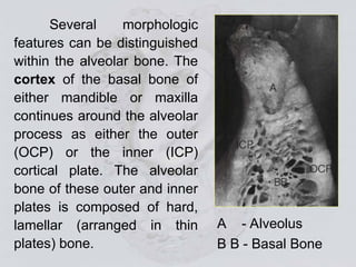
Several morphologic
features canbe distinguished
within the alveolar bone. The
cortex of the basal bone of
either mandible or maxilla
continues around the alveolar
process as either the outer
(OCP) or the inner (ICP)
cortical plate. The alveolar
bone of these outer and inner
plates is composed of hard,
lamellar (arranged in thin
plates) bone.
A - Alveolus
B B - Basal Bone
29.
Numerous blood vesselsand
nerve fibers pass through the
cribriform plate, providing unlimited
access between the alveolar bone
and tissues of the periodontal
ligament.
The actual tooth socket, is composed of a
slightly different type of hard bone called the
“cribriform (sieve like) plate”.
30.
The cribriform plateitself is made up of two
types of bony layers:
The Hard, lamellar type.
Bundle Bone oriented toward the
periodontal ligament and thick enough to
anchor the periodontal fibers within it.
31.
The periodontal
fibers visiblein the bundle
bone are termed
“Sharpey’s Fibers”. The
fibers of the periodontal
ligament function like a
miniature shock absorber
system that acts to
dissipate the forces of
occlusion through the
trabecular pattern of the
alveolar bone.
32.
The cribriform plate(alveolar bone proper)
forms the sockets, or alveoli, around each single- or
multiple- rooted tooth and follows the configuration
of the root(s) with precision, leaving only a small
space of less than 0.2 mm between the root and
bone occupied by the suspensory periodontal
ligament. Between the alveoli of adjacent teeth, the
alveolar bone forms an interdental septum
composed of adjacent cribriform plates and at times
interposed spongy bone. The roots of multirooted
teeth are separated from one another by
interradicular septum, and are composed of
adjacent cribriform plates and spongy bone
between them.
33.
When viewed witha dental radiograph
(IOPA), the cribriform plate surrounding each tooth
root appears as a dense white line called the
“Lamina dura”. We might assume that the
alveolar bone surrounding the tooth is denser or
more highly calcified than the other bone.
But this is not clearly the
case and is a radiographic
artifact based on the
geometry of the cribriform
plate.
34.
Between the corticaland cribriform plates of the
alveolar bone, another type of bone termed “Spongy
Bone” may be present. This spongy bone contains
small amounts of bony trabeculae (supporting beams)
surrounded by bone marrow that is of either the RED
(forming blood - in young adults) or YELLOW (fatty - in
older individuals). The response of the alveolar bone
to occlusal forces transmitted by the tension of the
periodontal fiber bundles is to form a trabecular
pattern that is both parallel and perpendicular to the
functional forces applied.
35.
On the labialsurfaces
of anterior teeth, the
outer cortical plate of
alveolar bone is very
thin and fused to the
cribriform plate, and
spongy bone is notably
absent.
36.
Alveolar bone isa plastic tissue, able to
respond and adapt to both functional occlusal
forces and changes in tooth position. E.g. “mesial
drift”, which is a nonpathologic, slow mesial tooth
migration due to enamel wear between the contact
areas of some or all adjacent natural teeth. For this
changes to occur in each alveoli of the teeth, both
Osteoblasts and Osteoclasts must be active.
Throughout the life of the individual, age-
related tooth wear, changes in occlusal loading and
pathologic changes in the teeth and supporting
tissues keep the trabecular pattern of alveolar bone
in a constant state of remodeling activity.
37.
CLASSIFICATION OF RESIDUALRIDGE
RESORPTION
According to Branemark et al in 1985, ridges
were classified on the basis of bone quantity
bone quantity and
bone quality
bone quality by radiographic means
BONE QUANTITY
BONE QUANTITY
Class A : Most of the alveolar bone is present.
Class B : Moderate residual ridge resorption occurs.
Class C : Advanced residual ridge resorption occurs.
Class D:Moderate resorption of the basal bone is
present.
Class E : Extreme resorption of the basal bone.
38.
BONE QUALITY
BONE QUALITY
Class1: Almost the entire jaw is composed of
homogenous compact bone.
Class 2: A thick layer of compact bone surrounds a
core of dense trabecular bone.
Class 3: A thin layer of cortical bone surrounds a core
of dense trabecular bone.
Class 4: A thin layer of cortical bone surrounds a core
of low density trabecular bone.
39.
ATWOODS CLASSIFICATION
Order I:pre-extraction.
Order II: post extraction.
Order III: high, well rounded.
Order IV: knife edge.
Order V: low, well rounded.
Order VI: depressed.
40.
CLASSIFICATION ACCORDING TOTHE
MANDIBULAR CANAL RESORPTION
RADIOGRAPHICALLY
Grade 0: the crest of the residual ridge above both
the mental foramen and the mandibular canal.
Grade I: the crest of residual ridge above both the
mandibular canal and the mental foramen at the top
of the residual ridge with or without a partially
resorbed ridge.
Grade II: the superior border of the mandibular canal
at the top of the residual ridge and the mental
foramen with or without a partially resorbed border.
41.
MERCIER’S CLASSIFICATION
Group I:High Crestal muscles and non resorbed ridge.
Group II: Painful atrophic ridge.
Group III: Absence of residual ridge.
42.
ZELTSER’S CLASSIFICAION
ZELTSER’S CLASSIFICAION
Group1: high muscle attachment and minimal
residual ridge resorption
Group 2: severe residual ridge resorption with pain.
Group 3: absence of residual ridge.
Group 4: severe resorption of basal bone.
43.
MISCH’S CLASSIFICATION
MISCH’S CLASSIFICATION
(basedon bone density)
(based on bone density)
D1: dense cortical bone
D2: thick dense to porous cortical bone on the crest
and coarse trabecular bone within.
D3: thin porous cortical bone on crest and fine
trabecular bone within.
D4: fine trabecular bone
D5: immature, non-mineralized bone.
44.
CLASSIFICATION ACCORDING TOTHE
CLASSIFICATION ACCORDING TO THE
AMERICAN COLLEGE OF
AMERICAN COLLEGE OF
PROSTHODONTISTS:
PROSTHODONTISTS:
Based on bone height (mandible only)
Based on bone height (mandible only)
Type I: Residual bone height of 21mm or greater
measured at the least vertical height of the mandible.
Type II: Residual bone height of 16-20mm measured
at the least vertical height of the mandible.
Type III: Residual alveolar bone height of 11-15mm
measured at the least vertical height of the mandible.
Type IV: Residual alveolar bone height of 10mm or
less measured at the least vertical height of the
mandible.
EDENTULOUS INTRAORAL
EDENTULOUS INTRAORAL
BONYCHANGES
BONY CHANGES
The loss of teeth means not only the loss
of the clinical crown but also the supporting
tissues, the periodontal ligament and alveolar
bone. When the alveolar bone is lost, the
resultant residual ridge is progressively resorbed
throughout the life of the individual (Atwood,
1971).
47.
It is criticalto successful dental practice that
the dentist understands the anatomy of the mouth
in both the dentulous and edentulous states and
the results of that transition in the individual.
Edentulous bony anatomy include:
Profound bone loss
Remodeling changes occurin the mandible that
account for the typical edentulous facial anatomy.
The overall length of the mandible does not
decrease but may in fact increase as new bone is
added to the mental protuberance, thus
accentuating the chin point.
51.
There isan anterior displacement of the
mandible (protrusive position) because of
residual ridge reduction, mandibular rotation
(Change in the angulation of the body relative to
the mandibular ramus), and deposition of bone
in the mental region.
Reduction in the residual ridges occurs in an
inferior direction in the molar and premolar
areas, but in both an inferior and lingual
direction in the incisor region.
There is generalized thinning of the anterior and
posterior aspects of the mandibular ramus.
53.
ALVEOLAR RIDGES
1. DevelopmentalStructure: The individual
variation in bone size and its degree of
calcification.
2. The size of the natural teeth: The teeth like the
bone show wide variation in size. Large teeth are
usually supported by bulky ridges, small teeth by
narrow ones.
The alveolar ridges vary greatly in size and
shape and their ultimate form is dependent on the
following factors:
54.
3. The amountof bone lost prior to the extraction
of the teeth: Periodontal disease is a chronic
inflammation of the supporting structures of the
teeth and results in the destruction of the alveolar
process. If the natural teeth are retained until
gross alveolar loss has occurred the resultant
alveolar ridges will be narrow and shallow.
4. The amount of alveolar process removed
during the extraction of the teeth: During
extraction with forceps the buccal alveolar plate is
sometimes fractured and removed with the tooth.
The commonest sites for this occurrence are the
upper and lower canine and first molar regions.
When teeth are removed by surgical dissection
some alveolar is always destroyed.
55.
5. The rateand degree of resorption: During the
first six weeks after the extraction of the teeth the
rate of resorption is rapid. During the second six
weeks it is fast but begins to slow down. At the
end of three months, on average, the immediate
post-extraction resorption is complete and
thereafter it continues throughout life at an ever-
decreasing pace.
6. The effect of previous dentures: ill-fitting
dentures, or dentures occluding with isolated
groups of natural teeth, may cause rapid
resorption of the alveolar process in the areas
where they cause excessive load or lateral stress.
56.
MAXILLARY DENTURE-BEARING AREA
Well-developedbut not abnormally thick ridges
and a palate with a moderate vault.
This is a favorable formation because:
The center of the palate presents an almost flat
horizontal area and this will aid adhesion.
The roomy sulcus allows for the development of
a good peripheral seal.
The well-developed ridges resist lateral and
antero-posterior movement of the denture.
57.
High V-shaped palateusually associated
with thick bulky ridges.
This may be an unfavorable
unfavorable formation because:
The forces of adhesion and cohesion are not at right
angles to the surface when counteracting the normal
displacing forces of gravity and so peripheral seal is
essential.
58.
Flat palate withsmall ridges and shallow
sulci.
This may be an unfavorable formation because:
The ill-developed or resorbed ridges do not resist
lateral and antero-posterior movement of the
denture.
The sulci being shallow do not form a good
peripheral seal, unless the width of the denture
periphery is adequate.
59.
Ridges exhibiting undercutareas.
These are unfavorable because:
frequently the flanges of the denture need to be
trimmed in order to be able to insert it and this
may reduce the effectiveness of the peripheral
seal.
60.
MANDIBULAR DENTURE-BEARING AREA
Broadand well developed ridges.
This is a favorable formation because:
It provides a large area on which to rest the denture
and prevents lateral and anteroposterior
movement.
The surface presented for adhesion is as large as it
can ever be in a lower jaw.
The lingual, labial and buccal sulci are satisfactory
for developing a close peripheral seal.
61.
Ridges exhibiting undercutareas.
These are unfavorable because:
If the denture is not eased away from the undercuts
pain and soreness will result and if it is eased, food
will lodge under the denture.
The easing of the periphery will reduce the surface
area of mucosal contact and will affect the
peripheral seal adversely.
62.
Well developed butnarrow or knife like ridges
These are unfavorable because:
The pressure of the denture during clenching and
mastication on the sharp ridge will cause pain.
Adhesive and cohesive forces are negligible
63.
Flat and atrophicridges.
These are unfavorable because:
No resistance is offered to anteroposterior or
lateral movements.
Frequently found to have resorbed to the level of
attachments of the mylohyoid, genioglossus and
buccinator muscles and if the denture base is
made sufficiently narrow not to encroach on
these structures, its area is too small for the
denture to function correctly.
When the area is increased to encroach on the
muscles they may move the dentures when they
contract.
65.
DIETARY GUIDELINES FORPATIENTS AT
RISK OF LOSING BONE
Maintain a high daily calcium intake
Obtain four servings of low fat dairy foods or obtain
equivalent amounts of calcium from green gram,
canned fish.
Take calcium supplements if dietary intake is low
66.
Prevent negative calciumbalance
Limit daily alcohol (2 glasses) and caffeine (2
cups) intake
Consume about 6 ounces of protein from
meat, poultry and fish
Use small amounts of processed foods high in
sodium
67.
Obtain 4000 I.Uof Vitamin D daily
•Spend 15 minutes in the sun 3 times a week
•Choose a multivitamin or calcium supplement
that contains 4000 I.U of Vitamin D.
Discuss calcium or drug interactions that interface
with calcium bioavailability with the physician
The edentulous areaswhere a fixed prosthesis is
to be provided may be overlooked during the
treatment planning phase. Unfortunately, any
deficiency or potential problem that may arise
during the fabrication of a pontic is often identified
only after the teeth have been prepared or even
when the master cast is ready to be sent to the
laboratory.
RESIDUAL RIDGE CONTOUR
70.
Proper preparation includesa careful
analysis of the critical dimensions of the
edentulous areas:
Mesiodistal width.
Mesiodistal width.
Buccolingual diameter.
Buccolingual diameter.
Occlusocervical distance.
Occlusocervical distance.
Location of the residual ridge.
Location of the residual ridge.
71.
The contour ofthe edentulous ridge should be
carefully evaluated during the treatment planning
phase. An ideally shaped ridge has a smooth,
regular surface of attached gingiva, which
facilitates maintenance of a plaque-free
environment. Its height and width should allow
placement of a pontic that appears to emerge
from the ridge and mimics the appearance of the
neighboring teeth. Facially, it must be free of
frenum attachment and of adequate facial height
to sustain the appearance of interdental papillae.
72.
Siebert
Siebert has classifiedresidual ridge
deformities into three categories:
1. Class I defects- faciolingual loss of tissue
width with normal ridge height.
2. Class II defects- loss of ridge height with
normal ridge width.
3. Class III defects- a combination of loss in
both dimensions.
73.
Loss of residualridge contour may lead to
unesthetic open gingival embrasures (“Black
triangles”), food impaction, and percolation of
saliva during speech.
74.
Surgical Modification
Although residualridge width may be
augmented with hard tissue grafts, this is
usually not indicated unless the
edentulous site is to receive an implant.
1. Roll technique uses soft tissue from the
lingual side of the edentulous site. The
epithelium is removed, and the tissue is
thinned and rolled back, thereby
thickening the facial aspect of the
residual ridge.
75.
2. Pouches maybe prepared in the facial
aspect of the residual ridge, into which
subepithelial or submucosal grafts may
be inserted.
76.
3. Interpositional graftis a wedge-shaped
connective tissue graft which is inserted
into a pouch preparation on the facial
aspect of the residual ridge.
AVAILABLE BONE
Available bonedescribes the amount of bone in the
edentulous area considered for implantation and is
measured in: Height.
Width.
Length
Angulation.
Crown-Implant body ratio.
79.
AVAILABLE BONE HEIGHT
Theheight of available bone is measured
from the crest of the edentulous ridge to the
opposing landmark, such as maxillary sinus,
mandibular canal, maxillary nares, inferior
border of the mandible, maxillary canine
eminence region etc.
80.
The minimum heightof the available bone
for endosteal implants is in part related to the
density of the bone. The more dense bone may
accommodate a shorter implant. The minimum
bone height for a predictable long-term endosteal
implant survival is 10mm.
82.
AVAILABLE BONE WIDTH
Widthis measured between the facial and
lingual plates at the crest of the potential implant site.
The crest is supported by a wider base. The root
form implants of 4.0 mm crestal diameter usually
require more than 5.0 mm of bone width to ensure
sufficient bone thickness and blood supply around
the implant for predictable survival. These
dimensions provide more than 0.5 mm bone on each
side of the implant at the crest.
84.
AVAILABLE BONE LENGTH
Themesio-distal length of available bone in
an edentulous area is often limited by adjacent
teeth or implants. The root form implants of 4.0
mm crestal diameter usually require a minimum
mesio-distal length of 7 mm.
85.
AVAILABLE BONE ANGULATION
Ideallythe bone angulation should be such
that the long axis of the implant can be placed
parallel to the long axis of the Prosthodontic
restoration. In edentulous areas with wide ridge,
and wider root form implants a modification upto
30 degrees can be achieved.
86.
CROWN-IMPLANT BODY RATIO
Thecrown height is measured from the
occlusal or incisal plane to the crest of the ridge
and the endosteal implant height from the crest
of the ridge to its apex. The greater the crown
height, the greater the lever arm with any lateral
force.
VARIABLE BONE DENSITY-WHY?
Cortical and trabecular bone are constantly modified
by either Modeling
Modeling or Remodeling.
Remodeling.
In bone
bone modeling
modeling there is independent sites of
formation and resorption and results in the change of
the shape or size of bone.
In bone
bone remodeling
remodeling the resorption and formation
are at the same site that replaces previously existing
bone and primarily affects the internal turnover of
bone.
94.
These adaptive phenomenaof modeling and
remodeling of bone have been associated with the
alteration of the mechanical stress environment
alteration of the mechanical stress environment
within the host bone.
within the host bone.
MacMillan
MacMillan and Parfitt
Parfitt noted that
Bone is most dense around the teeth (Cribriform
Plate).
Density of bone around the crest region is more
compared to the regions around the apices.
Generalized trabecular bone loss occurs in
regions around a tooth from a decrease in
mechanical stress.
95.
Frost
Frost reported amodel of four zones for compact
bone as it is related to mechanical adaptation to
stress
Pathologic overload zone
Mild overload zone
Adapted window zone
Acute disuse window zone
97.
ACUTE DISUSE WINDOWZONE
The bone loses mineral density, and disuse
atrophy occurs because modeling for new
bone is inhibited and remodeling is
stimulated, with a gradual net loss of bone.
0 - 50 Microstrain
This phenomenon is also seen in
microgravity environments in outer space.
98.
ADAPTED WINDOW ZONE
Representsan equilibrium of modeling &
remodeling, and bone conditions are
maintained at this level.
Bone remains in a steady state.
Histologically – Lamellar or load-bearing
bone.
50-1500 microstrain ideally desired around
and endosteal implant.
99.
MILD OVERLOAD ZONE
Bonemodeling stimulation and remodeling
inhibition.
Bone density and strength may eventually
decrease.
Histologically – Woven or repair bone.
Low density bone resulting from overloaded
implant
1500-3000 microstrain
100.
PATHOLOGIC OVERLOAD ZONE
Thebone resorb and only woven bone
formation is seen because of sustained
repair.
Microstrain greater than 3000.
Cortical fracture takes place at 10000-20000
microstrain
The crestal bone loss often evidenced
during early implant loading is a result of the
bone in the pathologic overload zone
101.
MISCH BONE DENSITY
CLASSIFICATION
D1Dense cortical bone
D2 Thick dense to porous cortical bone on
crest and coarse trabecular bone within
D3 Thin porous cortical bone on crest and
fine trabecular bone within
D4 Fine trabecular bone
D5 Immature, nonmineralized bone
RADIOGRAPHIC BONE DENSITY
CTscan can determine bone density
precisely.
Each CT image has pixels and each pixel
has a CT number (Housefield unit). Higher
the Housefield unit, denser the tissue.
D1 >1250 Housefield units
D2 850-1250 Housefield units
D3 350-850 Housefield units
D4 150-350 Housefield units
D5 < 150 Housefield units
HISTIOCYTOSIS X
Bone lesionappears as sharply “Punched-
out” lytic defect, often with irregular
margins.
The posterior mandible is the most
common site.
Mild dull pain is commonly present.
Alveolar bone involvement leads to severe
horizontal bone loss.
CLEIDOCRANIAL DYSPLASIA
Skulland clavicles are chief sites of disorder.
Large head with bulging of frontal bone.
Unusual mobility of shoulders.
Narrow high arched palate, prolonged retention
of deciduous teeth and delay or failure of
eruption of permanent teeth.
OSTEOPOROSIS
Osteoporosis is asystemic disease in the elderly.
Osteoporosis shows a decrease in the skeletal mass
without alteration in the chemical composition of
bone. Loss of the spongy spicules of bone that
support the weight bearing parts of the skeleton can
be seen in radiographs of regions of the skeleton that
bear heavy loads, such as the vertebral column,
epiphysis of long bones, the mandible and the
fingers.
In edentulous patients, reduction of the residual ridge
is one of the most important factors affecting denture
support, retention, stability, and masticatory function.
117.
SEVERITY OF OSTEOPOROSIS
JIKEI’SCLASSIFICATION
Class I – Horizontal trabeculae are decreased and
vertical trabeculae are prominent.
Class II – Decreasing of horizontal trabeculae is
more prominent and vertical trabeculae are sparse.
Class III – Horizontal trabeculae almost disappear
and vertical trabeculae are found to be indistinct.
118.
REFERENCES
1. Human OralEmbryology and Histology.
I. A. Mjor & O. Fejerskov.
2. A Colour Atlas and Textbook Of Oral Anatomy.
B.K.B. Berkovitz.
3. An Introduction to Dental Anatomy & Esthetics.
Robert P. Renner.
4. Sicher & Dubrul’s Oral Anatomy (8th
Edition)
E. Lloyd Dubrul.
5. Dental Histology & Embryology.
Dr. A. N. Radhakrishnan.
119.
6. Essentials ofComplete Denture Prosthodontics (2nd
edition)
Sheldon Winkler.
7. Contemporary Implant Dentistry (2nd
edition)
Carl E. Misch.
8. Clinical Removable Partial Prosthodontics (2nd
edition)
Kenneth L. Stewart
9. Boucher’s Prosthodontic Treatment for Edentulous
Patients (11th
edition)
George A. Zarb
10. Fundamentals of Fixed Prosthodontics (3rd
edition)
Herbert T. Shillingburg


![ALVEOLAR BONE IN HEALTH AND DISEASE [Autosaved].ppt](https://cdn.slidesharecdn.com/ss_thumbnails/alveolarboneinhealthanddiseaseautosaved-231125051936-9d7ab2b3-thumbnail.jpg?width=640&height=640&fit=bounds)


