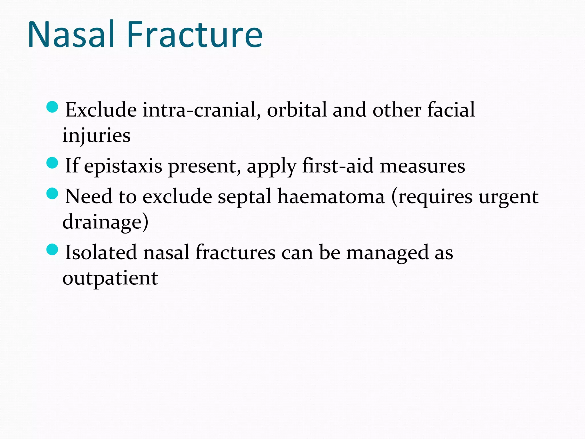
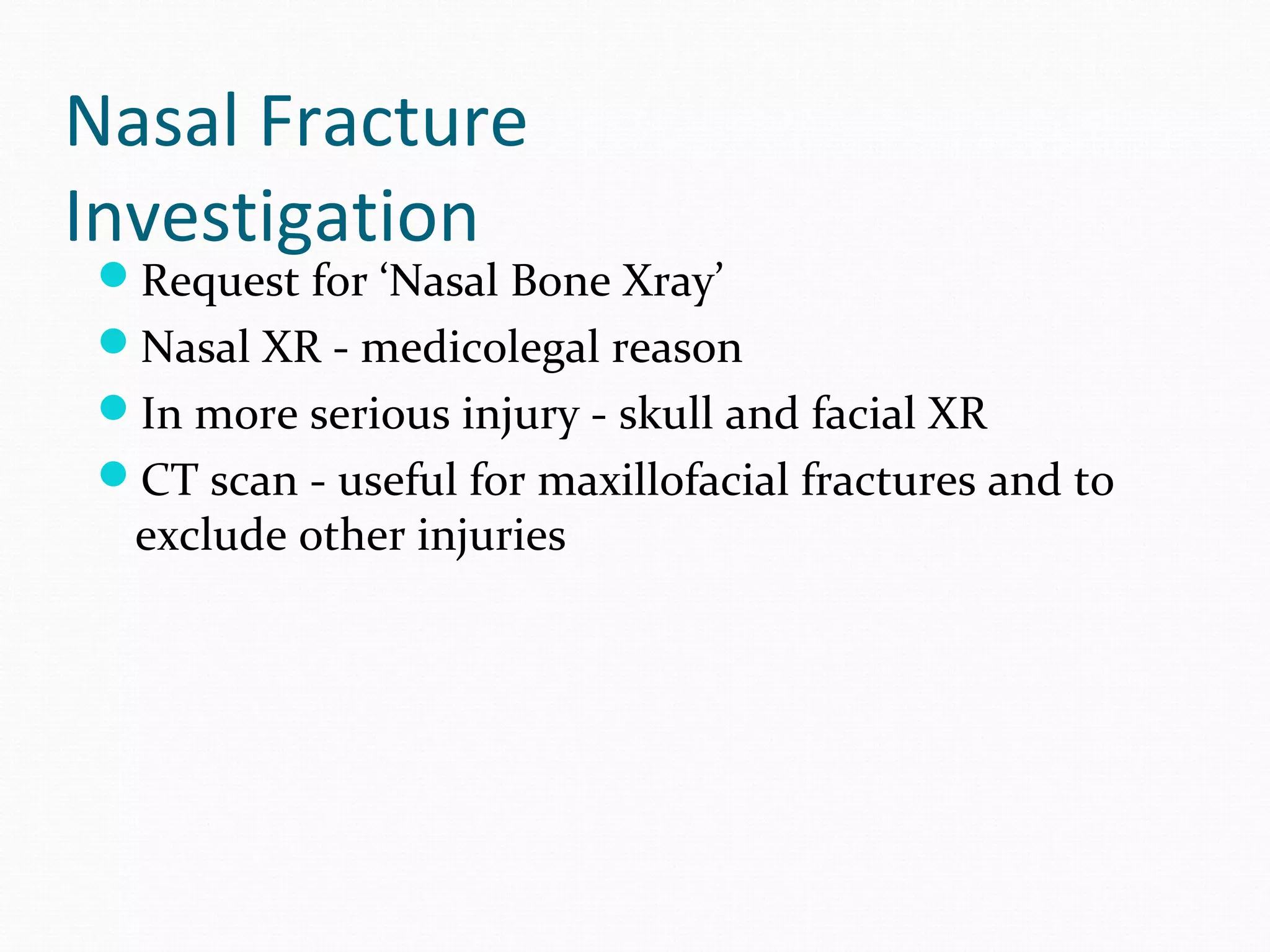
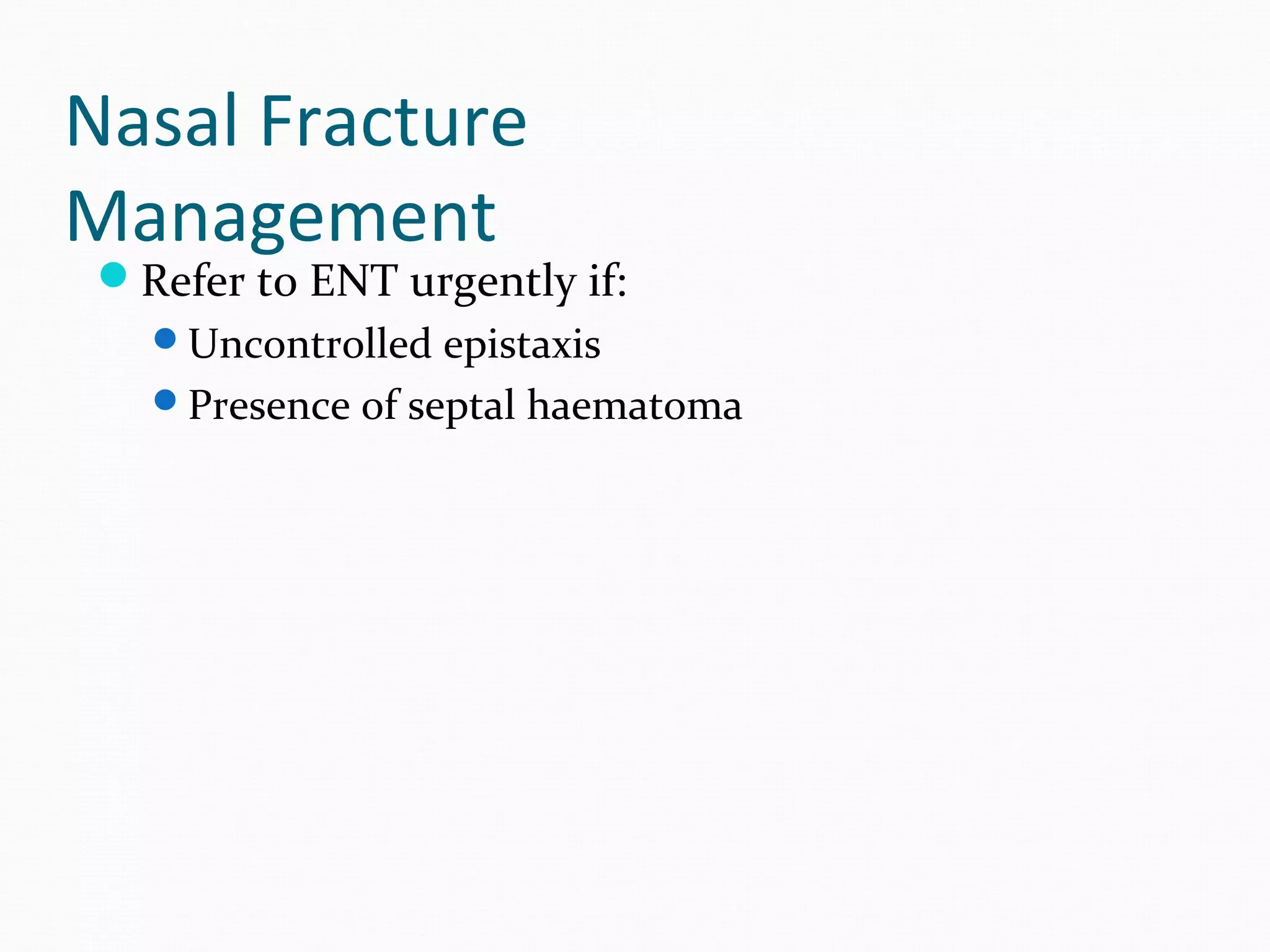
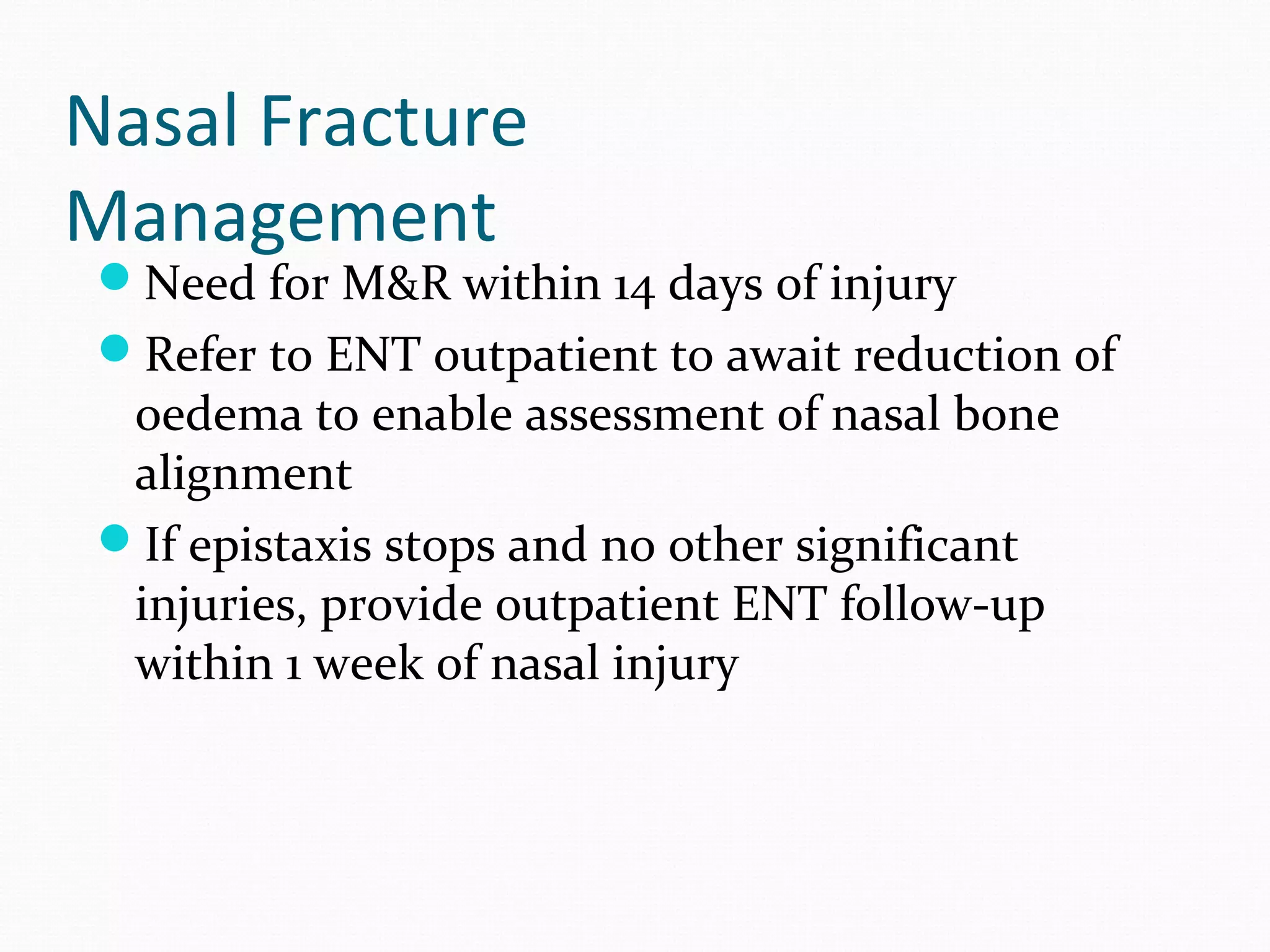
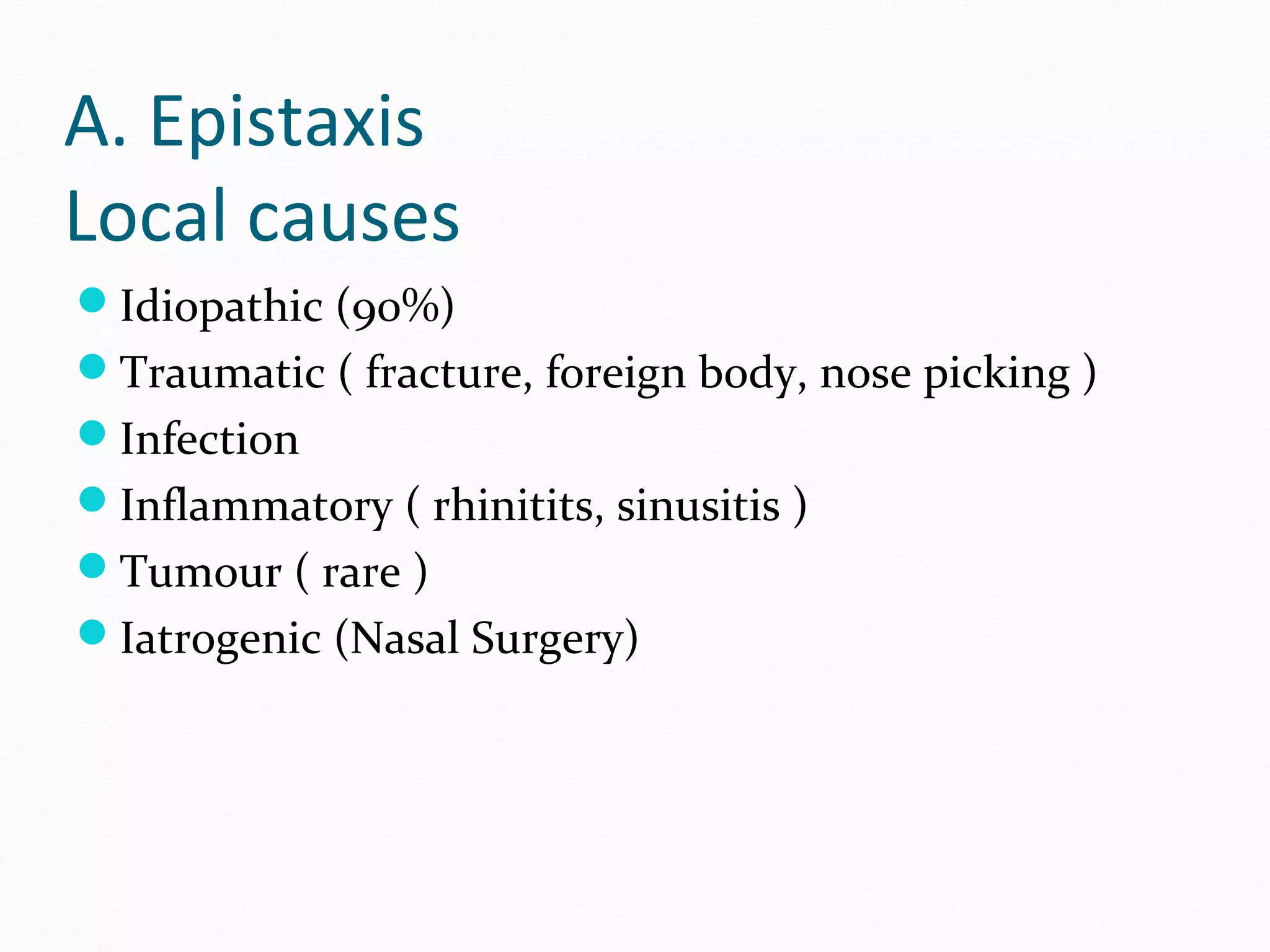
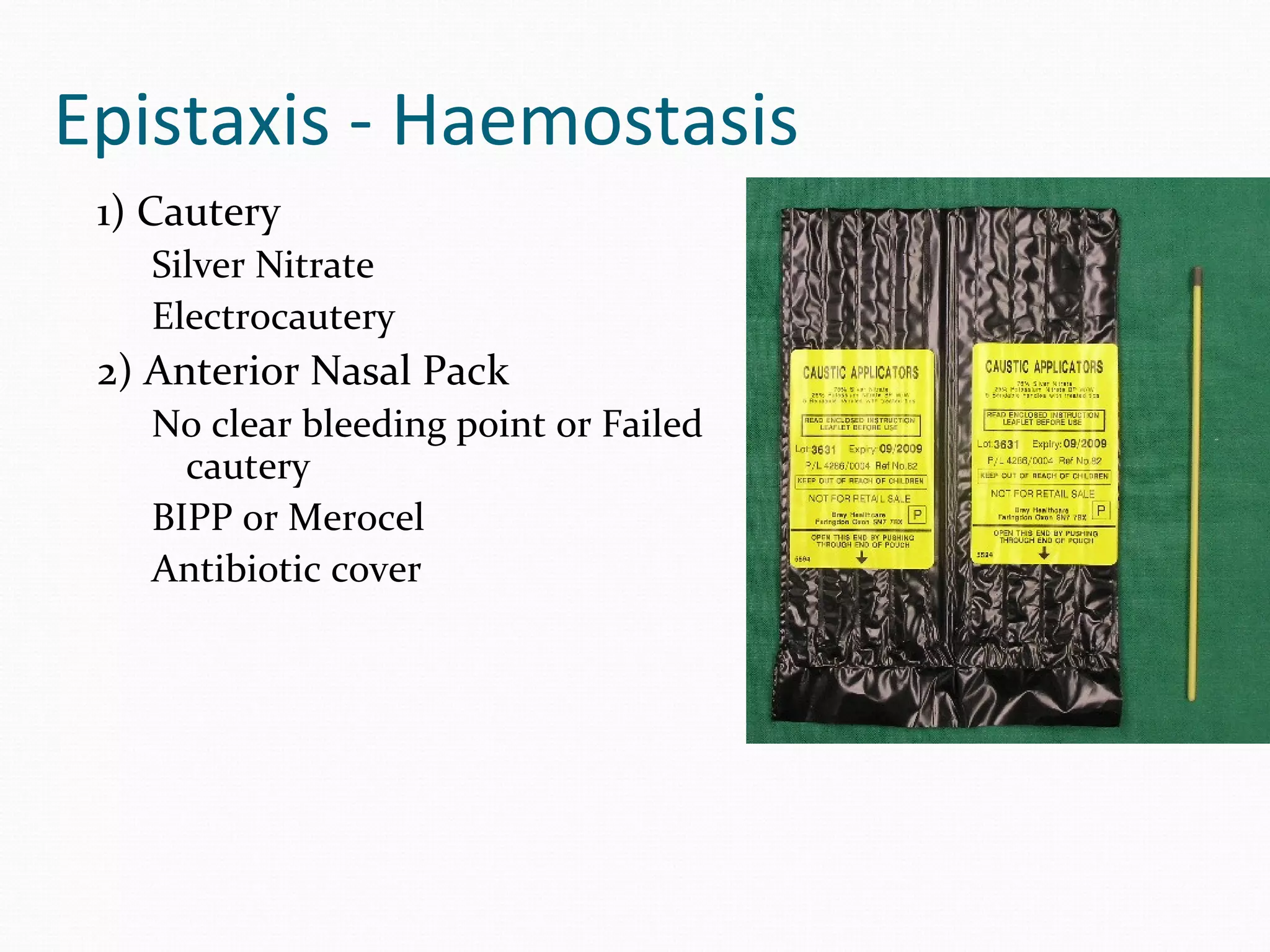
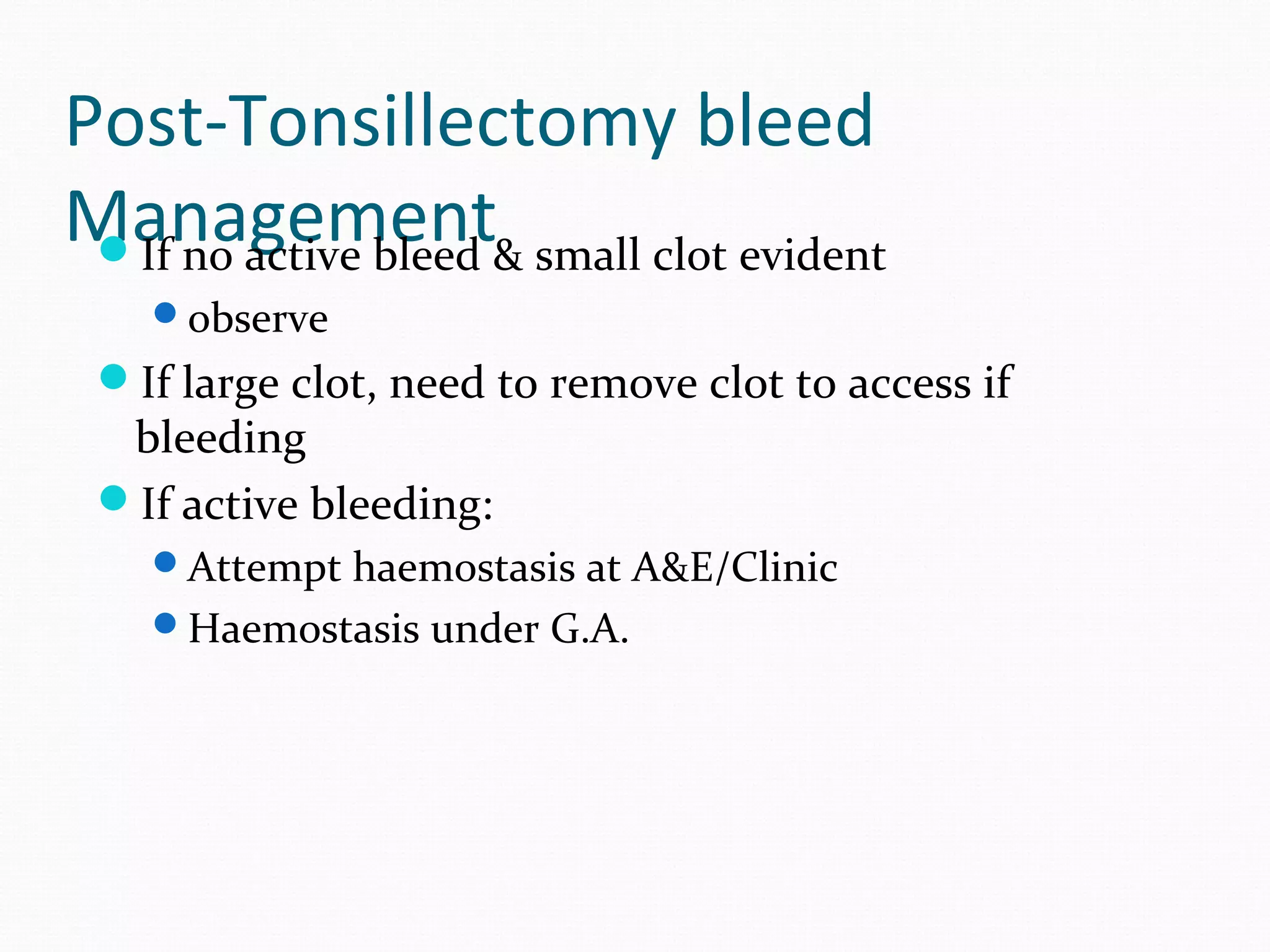
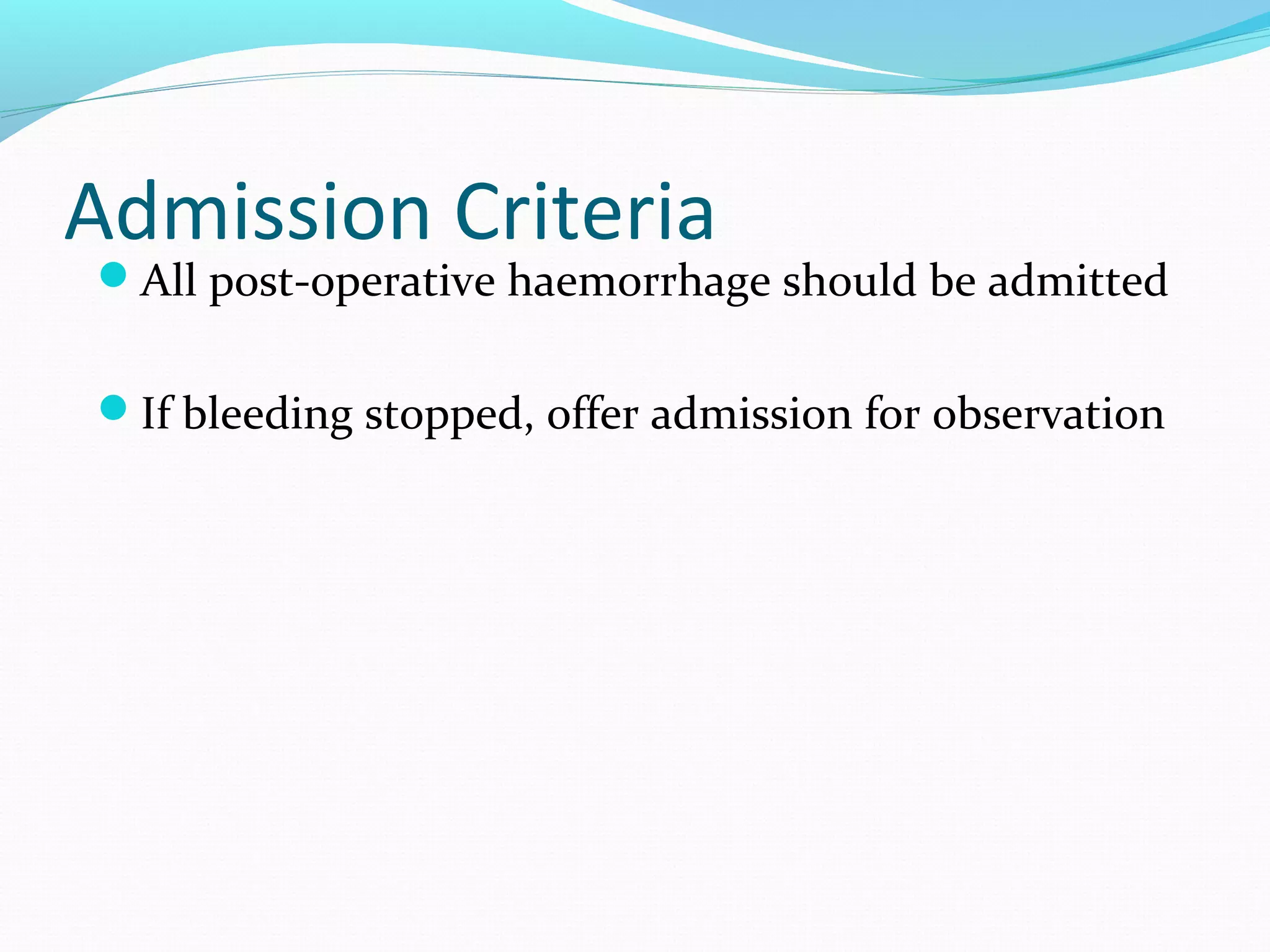
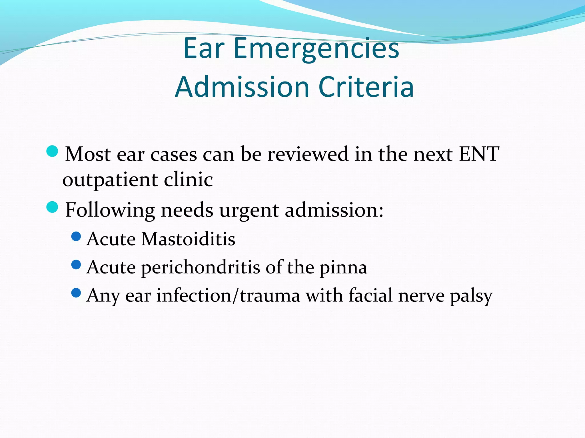
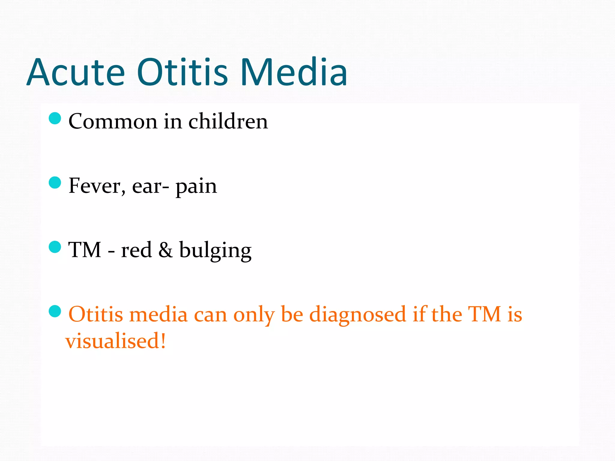
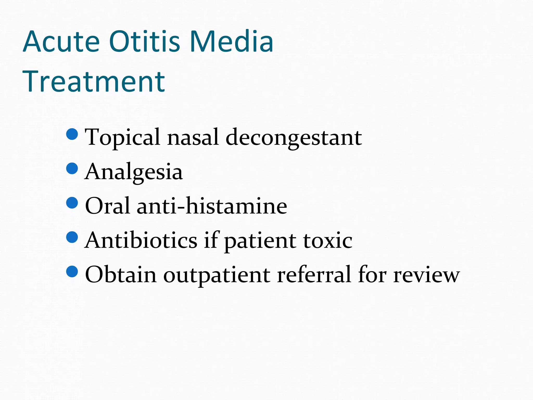
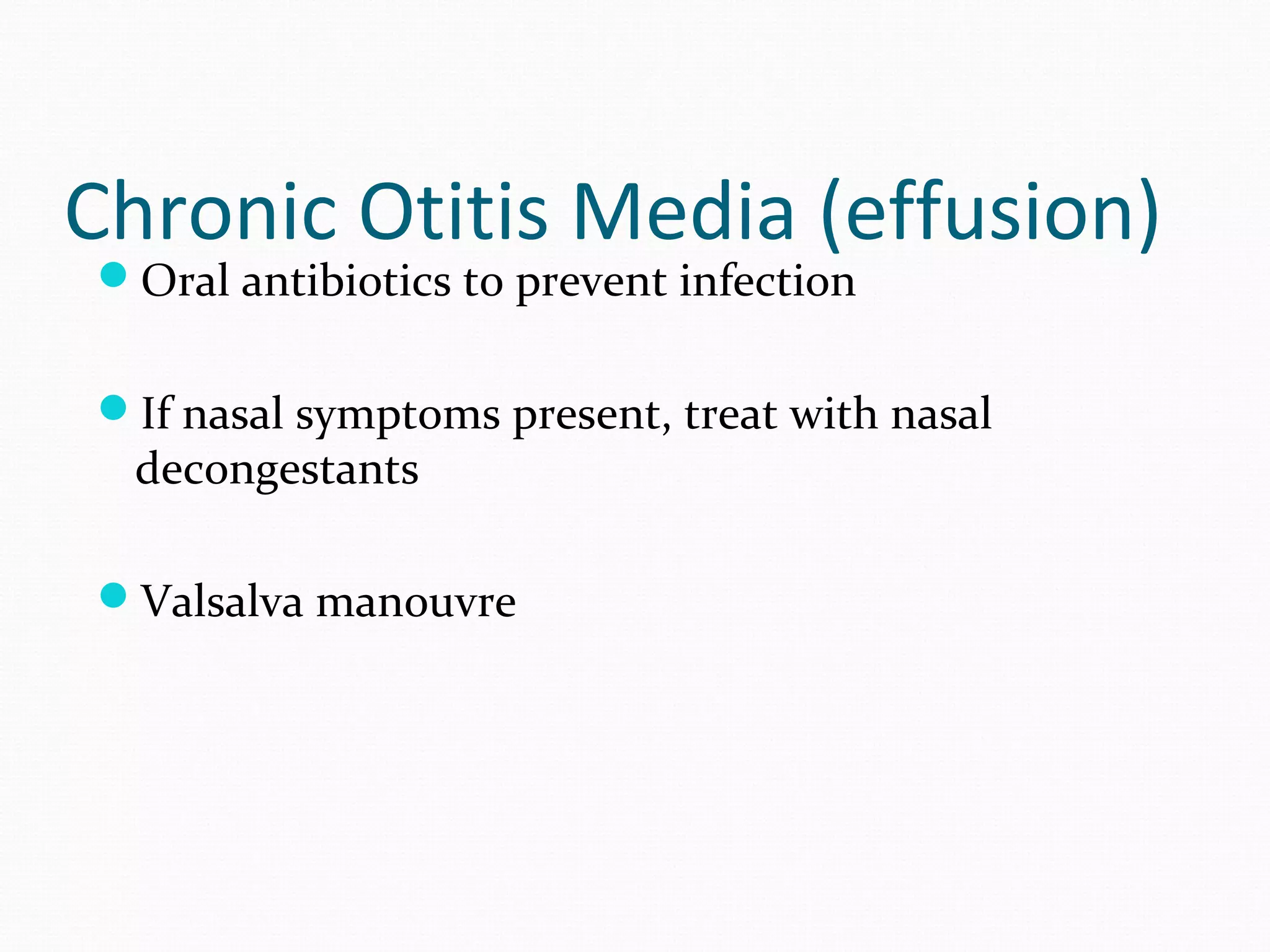
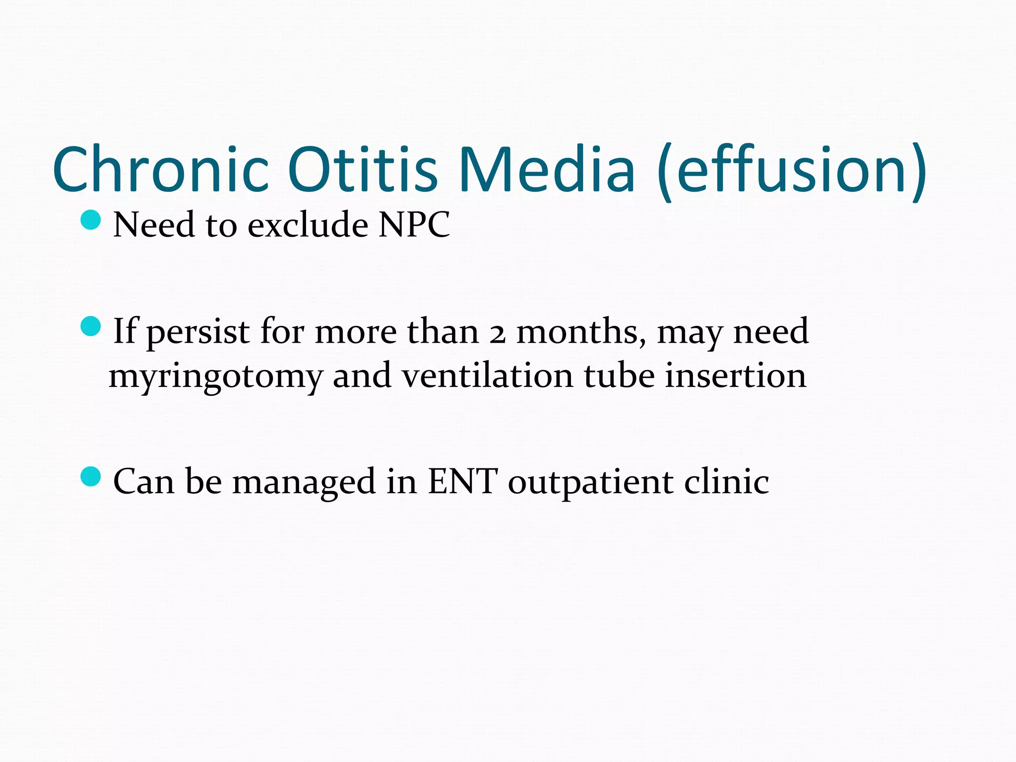
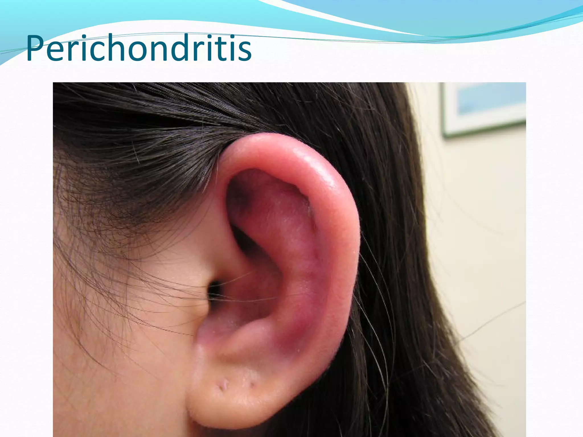
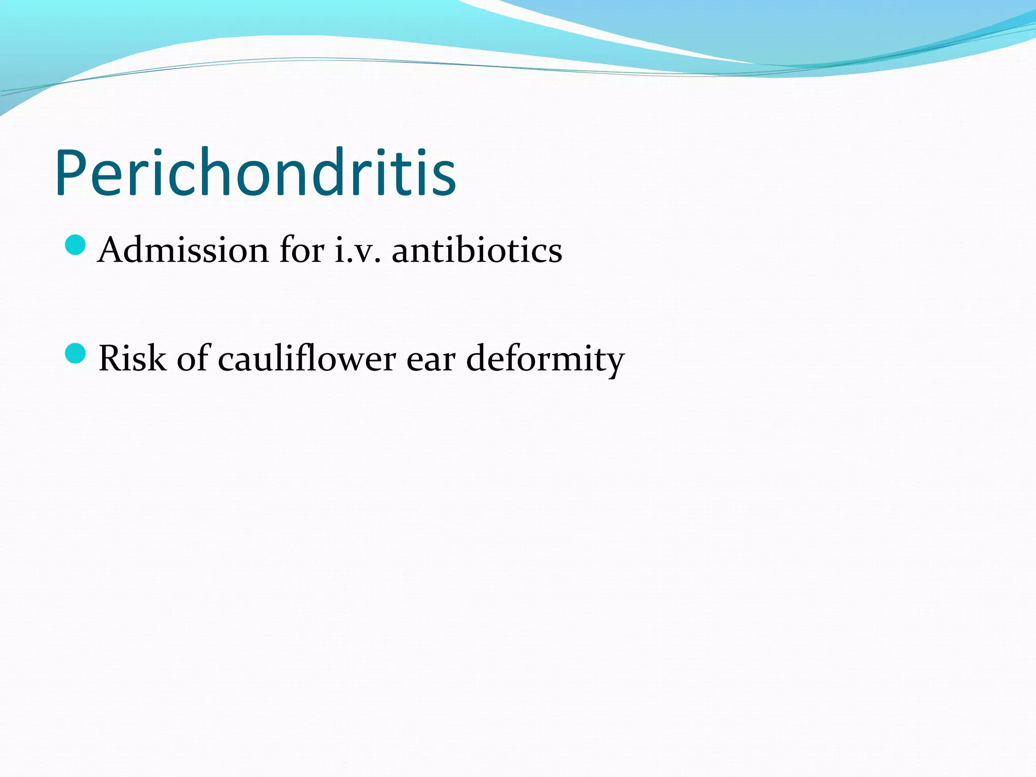
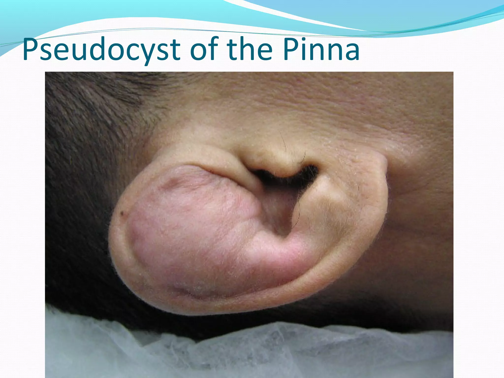
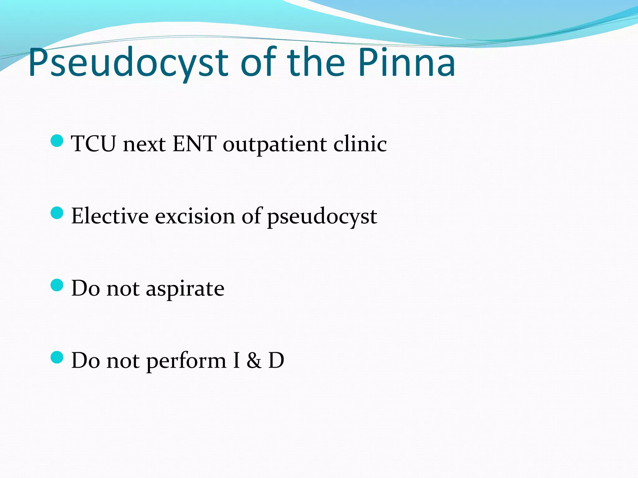
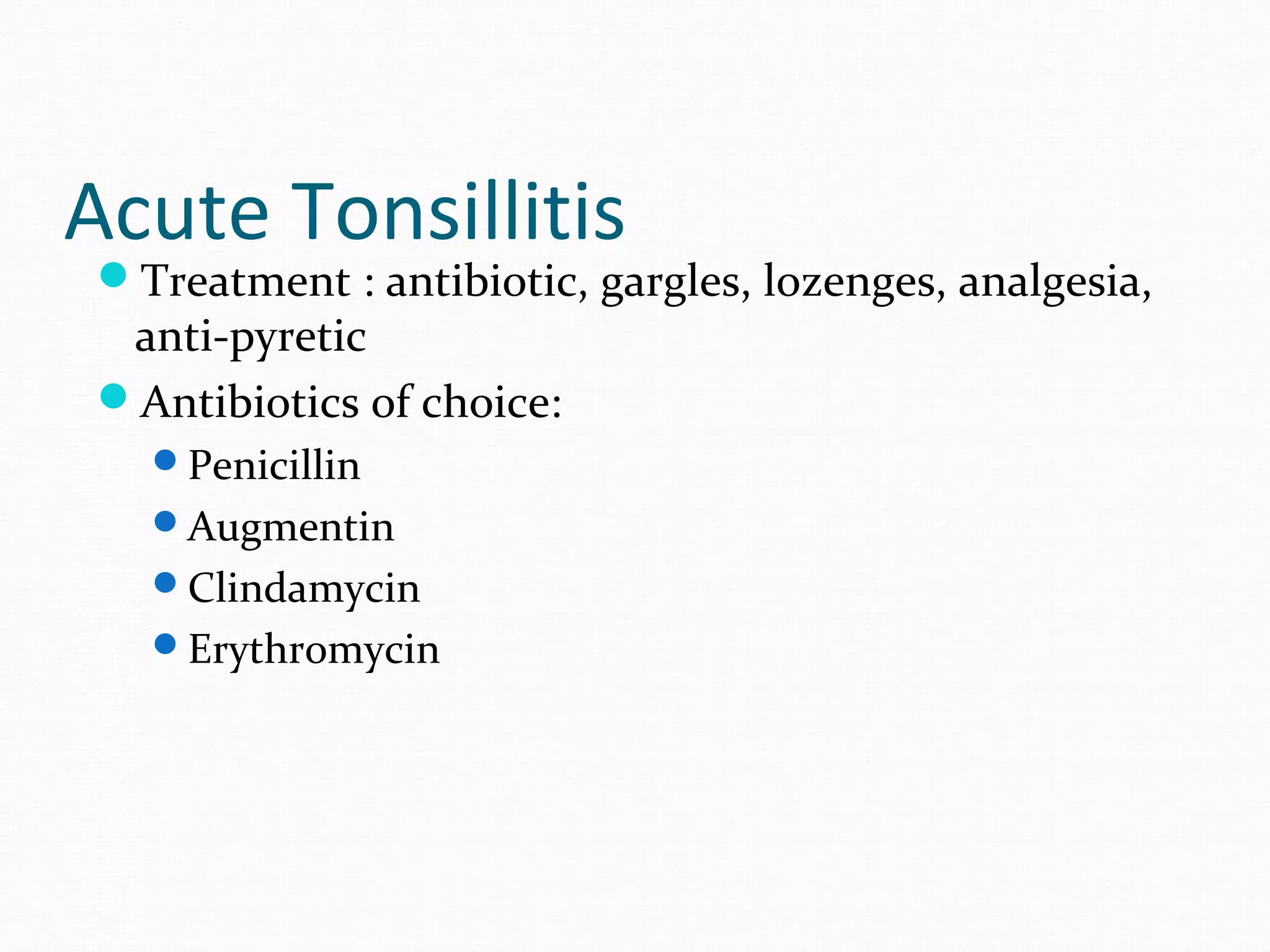
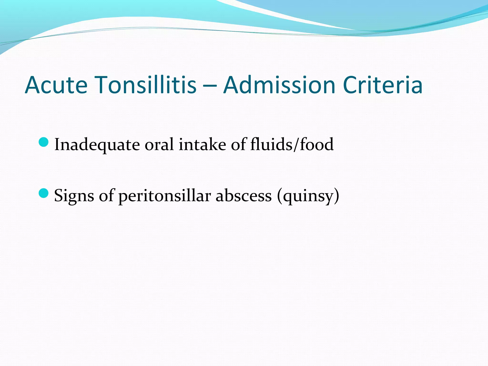
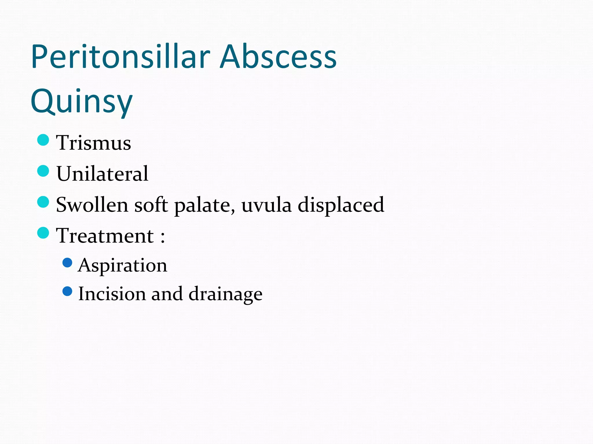
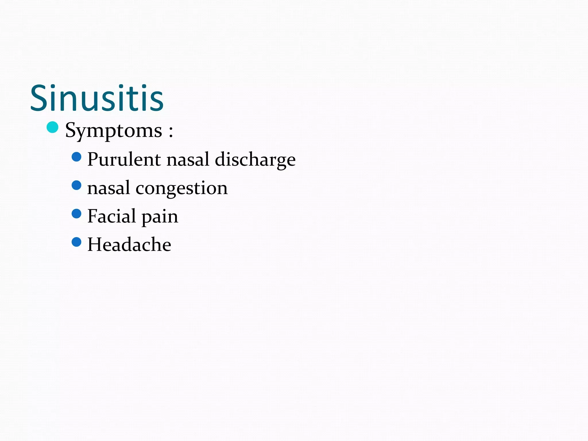
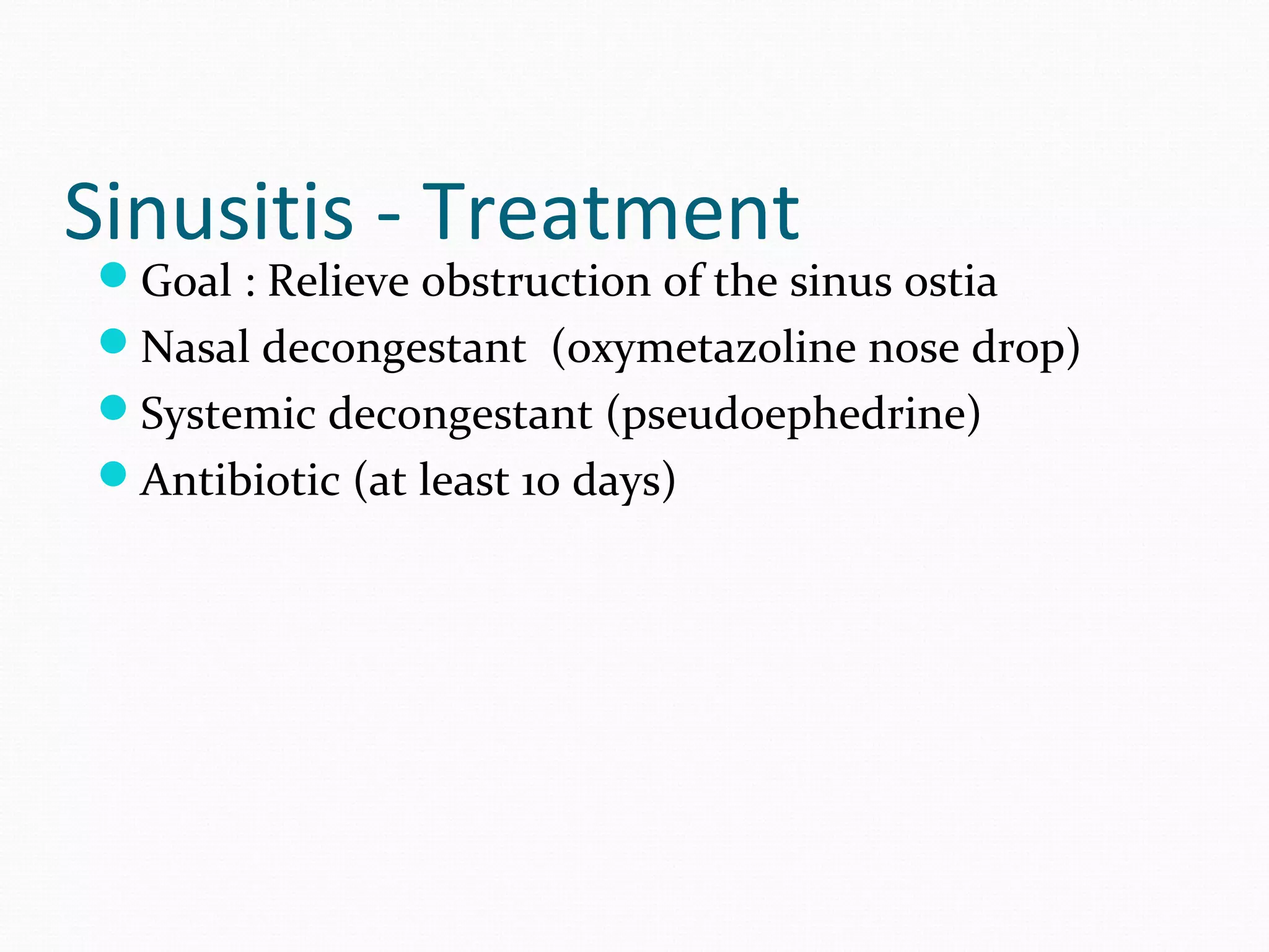
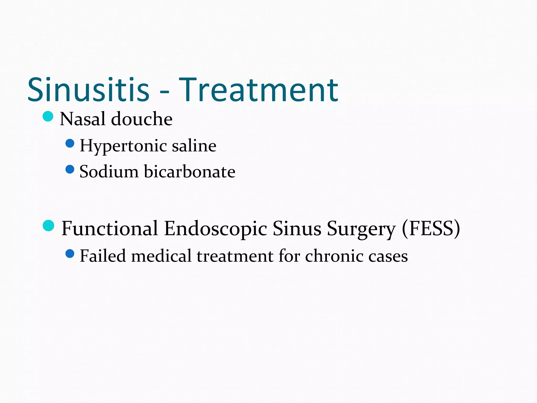
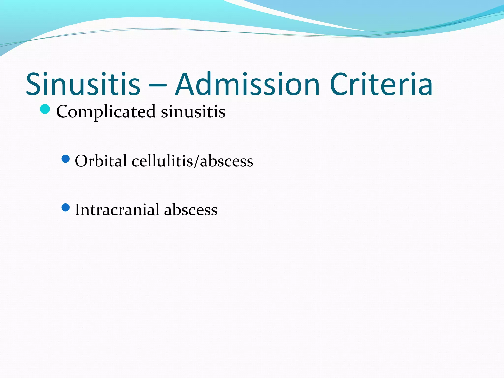
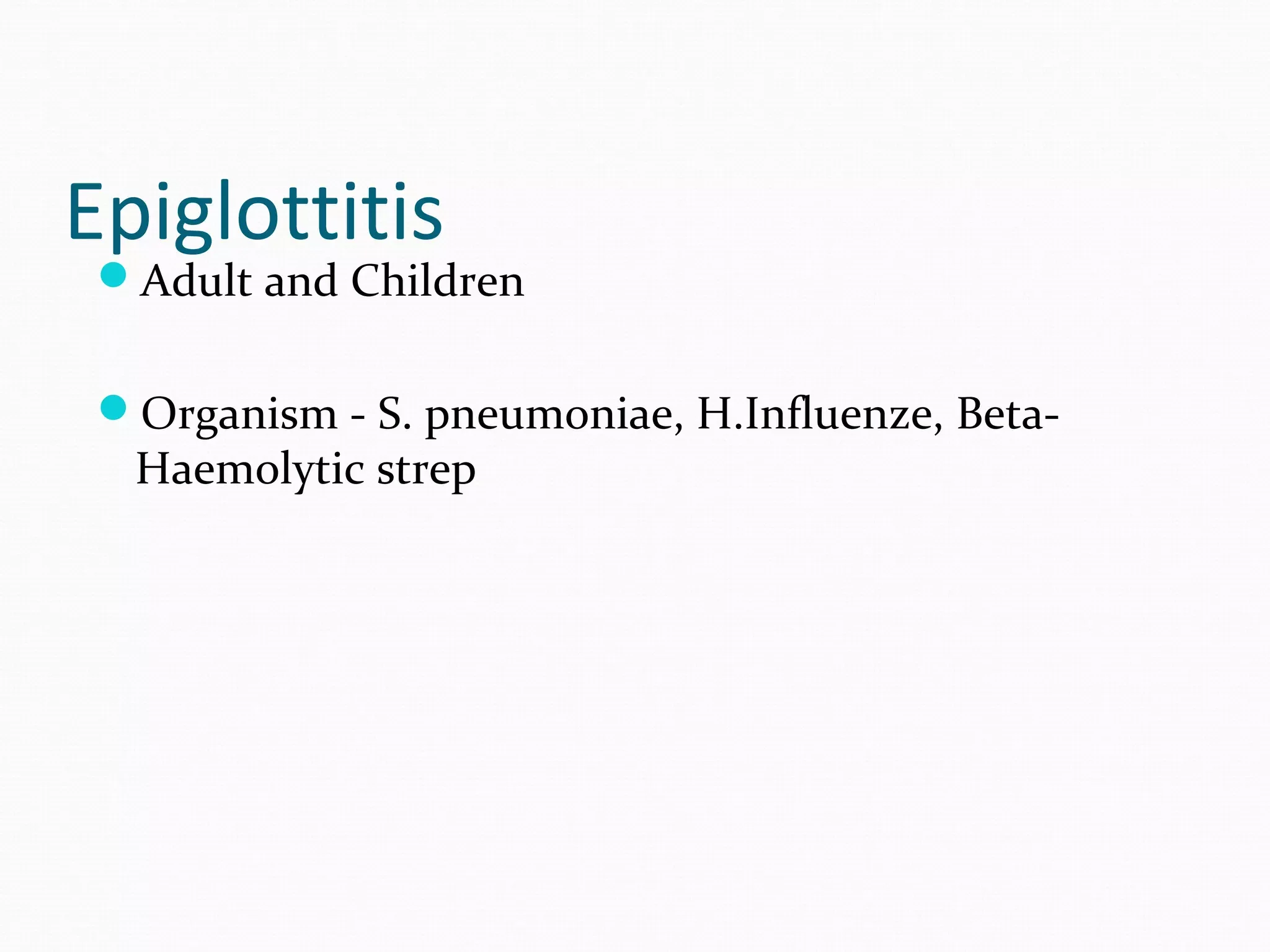
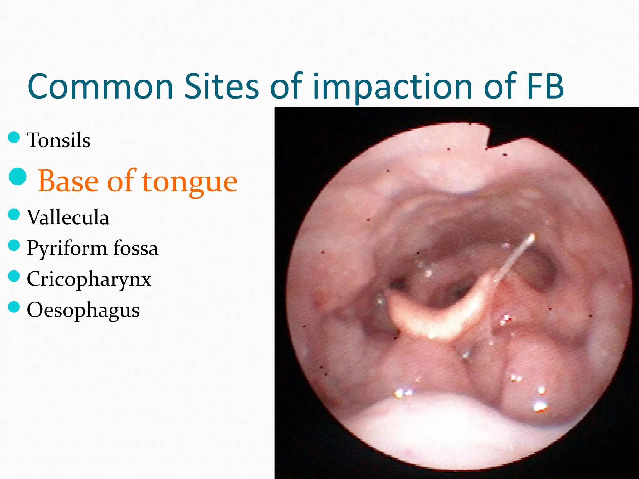
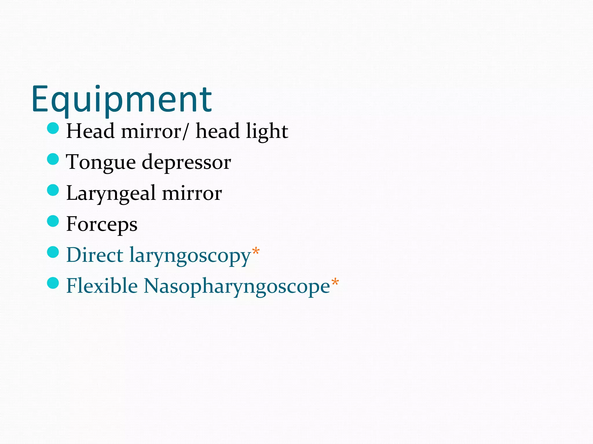
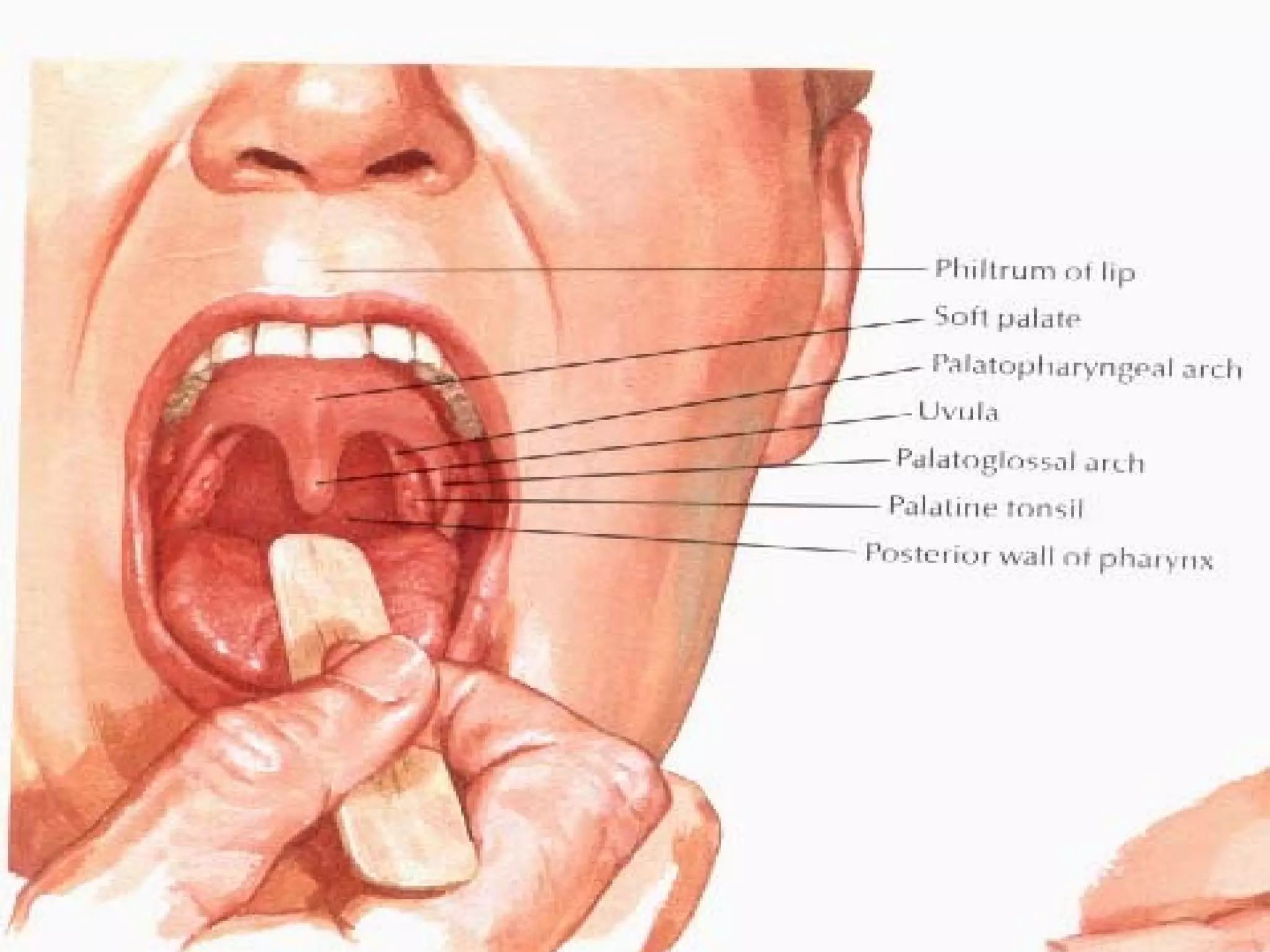
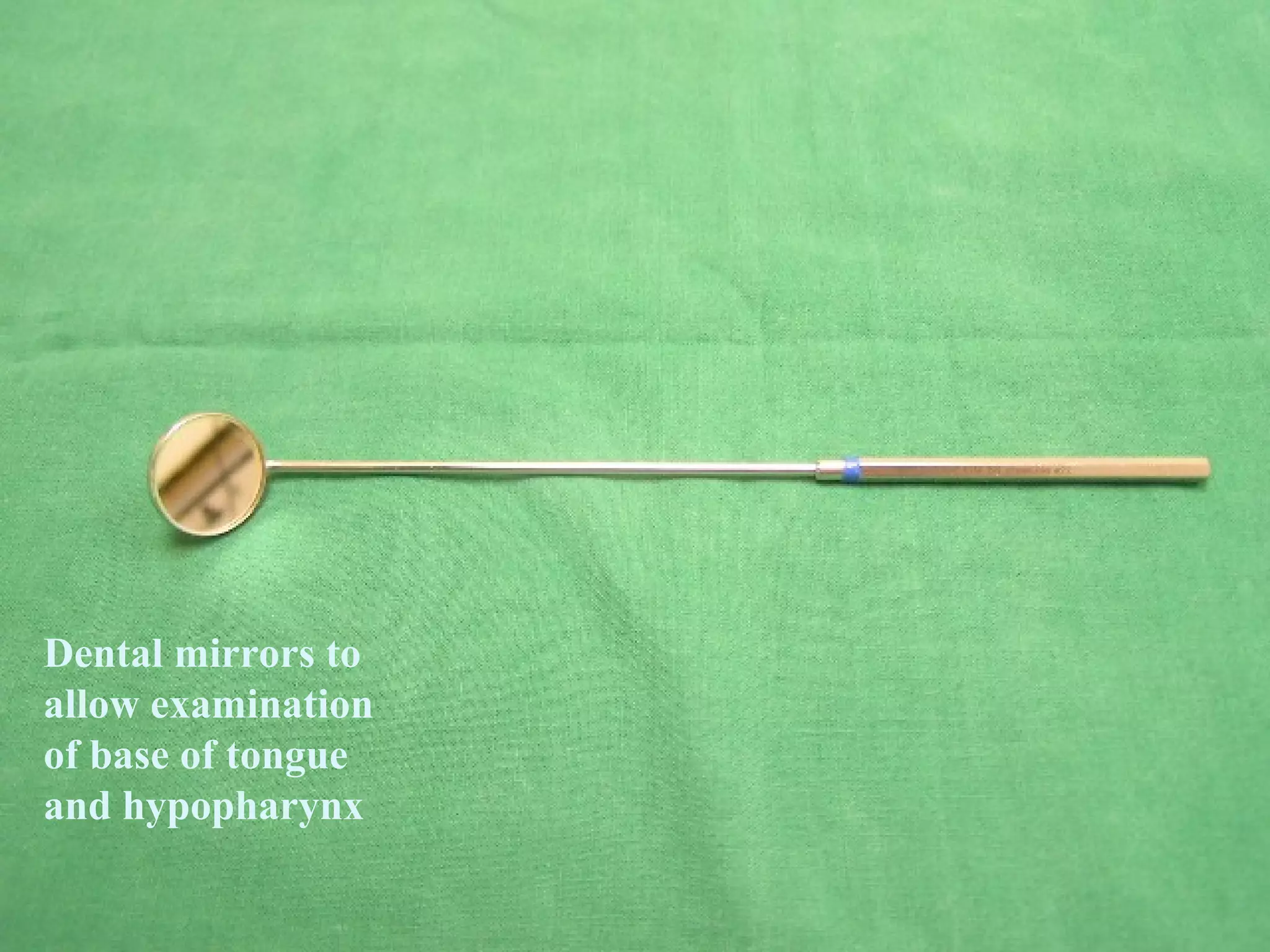
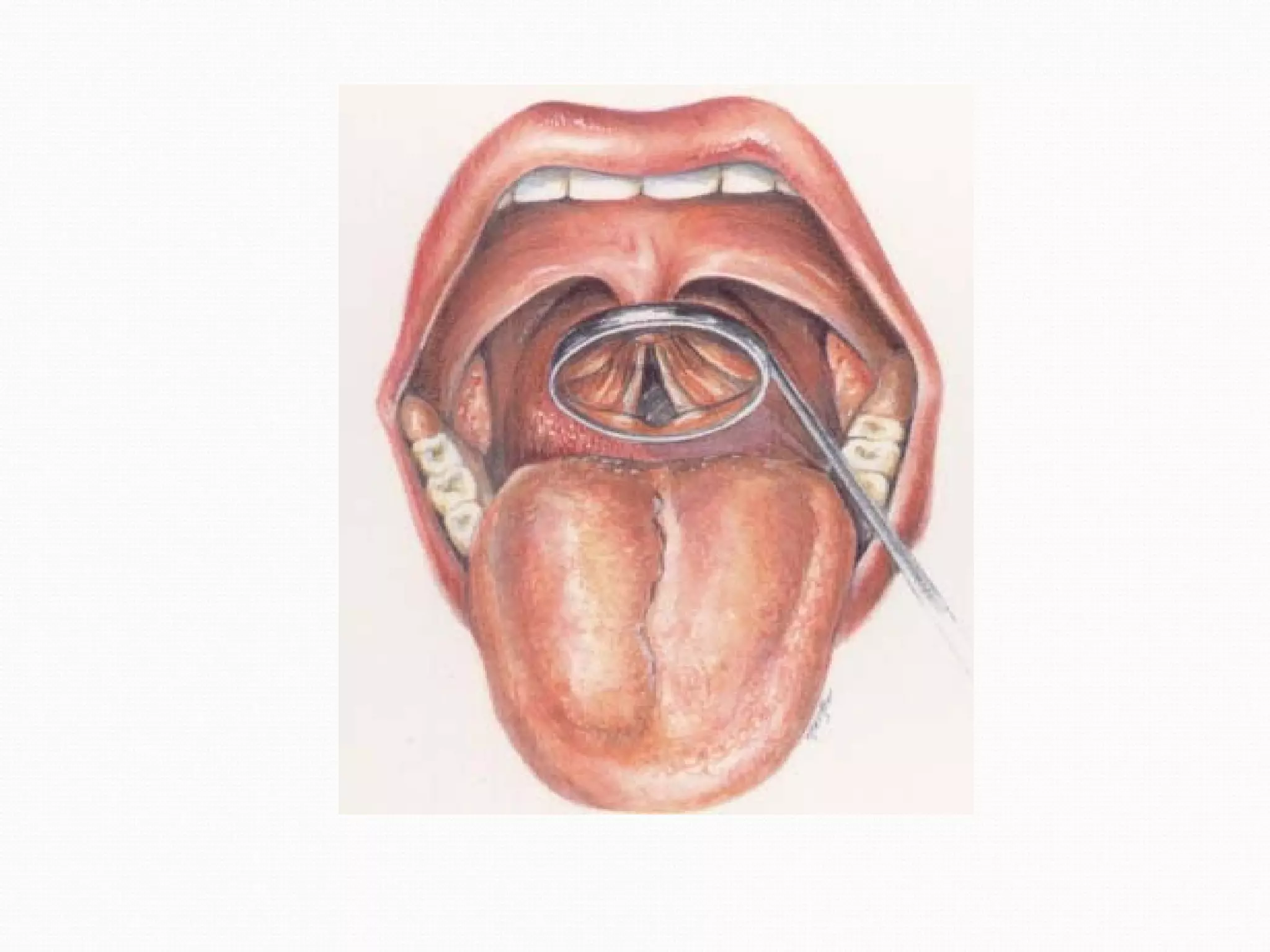
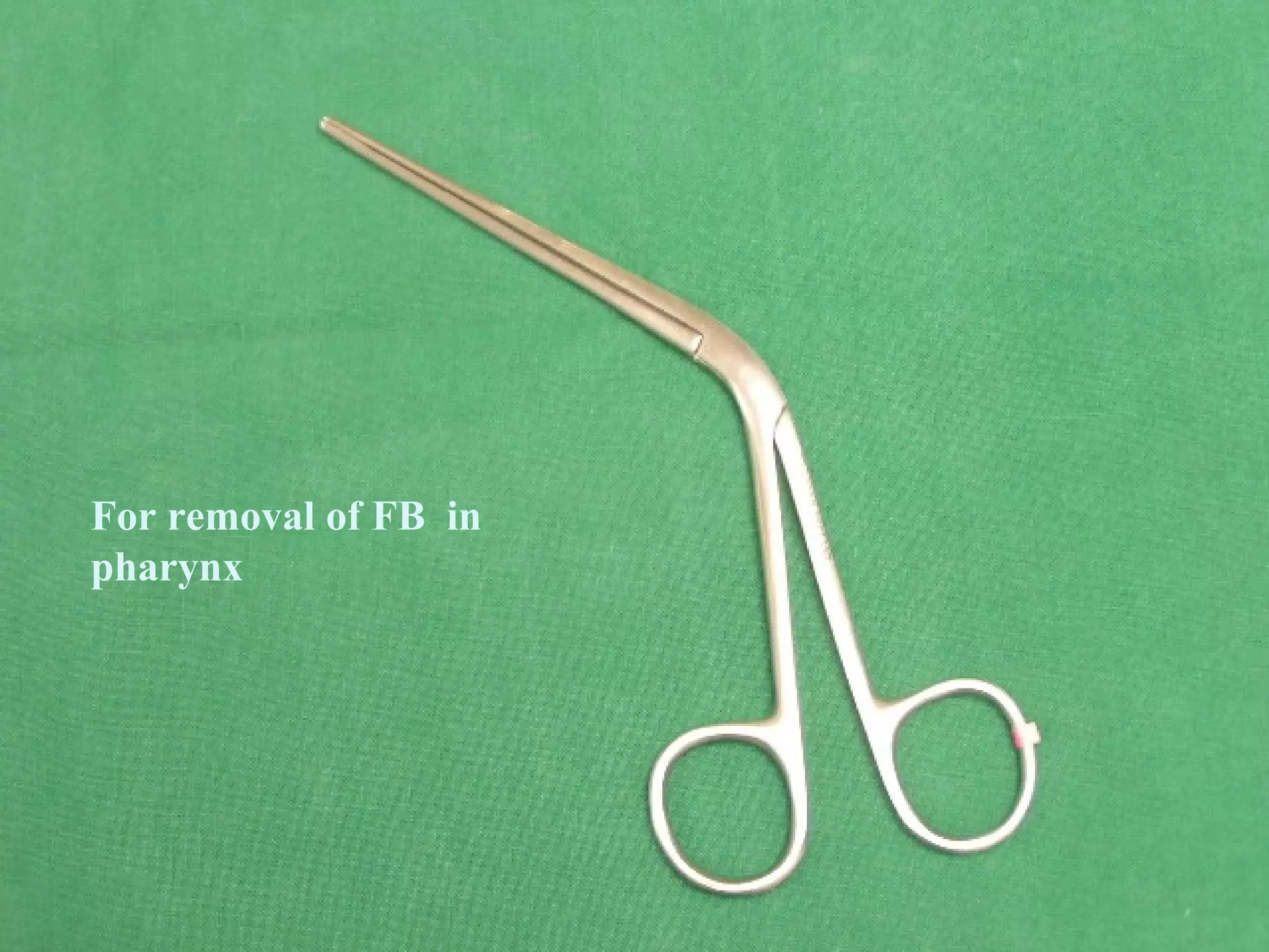
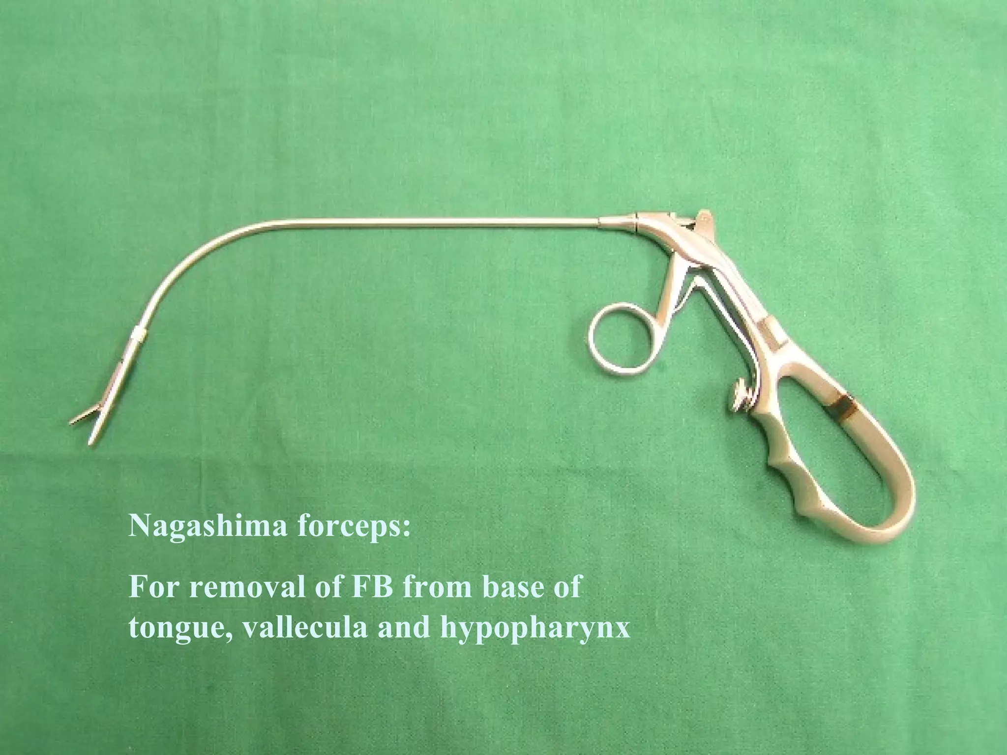
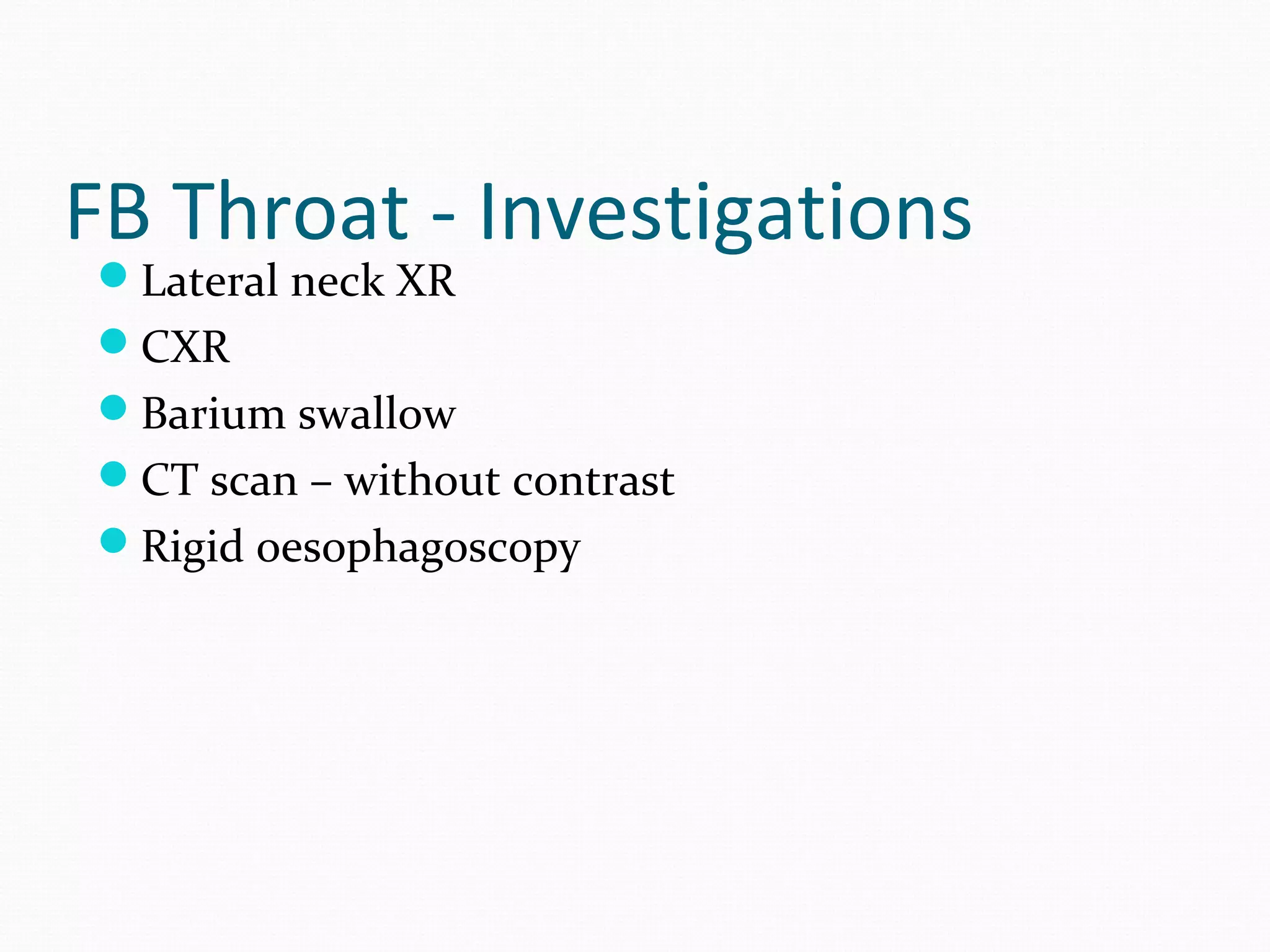
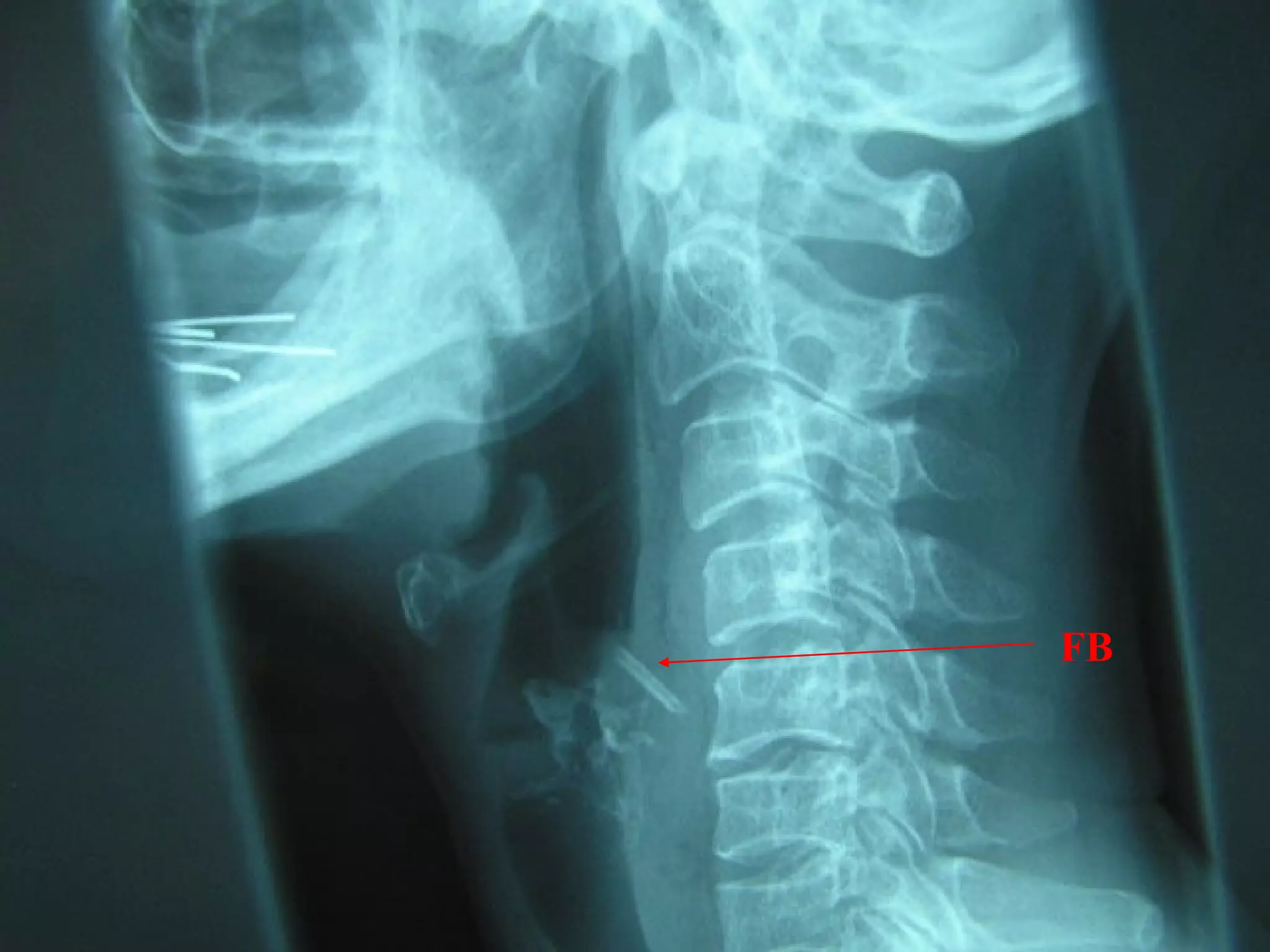
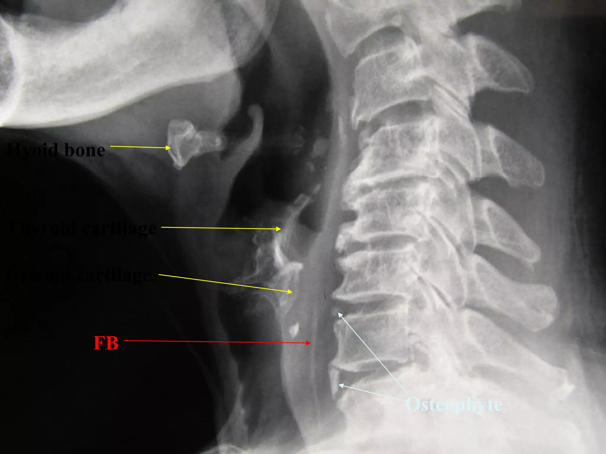
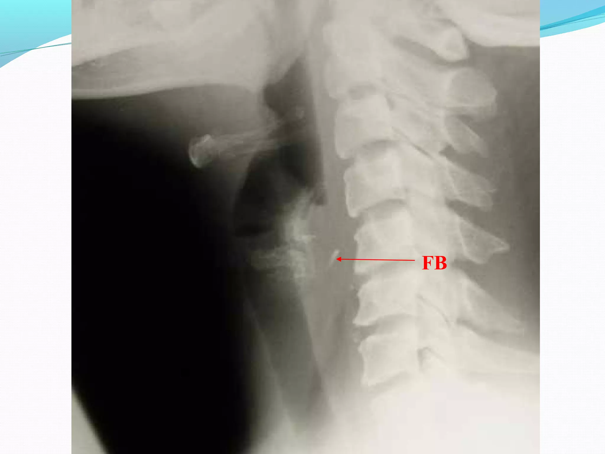
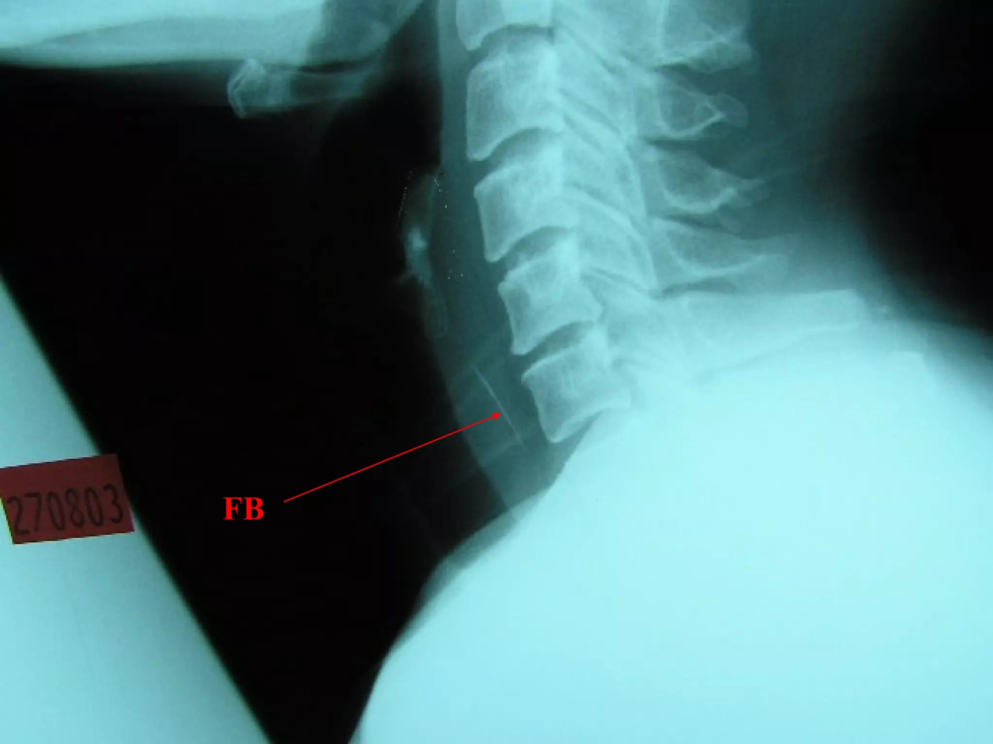
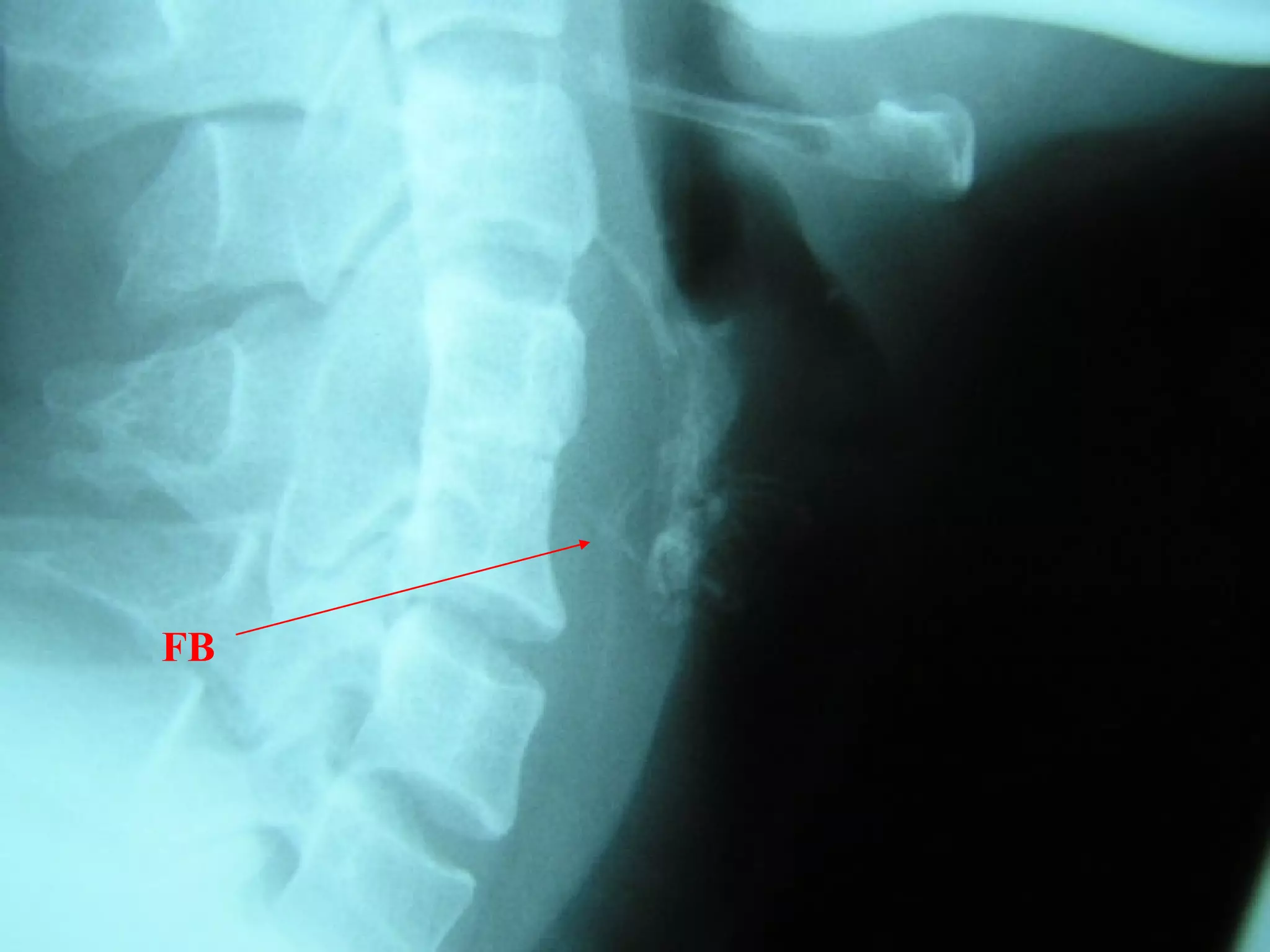
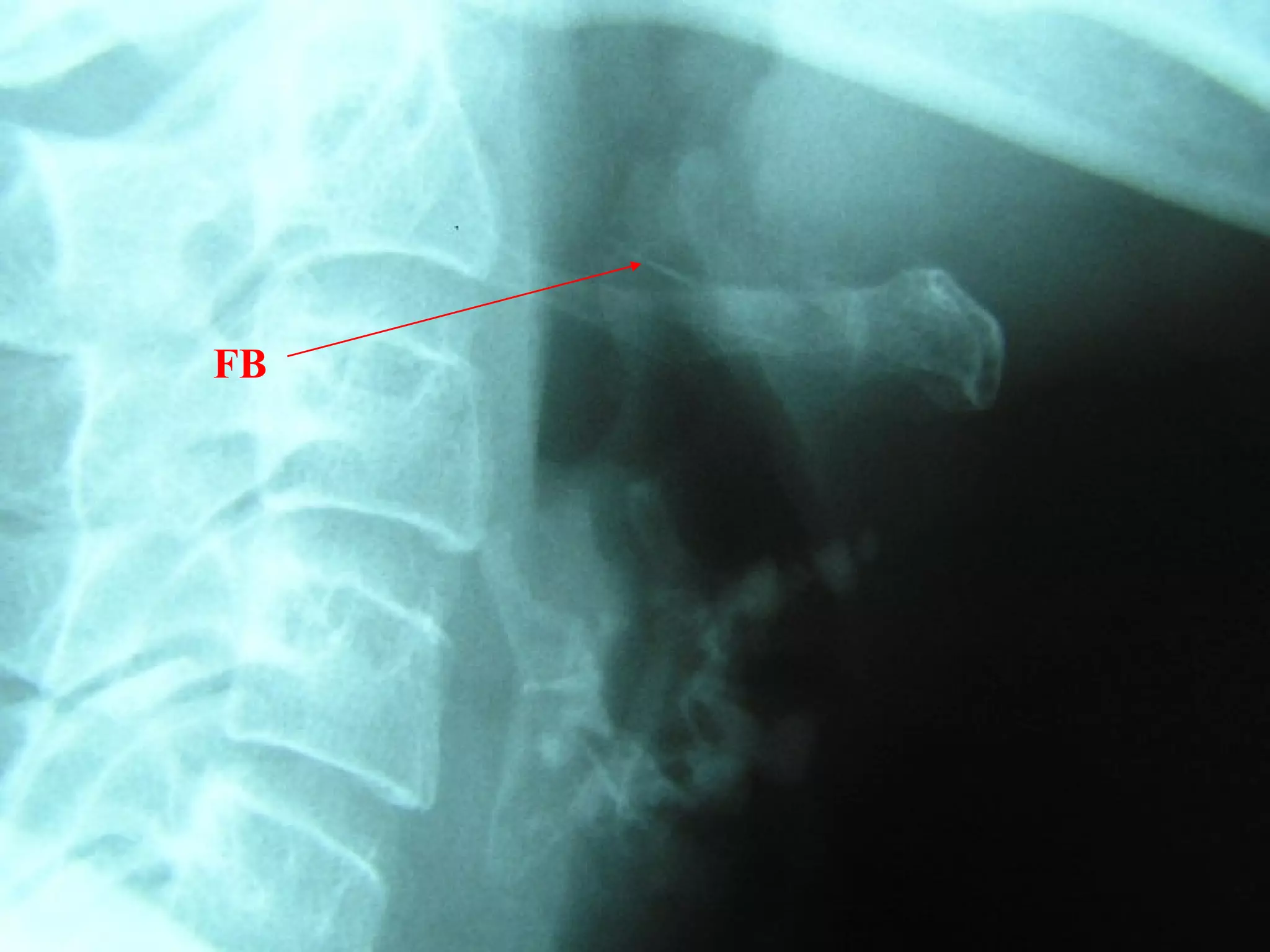
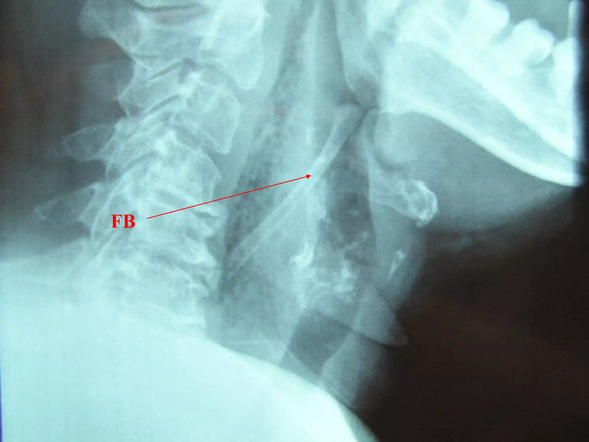
Topics:
Nasal Fracture
Haemorrhage - Epistaxis, Post-op Bleed
Ear Emergencies
Head & Neck Infections
Ingested Foreign Body
Nasal Fracture
Exclude intra-cranial, orbital and other facial

injuries
If epistaxis present, apply first-aid measures
Need to exclude septal haematoma (requires urgent
drainage)
Isolated nasal fractures can be managed as
outpatient
Nasal Fracture
Investigation

Request for ‘Nasal Bone Xray’
Nasal XR - medicolegal reason
In more serious injury - skull and facial XR
CT scan - useful for maxillofacial fractures and to

exclude other injuries
Nasal Fracture
Management

Refer to ENT urgently if:
Uncontrolled epistaxis
Presence of septal haematoma
Nasal Fracture
Management

Need for M&R within 14 days of injury
Refer to ENT outpatient to await reduction of

oedema to enable assessment of nasal bone
alignment
If epistaxis stops and no other significant
injuries, provide outpatient ENT follow-up
within 1 week of nasal injury
A. Epistaxis
Local causes
Idiopathic (90%)
Traumatic ( fracture, foreign body, nose picking )
Infection
Inflammatory ( rhinitits, sinusitis )
Tumour ( rare )
Iatrogenic (Nasal Surgery)
Epistaxis
General causes
Coagulopathy (Dengue, anticoagulant)
Hypertension
Hereditary Haemorrhagic telangiectasia
Raised venous pressure (whooping cough,

pneumonia)
Epistaxis

Majority are self limiting, esp. in children
90% bleed from Kiesselbach’s plexus (Little’s area)
Epistaxis
Kiesselbach plexus
Located on exposed anterior part of septum
Upper portion
ICA ( anterior and posterior ethmoidal arteries )

Lower portion
ECA ( Greater palatine, sphenopalatine, superior labial

arteries )
Epistaxis - First Aid
Sit up with head forward

Pinch the nose firmly with thumb and fingers for >

5min (Cartilaginous part)
Breathe through mouth
Ice pack on forehead
Ice cubes to suck
Epistaxis - Management
Assess blood loss

Resuscitation, i.v. access
Base line blood investigation
GXM
Medication - sedative or anti-hypertensive
Epistaxis - Management
Treat underlying cause
Reverse coagulopathy
Control hypertension
Allergic rhinitis
Sinusitis
Nasal hygiene
Haemostasis
Epistaxis - Haemostasis
1) Cautery
Silver Nitrate
Electrocautery

2) Anterior Nasal Pack
No clear bleeding point or Failed
cautery
BIPP or Merocel
Antibiotic cover
Epistaxis – Admission Criteria
Uncontrolled bleeding
Nasal packing done
Post-operative cases
Haemodynamically unstable
Poor premorbid conditions
Severe bleeds
B. Post-Tonsillectomy bleed
Resuscitation! & i.v. access

Assess for symptoms/signs of shock
Baseline blood including coagulation profile
GXM
NBM
Post-Tonsillectomy bleed
Management

First aid measures, e.g. ice gargle
Pressure (adrenaline gauze)
Silver nitrate
Electrocautery
Ligation of local bleeder
Post-Tonsillectomy bleed
Management& small clot evident
If no active bleed
observe

If large clot, need to remove clot to access if

bleeding
If active bleeding:

Attempt haemostasis at A&E/Clinic
Haemostasis under G.A.
Admission Criteria

All post-operative haemorrhage should be admitted
If bleeding stopped, offer admission for observation
Ear Emergencies
Admission Criteria
Most ear cases can be reviewed in the next ENT

outpatient clinic
Following needs urgent admission:
Acute Mastoiditis
Acute perichondritis of the pinna

Any ear infection/trauma with facial nerve palsy
Ear Cases Seen at A&E
1.

2.
3.
4.
5.
6.
7.

Impacted ear wax
Traumatic TM Perforation
Otitis Externa
Otitis Media
Sudden Sensorineural Hearing Loss
Foreign Body Ear
Miscellaneous
Impacted Ear Wax

Prescribe wax softeners (e.g. olive oil ear drops)
Obtain outpatient referral for review
Traumatic TM Perforation
If no other serious head injuries, can be followed up

as outpatient
1 week TCU
Keep ears dry
Antibiotics not required
Obtain outpatient referral for review
Otitis Externa
Treatment :
Aural toilet
Topical antibiotic ± steroid ear drops
Oral antibiotic for severe cases
Obtain outpatient referral for review
Acute Otitis Media
Common in children
Fever, ear- pain
TM - red & bulging
Otitis media can only be diagnosed if the TM is

visualised!
Acute Otitis Media
Treatment
Topical nasal decongestant
Analgesia
Oral anti-histamine
Antibiotics if patient toxic
Obtain outpatient referral for review
Chronic Otitis Media (effusion)
Oral antibiotics to prevent infection

If nasal symptoms present, treat with nasal

decongestants

Valsalva manouvre
Chronic Otitis Media (effusion)
Need to exclude NPC

If persist for more than 2 months, may need

myringotomy and ventilation tube insertion

Can be managed in ENT outpatient clinic
Chronic Suppurative Otitis Media
(CSOM)
Aural toilet
Topical ± oral antiobiotics
Keep ears dry
Elective Myringoplasty if perforation does not heal
Can be managed in ENT outpatient clinic
Sudden Sensorineural Hearing Loss
Loss of hearing of > 30 dB over 3 days, over at

least 3 frequencies
Sudden onset of hearing loss
Normal ear examination
Diagnose SNHL with tuning fork tests or puretone audiogram
Sudden Sensorineural Hearing Loss
Refer to next ENT outpatient clinic
Cover with oral prednisolone 1mg/kg if no

contraindication
Acyclovir 800mg 5x/day for 5 days, if onset within 1
week
Sudden Sensorineural Hearing Loss Causes
 Idiopathic – 85%
 Meniere’s disease
 Acoustic Neuroma
 Cerebellar-pointine angle tumours
 Ototoxicity
 Noise-induced
 Trauma
 Viral infection
 Vascular - impairment of cochlear blood supply
 Syphillis
 Immunological disorders
FB - Ear
Crocodile forceps-

cotton,paper,foam ,
sponge
Blunt hook – round
objects
Suction – fluid
FB - Ear
Syringing – C/I organic material
Insects
Killed by alcohol/lignocaine/olive oil
Removed

Can be managed in ENT outpatient clinic
Perichondritis
Perichondritis
Admission for i.v. antibiotics
Risk of cauliflower ear deformity
Pseudocyst of the Pinna
TCU next ENT outpatient clinic
Elective excision of pseudocyst
Do not aspirate
Do not perform I & D
Giddiness

≠

ENT Referral

Always exclude central causes first
Peripheral causes not life-threatening
Head & Neck Infections
Acute Tonsillitis

Peritonsillar Abscess (Quinsy)
Sinusitis
Epiglottitis
Deep Neck Infection
Acute Tonsillitis

Sorethroat, Fever, Odynophagia
Bilateral tonsils - enlarged, injected, swollen,

purulent exudates

Diptheria, Infectious Mononucleosis
Acute Tonsillitis

Treatment : antibiotic, gargles, lozenges, analgesia,

anti-pyretic
Antibiotics of choice:
Penicillin
Augmentin
Clindamycin
Erythromycin
Acute Tonsillitis – Admission Criteria
Inadequate oral intake of fluids/food
Signs of peritonsillar abscess (quinsy)
Peritonsillar Abscess
Quinsy
Trismus
Unilateral
Swollen soft palate, uvula displaced
Treatment :
Aspiration
Incision and drainage
Sinusitis

Symptoms :
Purulent nasal discharge
nasal congestion
Facial pain
Headache
Sinusitis - Diagnosis
Clinical
History
Sinus X-ray - not reliable, not necessary
Mucopus seen on nasal endoscopy
Sinusitis - Treatment

Goal : Relieve obstruction of the sinus ostia
Nasal decongestant (oxymetazoline nose drop)
Systemic decongestant (pseudoephedrine)
Antibiotic (at least 10 days)
Sinusitis - Treatment
Nasal douche

Hypertonic saline
Sodium bicarbonate

Functional Endoscopic Sinus Surgery (FESS)
Failed medical treatment for chronic cases
Sinusitis – Admission Criteria
Complicated sinusitis

Orbital cellulitis/abscess
Intracranial abscess
Epiglottitis

Adult and Children
Organism - S. pneumoniae, H.Influenze, Beta-

Haemolytic strep
Epiglottitis

Severe sore throat, odynophagia, high fever
Muffled voice, Difficulty in breathing
Sit erect and bend forward
Salivating
Epiglottitis

Indirect Laryngoscopy
Flexible Fiberoptic nasopharyngoscope
Lateral neck X-ray - thumb sign
Normal
epiglottis
Swollen
epiglottis
Epiglottitis - Treatment
Airway management
Monitor Closely
Intubation, Cricothyroidectomy, Tracheostomy
Oxygen
Antibiotic
Epinephrine, steroids
Deep Neck Infection
Neck swelling

Sore throat, odynophagia, trismus
Immunocompromised
Fever, unwell
Lateral neck XR
Airway control
Admission for CT, KIV I&D
Deep Neck
Infection
•Normal retropharyngeal
space on lateral neck XR is
up to 1 vertebral body
width from C5 and below.

Widened
retropharyngeal space
on lateral neck XR

•Up to half a vertebral
body width from C1 to C4
is normal
FB - Throat
History
Localised
Below post-cricoid region - midline
Mouth/ oropharynx – localised to side

Time of ingestion
FB - Throat
History
High risk
Sensory deprivation eg dentures (adults )

Otalgia, neck tenderness, fever, chest or back pain,

haemetamesis
FB - Throat
Examination
Distress
Unable to swallow saliva
Tracheal rock positive
Swallow test positive
Common Sites of impaction of FB
Tonsils
Base of tongue
Vallecula
Pyriform fossa
Cricopharynx
Oesophagus
Common Sites of impaction of FB
Tonsils
Base of tongue
Vallecula
Pyriform fossa
Cricopharynx
Oesophagus
Common Sites of impaction of FB
Tonsils

Base of tongue
Vallecula
Pyriform fossa
Cricopharynx
Oesophagus
Equipment

Head mirror/ head light
Tongue depressor
Laryngeal mirror
Forceps
Direct laryngoscopy*
Flexible Nasopharyngoscope*
Tongue depressors
to allow
examination of
tonsils
Dental mirrors to
allow examination
of base of tongue
and hypopharynx
For removal of FB in
pharynx
Nagashima forceps:
For removal of FB from base of
tongue, vallecula and hypopharynx
FB Throat - Investigations
Lateral neck XR
CXR
Barium swallow
CT scan – without contrast
Rigid oesophagoscopy
FB
FB
Hyoid bone

Thyroid cartilage
Cricoid cartilage
FB
Osteophyte
FB
FB
FB
FB
FB
FB Throat

>50% of ingested FB cannot be found!
Discharge with symptomatic treatment
Cover with antibiotics if diabetic patient or

immunocompromised
FB advice

Chest pain, fever, increasing symptoms
FB Throat
Can be seen in next ENT clinic if:
No FB found on detailed examination
No chest pain
Symptoms mild
Emergencies in ENT

Emergencies in ENT