
Recommended
More Related Content
What's hot
What's hot (19)
Viewers also liked
Viewers also liked (20)
Similar to Efm dunn with recording
Similar to Efm dunn with recording (20)
Recently uploaded
Recently uploaded (20)
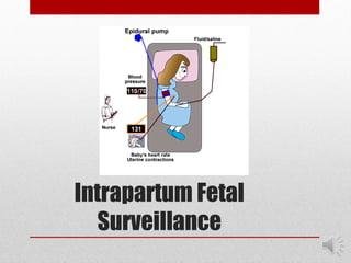
Efm dunn with recording
- 2. Electronic Fetal Monitoring (EFM) • Ultrasound (US) • Tocotransducer (TOCO) • Internal Fetal Scalp Electrode (IFSE) • Intrauterine Pressure Catheter (IUPC) • Doppler US
- 3. • The primary objective of EFM is to provide information about fetal oxygenation and prevent fetal injury that could result from impaired fetal oxygenation during labor. • This is achieved by detecting fetal heart rate changes early before they are prolonged and profound. EFM
- 4. • Adequate fetal oxygenation requires five related factors: • Adequate maternal blood flow and volume to the placenta. • Adequate oxygen saturation in maternal blood. • Adequate exchange of oxygen and carbon dioxide in the placenta. • An open circulatory path between the placenta and the fetus through vessels in the umbilical cord. • Adequate fetal circulatory and oxygen-carrying functions. Fetal Oxygenation
- 5. • Review: Oxygen rich and nutrient rich blood from the mother enters the intervillous spaces of the placenta through the spiral arteries. • Oxygen and nutrients in the maternal blood pass into the fetal blood that circulates in capillaries in the intervillous spaces. • CO2 and other waste products pass from the fetal blood into the maternal blood at the same time. • Maternal blood carrying fetal waste products drains from the intervillous spaces through endometrial veins and returns to the mother’s circulation for elimination. Uteroplacental Exchange
- 6. • Substances pass back and forth between mother and fetus without mixing of maternal and fetal blood if fetal capillaries remain intact. • During labor contractions gradually compress the spiral arteries, temporarily stopping maternal blood flow into the intervillous spaces. • Thus during contractions the fetus depends on the oxygen supply already present in body cells, along with intervillous spaces. Oxygen supply in these areas is enough for about 1 to2 minutes. Exchange continued:
- 7. • The average heart rate measured over a 2 minute period within a 10 minute window • Normal=110-160 • Brady=<110 for 10 minutes • Tachy=>160 for 10 minutes • Pre-term may have >160 due to immature parasympathetic NS Baseline FHR
- 8. • Most important component of the FHR • Irregular fluctuations in the baseline fetal heart rate. • Measured as the amplitude of the peak to trough in bpm • Evaluates fetal autonomic nervous system, adequate 02 status promotes normal FX • Long-term variability(LTV) broader fluctuations of the FHR • Short-term variability(STV) beat-to-beat variability, only with IFSE • No variability=fetal compromise Variability
- 10. Accelerations 15 bpm X 15 sec
- 11. Moderate variability = 6=25 bpm variation
- 12. Minimal variability = <5 bpm variation
- 13. • Accelerations (accels) – transitory abrupt increases in the FHR above the baseline,15 bpm above baseline FHR greater than 15 sec less than 2 minutes. Reassuring; thus denoting fetal movement and fetal well-being and are the basis for nonstress testing. • Decelerations- transient fall in FHR caused by stimulation of the parasympathetic NS. • Early=head compression • Late=utero-placental insufficiency • Variable=cord compression Periodic changes
- 14. Early decelerations = head compression
- 15. • Deceleration with each contraction • Mirror image when contraction begins the heart rate begins to drop • Action – prepare for birth Head compression
- 16. Late decelerations = uteroplacental insufficiency
- 17. • Deceleration with each contraction • Late onset, usually see a decrease in variability • Action=intra-uterine resuscitation • If on Pitocin – turn it OFF • Increase mainline fluid volume, bolus with LR • O2 via face mask at 8-10 L • Turn to left side, or whatever position best for blood flow • Scalp stimulation? Late decelerations
- 18. Variable decelerations = cord compression
- 19. • Occurs randomly • Not a problem if occasional and returns to baseline • If frequent, long return, < variability=intrauterine resuscitation. • Turn to left side • Pitocin off and increase fluids • O2 via face mask at 8- 10 L • Amnioinfusion – warm NS – only with variables!!!! Variable deceleration
- 20. • 1.If Pitocin is on turn it off! • 2. Turn the patient • 3. IV bolus • 4. Oxygen administration. 10 Liters per face mask. Non rebreather. Intrauterine Resusutation
- 21. Prolapsed cord • Do not remove fingers after exam • Relieve pressure from presenting part • Call for help, prepare for emergency C/S • Remain calm
- 23. • Reduced mobility • May require frequent adjustment of equipment to obtain continuous tracing • Best identifies the well oxygenated fetus…does not reliably identify the compromised fetus • Increased operative intervention EFM Limitations
- 24. • Intra-uterine resuscitation • Measures create anxiety, fear, and loss of control • Remain calm, educate patient and family, and choose words wisely • Report all interventions to MD Interventions
- 25. • Supplies more data, becomes part of the permanent record • Shows FHR in relation to stimuli • Provides a feeling of safety for the parent • Allows nurse/patient ratio to be 1:2 with central monitoring EFM Advantages
- 26. Fetal Scalp Electrode (FSE)
- 27. Intrauterine Pressure Catheter (IUPC)
- 28. • Indications: persistent, deep variable decelerations, meconium stained amniotic fluid. • Requires IUPC • Warm NS fluid of choice • Infusion pump • Weigh all chux and record output Amnioinfusion