
Recommended
More Related Content
What's hot
What's hot (20)
Similar to Fetal Monitoring Guide: Understanding Cardiotocography Patterns
Similar to Fetal Monitoring Guide: Understanding Cardiotocography Patterns (20)
More from Rajani17
More from Rajani17 (20)
Recently uploaded
Recently uploaded (20)
Fetal Monitoring Guide: Understanding Cardiotocography Patterns
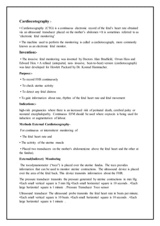
- 1. Cardiocotography:- • Cardiotocography (CTG) is a continuous electronic record of the fetal’s heart rate obtained via an ultrasound transducer placed on the mother’s abdomen • It is sometimes referred to as ‘electronic fetal monitoring’ • The machine used to perform the monitoring is called a cardiotocograph, more commonly known as an electronic fetal monitor. Invention:- • The invasive fetal monitoring was invented by Doctors Alan Bradfield, Orvan Hess and Edward Hon. • A refined (antepartal, non- invasive, beat-to-beat) version (cardiotocograph) was later developed for Hewlett Packard by Dr. Konrad Hammacher. Purpose:- • To record FHS continuously • To check uterine activity • To detect any fetal distress • To gain information about rate, rhythm of the fetal heart rate and fetal movement Indications:- high-risk pregnancies where there is an increased risk of perinatal death, cerebral palsy or neonatal encephalopathy. Continuous EFM should be used where oxytocin is being used for induction or augmentation of labour. Methods External Cardiotocography- For continuous or intermittent monitoring of • The fetal heart rate and • The activity of the uterine muscle • Placed two transducers on the mother's abdomen(one above the fetal heart and the other at the fundus). External(Indirect) Monitoring The tocodynamometer (“toco”) is placed over the uterine fundus. The toco provides information that can be used to monitor uterine contractions. The ultrasound device is placed over the area of the fetal back. This device transmits information about the FHR. The pressure transducer transmits the pressure generated by uterine contractions in mm Hg. •Each small vertical square is 5 mm Hg •Each small horizontal square is 10 seconds . •Each large horizontal square is 1 minute . Pressure Transducer Toco sensor Ultrasound transducer The ultrasound probe transmits the fetal heart rate in beats per minute. •Each small vertical square is 10 beats. •Each small horizontal square is 10 seconds . •Each large horizontal square is 1 minute .
- 2. Internal Cardiotocography- • Uses an electronic transducer connected directly to the fetal scalp through the cervical opening and is connected to the monitor. • Internal monitoring provides a more accurate. • Internal monitoring may be used when external monitoring of the fetal heart rate is inadequate. • It need some degree of cervical dilatation. Internal Monitoring Criteria for Internal Monitoring: Amniotic membranes must be ruptured Cervix dilated 2 cm. Presentation must be cephali c Presenting part down against the cervix Equipment:- • Cardiotocograph • Transducer(2):Toco and cardio • Conduction gel or paste • Abdominal binder (two belts) • Monitor paper • Tissue paper Preparation for CTG • Determine the indication for fetal monitoring • Explain the purpose, time required for test • Instruct the women for empty the bladder • Place the women in supine position • Uncover the abdomen Procedure:- • Place the toco sensor on the fundus of uterus and fix it with abdominal binder • Identify the presentation and position of the fetus • Localize the FHS and fix it with abdominal binder • Assure the recording of FHS and uterine contraction • Explain the mother to push the bottom when she feel any movements
- 3. • Turn off the monitor and replace • Read the CTG and immediately notify the doctor ,if any abnormality seen Interpretation:- • Uterine activity (contractions) • Baseline fetal heart rate (FHR) • Baseline FHR variability • Presence of accelerations • Periodic or episodic decelerations • Changes or trends of FHR patterns over time. Uterine activity (contraction) :- • Frequency- the amount of time between the start of one contraction to the start of the next contraction. • Duration :The amount of time from the start of a contraction to the end of the same contraction • Intensity (strongeness):a measure of how strong a contraction is. In early labour the contractions are weak, with amplitude of about 20 mm Hg and at the end of the first stage 60 mm Hg • Resting Tone- a measure of how relaxed the uterus is between contraction(between 4-10 mm Hg) • Interval- the amount of time between the end of one contraction to the beginning of the next contraction. • Record the number of contractions present in a 10 minute period - e.g. 3 in 10 • Each big square is equal to 1 minute, so look how many contractions occurred in 10 squares • Individual contractions are seen as peaks on the part of the CTG monitoring uterine activity Baseline fetal heart rate:- The mean level of the FHR when this is stable, excluding accelerations and decelerations. It is determined over a time period of 5 or 10 minutes and expressed in bpm. – Normal Baseline FHR 110–160 bpm – Moderate bradycardia 100–109 bpm – Moderate tachycardia 161–180 bpm – Abnormal bradycardia < 100 bpm – Abnormal tachycardia > 180 bpm Baseline Fetal Heart rate Normal Pattern • Baseline FHR = 110 – 160 bpm
- 4. Baseline Variability refers to the normal beat to beat changes in FHR. • Normal variability is between 5- 15 bpm. •Variability can be measured by analysing a one-minute portion of the CTG by estimating the difference in beats per minute between the highest peak and lowest trough of fluctuation in a one-minute segment of the trace 37 Accelerations • To be called an acceleration, the peak must be greater than or equal to 15 bpm, and the acceleration must last greater than or equal to 15 seconds from the onset to return to baseline. • Prolonged acceleration: is greater than or equal to 2 minutes but less than 10 minutes in duration. • Before 32 weeks of gestation, accelerations are defined as having a peak greater than or equal to 10 bpm and a duration of greater than or equal to 10 second Deceleration :- Decreases in fetal heart rate from the base line by at least 15b/m, lasting for at least 15 seconds. • EARLY : Head compression • LATE : U-P Insufficiency • VARIABLE : Cord compression Primary CNS dysfn 484/24/2016 Early Deceleration • Early Deceleration: Early begin at start of uterine contraction and end with conclusion of contraction. Early decelerations are not a sign of fetal problems. • In most cases the onset, nadir(lowest point), and recovery of the deceleration are coincident with the beginning, peak, and ending of the contraction, respective • Related to Head Compression • Intervention – No intervention necessary. Just continue to watch for any changes. • Early decelerations are a benign( kind/ gentle) finding caused by a vasovagal response as a result of fetal head compression by the contraction. • Pressure on the fetal skull alters the cerebral blood flow and this in turn stimulates the vagus nerve Variable Deceleration • Variable decelerations are variable in duration, intensity, and timing Variable decelerations Abrupt(sudden) decrease in FHR of > 15 beats per minute measured from the most recently determined baseline rate. The onset of deceleration to nadir is less than 30 seconds. The deceleration lasts > 15 seconds and less than 2 minutes.
- 5. • Related to cord compression • Intervention – Reposition – Amnioinfusion • The umbilical vein is often occluded first causing an acceleration in response • Then the umbilical artery is occluded causing a subsequent rapid deceleration • When pressure on the cord is reduced another acceleration occurs & then the baseline rate returns • Accelerations before & after a variable deceleration are known as the “shoulders of deceleration” • There presence indicates the foetus is not yet hypoxic & is adapting to the reduced blood flow. Late Deceleration Late Deceleration Gradual decrease in FHR with onset of deceleration to nadir >30 seconds. Onset of the declaration occurs after the beginning of the contraction, and the nadir of the deceleration occurs after the peak of the contraction. • Related to decreased uteroplacental perfusion • The fetal heart tones return to the baseline AFTER end of contraction Late Decelerations Management :- • Place patient on side •Administer O2 by tight face mask •Discontinue oxytocin. •Correct any hypotension •IV hydration. •If hyperstimulation is present consider terbutaline 0.25 mg SC •If late decelerations persist for more than 30 minutes despite the above maneuvers, fetal scalp pH is indicated. •Scalp pH > 7.25 is reassuring, pH 7.2-7.25 may be repeated in 30 minutes. •Deliver for pH < 7.2 or minimal baseline variability with late or prolonged decelerations and inability to obtain fetal scalp pH These maneuvers are primarily intended to alleviate "reflex" lates. Prolonged Deceleration :- A prolonged deceleration is present when there is a visually apparent decrease in FHR from the baseline that is greater than or equal to 15 bpm, lasting greater than or equal to 2 minutes, but less than 10 minutes.
- 6. • If it lasts between 2-3 minutes it is classed as Non- Reasurring • If it lasts longer than 3 minutes it is immediately classed as Abnormal • Action must be taken quickly – e.g. Foetal blood sampling / emergency C-section
- 7. Bibliography:- • Dutta, D.C. (2004).Text book of Obstetrics. Sixth edition, New Central book agency • Arias, F. Daftary, S.N. & Bhide, A. G.(2013). Practical guide to high risk pregnancy and delivery. Third edition, Elsiever • 4/24/2016 94Nirsuba Gurung MN 1st year • The Royal Australian and New Zealand College of Obstetricians and Gynaecologists (2006) Intrapartum Fetal Surveillance Clinical Guidelines. • Baker L, Beaves M, Trickey D and Wallace E. 2009. Fetal Surveillance: A Practical Guide. Southern Health and RANZCOG