SlideShare a Scribd company logo
A metaphase cell positive for the BCR/ABL rearrangement using FISH
Different Techniques Used in
Cytogenetics
Introduction & Definition
• Cytogenetics is a branch of genetics that is
concerned with the study of the structure and
function of the cell, especially the
chromosomes. It includes routine analysis of G-
banded chromosomes, other cytogenetic
banding techniques, as well as molecular
cytogenetics such as Fluorescent In
Situ Hybridization (FISH) and Comparative
Genomic Hybridization (CGH).
History
• Chromosomes were first observed in plant cells by Karl Wilhelm von Nägeli in
1842. Their behavior in animal (salamander) cells was described by Walther
Flemming, the discoverer of mitosis, in 1882. The name was coined by another
German anatomist, von Waldeyer in 1888.
• In science books, the number of human chromosomes remained at 48 for over
thirty years. New techniques were needed to correct this error. Joe Hin Tjio
working in Albert Levan’s lab was responsible for finding the approach:
• Using cells in culture
• Pre-treating cells in a hypotonic solution, which swells them and spreads the
chromosomes
• Arresting mitosis in metaphase by a solution of colchicine
• Squashing the preparation on the slide forcing the chromosomes into a single
plane
• Cutting up a photomicrograph and arranging the result into an indisputable
karyogram.It took until 1956 until it became generally accepted that the
karyotype of man included only 46 chromosomes.Rather interestingly,
the greatapes have 48 chromosomes. Human chromosome 2was formed by a
merger of ancestral chromosomes, reducing the number.
Techniques
1. Karyotyping
1. Slide preparation
2. Analysis
2. Fluorescent in situ hybridization
1. Slide preparation
2. Analysis
Techniques Karyotyping
• Routine chromosome analysis (Karyotyping) refers to analysis
of metaphase chromosomes which have been banded
using trypsin followed by Giemsa, Leishmanns, or a mixture of the
two.
Several chromosome-banding techniques are used in cytogenetics
laboratories.
Quinacrine banding (Q-banding) was the first staining method used to
produce specific banding patterns. This method requires a
fluorescence microscope and is no longer as widely used
as Giemsa banding (G-banding). Reverse banding, or R-banding,
requires heat treatment and reverses the usual black-and-white
pattern that is seen in G-bands and Q-bands. This method is
particularly helpful for staining the distal ends of chromosomes.
Other staining techniques include C-banding.
Slide Preparation
• Cells from bone marrow, blood, amniotic fluid, cord blood, tumor, and
tissues (including skin, umbilical cord, chorionic villi, liver, and many
other organs) can be cultured using standard cell culture techniques in
order to increase their number.
• A mitotic inhibitor (colchicine, colcemid) is then added to the culture.
This stops cell division at mitosis which allows an increased yield of
mitotic cells for analysis. The cells are then centrifuged and media and
mitotic inhibitor are removed, and replaced with a hypotonic solution.
This causes the white blood cells or fibroblasts to swell so that the
chromosomes will spread when added to a slide as well as lyses the
red blood cells. After the cells have been allowed to sit in hypotonic
solution, Carnoy's fixative (3:1 methanol to glacial acetic acid) is
added. This kills the cells and hardens the nuclei of the remaining
white blood cells. The cells are generally fixed repeatedly to remove
any debris or remaining red blood cells. The cell suspension is then
dropped onto specimen slides. After aging the slides in an oven or
waiting a few days they are ready for banding and analysis.
Analysis
Analysis of banded chromosomes is done at
a microscope by a clinical laboratory specialist in
cytogenetics (CLSp(CG)). Generally 20 cells are
analyzed which is enough to rule out mosaicism to
an acceptable level. The results are summarized and
given to a board-certified cytogeneticist for review,
and to write an interpretation taking into account
the patients previous history and other clinical
findings.
Fluorescent in situ hybridization
Fluorescent in situ hybridization refers to using
fluorescently labeled probe to hybridize to cytogenetic
cell preparations.
In addition to standard preparations FISH can also be
performed on:
1. bone marrow smears
2. blood smears
3. paraffin embedded tissue preparations
4. enzymatically dissociated tissue samples
5. uncultured bone marrow
6. uncultured amniocytes
7. cytospin preparations Fig of FSH
Slide preparation
• The slide is aged using a salt solution usually
consisting of 2X SSC (salt, sodium citrate). The slides
are then dehydrated in ethanol, and the probe
mixture is added. The sample DNA and the probe
DNA are then co-denatured using a heated plate
and allowed to re-anneal for at least 4 hours. The
slides are then washed to remove excess unbound
probe, and counterstained with 4',6-Diamidino-2-
phenylindole (DAPI) or propidium iodide.
Analysis
• Analysis of FISH specimens is done
by fluorescence microscopy by a clinical
laboratory specialist in cytogenetics. For oncology
generally a large number of interphase cells are
scored in order to rule out low-level residual
disease, generally between 200 and 1,000 cells
are counted and scored. For congenital problems
usually 20 metaphase cells are scored.
Future of cytogenetics
• Advances now focus on molecular cytogenetics
including automated systems for counting the
results of standard FISH preparations and
techniques for virtual karyotyping, such as
comparative genomic hybridization arrays, CGH
and Single nucleotide polymorphism arrays.
Conclusion
• So far, no system can classify banded chromosomes as
robustly and accurately as a skilled cytogeneticist,
despite the millions of dollars that have been invested
in automated karyotype analysis since 1968. Currently
cytogenetics is paving its way into the molecular
approaches in deciphering the structure, function and
evolution of chromosomes. Still,conventional
cytogenetics where routine banding techniques are
employed remains a simple and popular technique to
get an overview of the human genome. Routine
banded karyotype analysiscan now be combined with
M-FISH and other molecular techniques leading to
moreprecise detection of various syndromes in
children.
Different techniques used in cytogenetics

More Related Content

What's hot

Demonstration of protein and nucleic acids
Demonstration of protein and nucleic acidsDemonstration of protein and nucleic acids
Demonstration of protein and nucleic acids
MAHMOUD IBRAHIM
 
Nucleic acid stains
Nucleic acid stainsNucleic acid stains
Nucleic acid stains
Sizan Thapa
 
Ppt on karyotyping, chromosome banding and chromosome painting.
Ppt on karyotyping, chromosome banding and chromosome painting.Ppt on karyotyping, chromosome banding and chromosome painting.
Ppt on karyotyping, chromosome banding and chromosome painting.
ICRISAT
 
Cytogenetic analysis
Cytogenetic analysisCytogenetic analysis
Cytogenetic analysis
Ikram Ullah
 
Presentation on Fluorescence in-Situ Hybridization (FISH)
Presentation on Fluorescence in-Situ Hybridization (FISH)Presentation on Fluorescence in-Situ Hybridization (FISH)
Presentation on Fluorescence in-Situ Hybridization (FISH)
Dr. Kaushik Kumar Panigrahi
 
Cryostat and frozen section
Cryostat and frozen sectionCryostat and frozen section
Cryostat and frozen section
SUNIL KUMAR PEDDANA
 
Fixation of tissues
Fixation of tissuesFixation of tissues
Fixation of tissues
SUNIL KUMAR PEDDANA
 
Flow cytometry
Flow cytometryFlow cytometry
Flow cytometry
Pradeep Singh Narwat
 
Fluorescent in situ hybridization (FISH)
Fluorescent in situ hybridization (FISH)Fluorescent in situ hybridization (FISH)
Fluorescent in situ hybridization (FISH)
Gauravrajsinh Vaghela
 
Mounting media
Mounting mediaMounting media
Mounting media
MAHMOUD IBRAHIM
 
Fixation & fixatives in histopathology, dr naveen reddy
Fixation & fixatives in histopathology, dr naveen reddyFixation & fixatives in histopathology, dr naveen reddy
Fixation & fixatives in histopathology, dr naveen reddy
Naveen Parvathareddy
 
Staining by hematoxylin and eosin
Staining by hematoxylin and eosinStaining by hematoxylin and eosin
Staining by hematoxylin and eosin
Sowmya Srinivas
 
Cytogenetic abnormalities
Cytogenetic abnormalities  Cytogenetic abnormalities
Cytogenetic abnormalities
dhanya89
 
Kell blood group system
Kell blood group systemKell blood group system
Kell blood group system
Rafiq Ahmad
 
Flow cytometry
Flow cytometryFlow cytometry
Flow cytometry
tashagarwal
 
Frozen section and cryostat
Frozen section and cryostatFrozen section and cryostat
Frozen section and cryostat
MAHMOUD IBRAHIM
 
Histochemistry
HistochemistryHistochemistry
Histochemistry
Saurabh Tiwary
 
Fixatives
Fixatives Fixatives
Fixatives
Aseem Jain
 
Histopatholgy staining by suchit kumar
Histopatholgy staining by suchit kumarHistopatholgy staining by suchit kumar
Histopatholgy staining by suchit kumar
suchitkumar24
 
Special stains bmlt
Special stains bmltSpecial stains bmlt
Special stains bmlt
Sizan Thapa
 

What's hot (20)

Demonstration of protein and nucleic acids
Demonstration of protein and nucleic acidsDemonstration of protein and nucleic acids
Demonstration of protein and nucleic acids
 
Nucleic acid stains
Nucleic acid stainsNucleic acid stains
Nucleic acid stains
 
Ppt on karyotyping, chromosome banding and chromosome painting.
Ppt on karyotyping, chromosome banding and chromosome painting.Ppt on karyotyping, chromosome banding and chromosome painting.
Ppt on karyotyping, chromosome banding and chromosome painting.
 
Cytogenetic analysis
Cytogenetic analysisCytogenetic analysis
Cytogenetic analysis
 
Presentation on Fluorescence in-Situ Hybridization (FISH)
Presentation on Fluorescence in-Situ Hybridization (FISH)Presentation on Fluorescence in-Situ Hybridization (FISH)
Presentation on Fluorescence in-Situ Hybridization (FISH)
 
Cryostat and frozen section
Cryostat and frozen sectionCryostat and frozen section
Cryostat and frozen section
 
Fixation of tissues
Fixation of tissuesFixation of tissues
Fixation of tissues
 
Flow cytometry
Flow cytometryFlow cytometry
Flow cytometry
 
Fluorescent in situ hybridization (FISH)
Fluorescent in situ hybridization (FISH)Fluorescent in situ hybridization (FISH)
Fluorescent in situ hybridization (FISH)
 
Mounting media
Mounting mediaMounting media
Mounting media
 
Fixation & fixatives in histopathology, dr naveen reddy
Fixation & fixatives in histopathology, dr naveen reddyFixation & fixatives in histopathology, dr naveen reddy
Fixation & fixatives in histopathology, dr naveen reddy
 
Staining by hematoxylin and eosin
Staining by hematoxylin and eosinStaining by hematoxylin and eosin
Staining by hematoxylin and eosin
 
Cytogenetic abnormalities
Cytogenetic abnormalities  Cytogenetic abnormalities
Cytogenetic abnormalities
 
Kell blood group system
Kell blood group systemKell blood group system
Kell blood group system
 
Flow cytometry
Flow cytometryFlow cytometry
Flow cytometry
 
Frozen section and cryostat
Frozen section and cryostatFrozen section and cryostat
Frozen section and cryostat
 
Histochemistry
HistochemistryHistochemistry
Histochemistry
 
Fixatives
Fixatives Fixatives
Fixatives
 
Histopatholgy staining by suchit kumar
Histopatholgy staining by suchit kumarHistopatholgy staining by suchit kumar
Histopatholgy staining by suchit kumar
 
Special stains bmlt
Special stains bmltSpecial stains bmlt
Special stains bmlt
 

Similar to Different techniques used in cytogenetics

Intorduction to Cytogenetics
Intorduction to CytogeneticsIntorduction to Cytogenetics
Intorduction to Cytogenetics
mohammadjamil37
 
Genetic Series Cytogenetics.pptx
Genetic Series Cytogenetics.pptxGenetic Series Cytogenetics.pptx
Genetic Series Cytogenetics.pptx
Mathew Joseph
 
Karyotyping.pptx
Karyotyping.pptxKaryotyping.pptx
Karyotyping.pptx
Mathew Joseph
 
Flow cytometry
Flow cytometryFlow cytometry
Flow cytometry
bilal musharaf
 
MOLECULAR AND CYTOGENETIC ANALYSIS -BMLS GENERAL &HBT-1.pptx
MOLECULAR AND CYTOGENETIC ANALYSIS -BMLS GENERAL &HBT-1.pptxMOLECULAR AND CYTOGENETIC ANALYSIS -BMLS GENERAL &HBT-1.pptx
MOLECULAR AND CYTOGENETIC ANALYSIS -BMLS GENERAL &HBT-1.pptx
AmosiRichard
 
Cytogenetics in diagnosis of disease
Cytogenetics in diagnosis of diseaseCytogenetics in diagnosis of disease
Cytogenetics in diagnosis of disease
jatadhar123
 
GENETICS.pptx
GENETICS.pptxGENETICS.pptx
GENETICS.pptx
ASHUTOSHSINGH399808
 
Flow cytometry for cell componenet analysis
Flow cytometry for cell componenet analysisFlow cytometry for cell componenet analysis
Flow cytometry for cell componenet analysis
RAJA GOPAL
 
Molecular testing techniques in cytology specimens
Molecular testing techniques in cytology specimensMolecular testing techniques in cytology specimens
Molecular testing techniques in cytology specimens
Sudipta Naskar
 
Ezhil Final. Ppt
Ezhil Final. PptEzhil Final. Ppt
Ezhil Final. Ppt
guest094207
 
Laboratory diagnosis of neoplasm
Laboratory diagnosis of neoplasmLaboratory diagnosis of neoplasm
Laboratory diagnosis of neoplasm
Sunita Patil
 
HLA typing.ppt
HLA typing.pptHLA typing.ppt
HLA typing.ppt
Tilak Saha
 
CYTOGENETICS cytogenetics abnormality.pptx
CYTOGENETICS cytogenetics abnormality.pptxCYTOGENETICS cytogenetics abnormality.pptx
CYTOGENETICS cytogenetics abnormality.pptx
mfasna35
 
Genotoxicity Study (Micronucleus Test).docx
Genotoxicity Study (Micronucleus Test).docxGenotoxicity Study (Micronucleus Test).docx
Genotoxicity Study (Micronucleus Test).docx
ShraddhaRaut43
 
Cytogenetics iscn
Cytogenetics iscnCytogenetics iscn
Cytogenetics iscn
Ruslan Bayramov
 
Applications of Flow Cytometry | Cell Analysis
Applications of Flow Cytometry | Cell AnalysisApplications of Flow Cytometry | Cell Analysis
Applications of Flow Cytometry | Cell Analysis
University of The Punjab
 
immunohistochemically and in situ hybridizati.pptx
immunohistochemically and in situ hybridizati.pptximmunohistochemically and in situ hybridizati.pptx
immunohistochemically and in situ hybridizati.pptx
Marwa972783
 
immunohistochemically and in situ hybridizati.pptx
immunohistochemically and in situ hybridizati.pptximmunohistochemically and in situ hybridizati.pptx
immunohistochemically and in situ hybridizati.pptx
Marwa972783
 
Liquid biopsy
Liquid biopsyLiquid biopsy
Liquid biopsy
Dr. Shashank Agrawal
 
Internship - FMRI, Gurgaon (Dec '18 - Jan '19)
Internship - FMRI, Gurgaon (Dec '18 - Jan '19) Internship - FMRI, Gurgaon (Dec '18 - Jan '19)
Internship - FMRI, Gurgaon (Dec '18 - Jan '19)
AryanDugar
 

Similar to Different techniques used in cytogenetics (20)

Intorduction to Cytogenetics
Intorduction to CytogeneticsIntorduction to Cytogenetics
Intorduction to Cytogenetics
 
Genetic Series Cytogenetics.pptx
Genetic Series Cytogenetics.pptxGenetic Series Cytogenetics.pptx
Genetic Series Cytogenetics.pptx
 
Karyotyping.pptx
Karyotyping.pptxKaryotyping.pptx
Karyotyping.pptx
 
Flow cytometry
Flow cytometryFlow cytometry
Flow cytometry
 
MOLECULAR AND CYTOGENETIC ANALYSIS -BMLS GENERAL &HBT-1.pptx
MOLECULAR AND CYTOGENETIC ANALYSIS -BMLS GENERAL &HBT-1.pptxMOLECULAR AND CYTOGENETIC ANALYSIS -BMLS GENERAL &HBT-1.pptx
MOLECULAR AND CYTOGENETIC ANALYSIS -BMLS GENERAL &HBT-1.pptx
 
Cytogenetics in diagnosis of disease
Cytogenetics in diagnosis of diseaseCytogenetics in diagnosis of disease
Cytogenetics in diagnosis of disease
 
GENETICS.pptx
GENETICS.pptxGENETICS.pptx
GENETICS.pptx
 
Flow cytometry for cell componenet analysis
Flow cytometry for cell componenet analysisFlow cytometry for cell componenet analysis
Flow cytometry for cell componenet analysis
 
Molecular testing techniques in cytology specimens
Molecular testing techniques in cytology specimensMolecular testing techniques in cytology specimens
Molecular testing techniques in cytology specimens
 
Ezhil Final. Ppt
Ezhil Final. PptEzhil Final. Ppt
Ezhil Final. Ppt
 
Laboratory diagnosis of neoplasm
Laboratory diagnosis of neoplasmLaboratory diagnosis of neoplasm
Laboratory diagnosis of neoplasm
 
HLA typing.ppt
HLA typing.pptHLA typing.ppt
HLA typing.ppt
 
CYTOGENETICS cytogenetics abnormality.pptx
CYTOGENETICS cytogenetics abnormality.pptxCYTOGENETICS cytogenetics abnormality.pptx
CYTOGENETICS cytogenetics abnormality.pptx
 
Genotoxicity Study (Micronucleus Test).docx
Genotoxicity Study (Micronucleus Test).docxGenotoxicity Study (Micronucleus Test).docx
Genotoxicity Study (Micronucleus Test).docx
 
Cytogenetics iscn
Cytogenetics iscnCytogenetics iscn
Cytogenetics iscn
 
Applications of Flow Cytometry | Cell Analysis
Applications of Flow Cytometry | Cell AnalysisApplications of Flow Cytometry | Cell Analysis
Applications of Flow Cytometry | Cell Analysis
 
immunohistochemically and in situ hybridizati.pptx
immunohistochemically and in situ hybridizati.pptximmunohistochemically and in situ hybridizati.pptx
immunohistochemically and in situ hybridizati.pptx
 
immunohistochemically and in situ hybridizati.pptx
immunohistochemically and in situ hybridizati.pptximmunohistochemically and in situ hybridizati.pptx
immunohistochemically and in situ hybridizati.pptx
 
Liquid biopsy
Liquid biopsyLiquid biopsy
Liquid biopsy
 
Internship - FMRI, Gurgaon (Dec '18 - Jan '19)
Internship - FMRI, Gurgaon (Dec '18 - Jan '19) Internship - FMRI, Gurgaon (Dec '18 - Jan '19)
Internship - FMRI, Gurgaon (Dec '18 - Jan '19)
 

Recently uploaded

waterlessdyeingtechnolgyusing carbon dioxide chemicalspdf
waterlessdyeingtechnolgyusing carbon dioxide chemicalspdfwaterlessdyeingtechnolgyusing carbon dioxide chemicalspdf
waterlessdyeingtechnolgyusing carbon dioxide chemicalspdf
LengamoLAppostilic
 
Eukaryotic Transcription Presentation.pptx
Eukaryotic Transcription Presentation.pptxEukaryotic Transcription Presentation.pptx
Eukaryotic Transcription Presentation.pptx
RitabrataSarkar3
 
EWOCS-I: The catalog of X-ray sources in Westerlund 1 from the Extended Weste...
EWOCS-I: The catalog of X-ray sources in Westerlund 1 from the Extended Weste...EWOCS-I: The catalog of X-ray sources in Westerlund 1 from the Extended Weste...
EWOCS-I: The catalog of X-ray sources in Westerlund 1 from the Extended Weste...
Sérgio Sacani
 
Randomised Optimisation Algorithms in DAPHNE
Randomised Optimisation Algorithms in DAPHNERandomised Optimisation Algorithms in DAPHNE
Randomised Optimisation Algorithms in DAPHNE
University of Maribor
 
Shallowest Oil Discovery of Turkiye.pptx
Shallowest Oil Discovery of Turkiye.pptxShallowest Oil Discovery of Turkiye.pptx
Shallowest Oil Discovery of Turkiye.pptx
Gokturk Mehmet Dilci
 
ESR spectroscopy in liquid food and beverages.pptx
ESR spectroscopy in liquid food and beverages.pptxESR spectroscopy in liquid food and beverages.pptx
ESR spectroscopy in liquid food and beverages.pptx
PRIYANKA PATEL
 
23PH301 - Optics - Optical Lenses.pptx
23PH301 - Optics  -  Optical Lenses.pptx23PH301 - Optics  -  Optical Lenses.pptx
23PH301 - Optics - Optical Lenses.pptx
RDhivya6
 
(June 12, 2024) Webinar: Development of PET theranostics targeting the molecu...
(June 12, 2024) Webinar: Development of PET theranostics targeting the molecu...(June 12, 2024) Webinar: Development of PET theranostics targeting the molecu...
(June 12, 2024) Webinar: Development of PET theranostics targeting the molecu...
Scintica Instrumentation
 
在线办理(salfor毕业证书)索尔福德大学毕业证毕业完成信一模一样
在线办理(salfor毕业证书)索尔福德大学毕业证毕业完成信一模一样在线办理(salfor毕业证书)索尔福德大学毕业证毕业完成信一模一样
在线办理(salfor毕业证书)索尔福德大学毕业证毕业完成信一模一样
vluwdy49
 
The debris of the ‘last major merger’ is dynamically young
The debris of the ‘last major merger’ is dynamically youngThe debris of the ‘last major merger’ is dynamically young
The debris of the ‘last major merger’ is dynamically young
Sérgio Sacani
 
aziz sancar nobel prize winner: from mardin to nobel
aziz sancar nobel prize winner: from mardin to nobelaziz sancar nobel prize winner: from mardin to nobel
aziz sancar nobel prize winner: from mardin to nobel
İsa Badur
 
11.1 Role of physical biological in deterioration of grains.pdf
11.1 Role of physical biological in deterioration of grains.pdf11.1 Role of physical biological in deterioration of grains.pdf
11.1 Role of physical biological in deterioration of grains.pdf
PirithiRaju
 
ESA/ACT Science Coffee: Diego Blas - Gravitational wave detection with orbita...
ESA/ACT Science Coffee: Diego Blas - Gravitational wave detection with orbita...ESA/ACT Science Coffee: Diego Blas - Gravitational wave detection with orbita...
ESA/ACT Science Coffee: Diego Blas - Gravitational wave detection with orbita...
Advanced-Concepts-Team
 
Applied Science: Thermodynamics, Laws & Methodology.pdf
Applied Science: Thermodynamics, Laws & Methodology.pdfApplied Science: Thermodynamics, Laws & Methodology.pdf
Applied Science: Thermodynamics, Laws & Methodology.pdf
University of Hertfordshire
 
8.Isolation of pure cultures and preservation of cultures.pdf
8.Isolation of pure cultures and preservation of cultures.pdf8.Isolation of pure cultures and preservation of cultures.pdf
8.Isolation of pure cultures and preservation of cultures.pdf
by6843629
 
The binding of cosmological structures by massless topological defects
The binding of cosmological structures by massless topological defectsThe binding of cosmological structures by massless topological defects
The binding of cosmological structures by massless topological defects
Sérgio Sacani
 
Compexometric titration/Chelatorphy titration/chelating titration
Compexometric titration/Chelatorphy titration/chelating titrationCompexometric titration/Chelatorphy titration/chelating titration
Compexometric titration/Chelatorphy titration/chelating titration
Vandana Devesh Sharma
 
The cost of acquiring information by natural selection
The cost of acquiring information by natural selectionThe cost of acquiring information by natural selection
The cost of acquiring information by natural selection
Carl Bergstrom
 
Katherine Romanak - Geologic CO2 Storage.pdf
Katherine Romanak - Geologic CO2 Storage.pdfKatherine Romanak - Geologic CO2 Storage.pdf
Katherine Romanak - Geologic CO2 Storage.pdf
Texas Alliance of Groundwater Districts
 
20240520 Planning a Circuit Simulator in JavaScript.pptx
20240520 Planning a Circuit Simulator in JavaScript.pptx20240520 Planning a Circuit Simulator in JavaScript.pptx
20240520 Planning a Circuit Simulator in JavaScript.pptx
Sharon Liu
 

Recently uploaded (20)

waterlessdyeingtechnolgyusing carbon dioxide chemicalspdf
waterlessdyeingtechnolgyusing carbon dioxide chemicalspdfwaterlessdyeingtechnolgyusing carbon dioxide chemicalspdf
waterlessdyeingtechnolgyusing carbon dioxide chemicalspdf
 
Eukaryotic Transcription Presentation.pptx
Eukaryotic Transcription Presentation.pptxEukaryotic Transcription Presentation.pptx
Eukaryotic Transcription Presentation.pptx
 
EWOCS-I: The catalog of X-ray sources in Westerlund 1 from the Extended Weste...
EWOCS-I: The catalog of X-ray sources in Westerlund 1 from the Extended Weste...EWOCS-I: The catalog of X-ray sources in Westerlund 1 from the Extended Weste...
EWOCS-I: The catalog of X-ray sources in Westerlund 1 from the Extended Weste...
 
Randomised Optimisation Algorithms in DAPHNE
Randomised Optimisation Algorithms in DAPHNERandomised Optimisation Algorithms in DAPHNE
Randomised Optimisation Algorithms in DAPHNE
 
Shallowest Oil Discovery of Turkiye.pptx
Shallowest Oil Discovery of Turkiye.pptxShallowest Oil Discovery of Turkiye.pptx
Shallowest Oil Discovery of Turkiye.pptx
 
ESR spectroscopy in liquid food and beverages.pptx
ESR spectroscopy in liquid food and beverages.pptxESR spectroscopy in liquid food and beverages.pptx
ESR spectroscopy in liquid food and beverages.pptx
 
23PH301 - Optics - Optical Lenses.pptx
23PH301 - Optics  -  Optical Lenses.pptx23PH301 - Optics  -  Optical Lenses.pptx
23PH301 - Optics - Optical Lenses.pptx
 
(June 12, 2024) Webinar: Development of PET theranostics targeting the molecu...
(June 12, 2024) Webinar: Development of PET theranostics targeting the molecu...(June 12, 2024) Webinar: Development of PET theranostics targeting the molecu...
(June 12, 2024) Webinar: Development of PET theranostics targeting the molecu...
 
在线办理(salfor毕业证书)索尔福德大学毕业证毕业完成信一模一样
在线办理(salfor毕业证书)索尔福德大学毕业证毕业完成信一模一样在线办理(salfor毕业证书)索尔福德大学毕业证毕业完成信一模一样
在线办理(salfor毕业证书)索尔福德大学毕业证毕业完成信一模一样
 
The debris of the ‘last major merger’ is dynamically young
The debris of the ‘last major merger’ is dynamically youngThe debris of the ‘last major merger’ is dynamically young
The debris of the ‘last major merger’ is dynamically young
 
aziz sancar nobel prize winner: from mardin to nobel
aziz sancar nobel prize winner: from mardin to nobelaziz sancar nobel prize winner: from mardin to nobel
aziz sancar nobel prize winner: from mardin to nobel
 
11.1 Role of physical biological in deterioration of grains.pdf
11.1 Role of physical biological in deterioration of grains.pdf11.1 Role of physical biological in deterioration of grains.pdf
11.1 Role of physical biological in deterioration of grains.pdf
 
ESA/ACT Science Coffee: Diego Blas - Gravitational wave detection with orbita...
ESA/ACT Science Coffee: Diego Blas - Gravitational wave detection with orbita...ESA/ACT Science Coffee: Diego Blas - Gravitational wave detection with orbita...
ESA/ACT Science Coffee: Diego Blas - Gravitational wave detection with orbita...
 
Applied Science: Thermodynamics, Laws & Methodology.pdf
Applied Science: Thermodynamics, Laws & Methodology.pdfApplied Science: Thermodynamics, Laws & Methodology.pdf
Applied Science: Thermodynamics, Laws & Methodology.pdf
 
8.Isolation of pure cultures and preservation of cultures.pdf
8.Isolation of pure cultures and preservation of cultures.pdf8.Isolation of pure cultures and preservation of cultures.pdf
8.Isolation of pure cultures and preservation of cultures.pdf
 
The binding of cosmological structures by massless topological defects
The binding of cosmological structures by massless topological defectsThe binding of cosmological structures by massless topological defects
The binding of cosmological structures by massless topological defects
 
Compexometric titration/Chelatorphy titration/chelating titration
Compexometric titration/Chelatorphy titration/chelating titrationCompexometric titration/Chelatorphy titration/chelating titration
Compexometric titration/Chelatorphy titration/chelating titration
 
The cost of acquiring information by natural selection
The cost of acquiring information by natural selectionThe cost of acquiring information by natural selection
The cost of acquiring information by natural selection
 
Katherine Romanak - Geologic CO2 Storage.pdf
Katherine Romanak - Geologic CO2 Storage.pdfKatherine Romanak - Geologic CO2 Storage.pdf
Katherine Romanak - Geologic CO2 Storage.pdf
 
20240520 Planning a Circuit Simulator in JavaScript.pptx
20240520 Planning a Circuit Simulator in JavaScript.pptx20240520 Planning a Circuit Simulator in JavaScript.pptx
20240520 Planning a Circuit Simulator in JavaScript.pptx
 

Different techniques used in cytogenetics

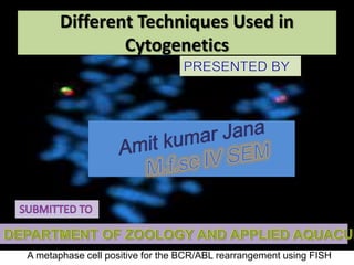
  • 1. A metaphase cell positive for the BCR/ABL rearrangement using FISH Different Techniques Used in Cytogenetics
  • 2. Introduction & Definition • Cytogenetics is a branch of genetics that is concerned with the study of the structure and function of the cell, especially the chromosomes. It includes routine analysis of G- banded chromosomes, other cytogenetic banding techniques, as well as molecular cytogenetics such as Fluorescent In Situ Hybridization (FISH) and Comparative Genomic Hybridization (CGH).
  • 3. History • Chromosomes were first observed in plant cells by Karl Wilhelm von Nägeli in 1842. Their behavior in animal (salamander) cells was described by Walther Flemming, the discoverer of mitosis, in 1882. The name was coined by another German anatomist, von Waldeyer in 1888. • In science books, the number of human chromosomes remained at 48 for over thirty years. New techniques were needed to correct this error. Joe Hin Tjio working in Albert Levan’s lab was responsible for finding the approach: • Using cells in culture • Pre-treating cells in a hypotonic solution, which swells them and spreads the chromosomes • Arresting mitosis in metaphase by a solution of colchicine • Squashing the preparation on the slide forcing the chromosomes into a single plane • Cutting up a photomicrograph and arranging the result into an indisputable karyogram.It took until 1956 until it became generally accepted that the karyotype of man included only 46 chromosomes.Rather interestingly, the greatapes have 48 chromosomes. Human chromosome 2was formed by a merger of ancestral chromosomes, reducing the number.
  • 4. Techniques 1. Karyotyping 1. Slide preparation 2. Analysis 2. Fluorescent in situ hybridization 1. Slide preparation 2. Analysis
  • 5. Techniques Karyotyping • Routine chromosome analysis (Karyotyping) refers to analysis of metaphase chromosomes which have been banded using trypsin followed by Giemsa, Leishmanns, or a mixture of the two. Several chromosome-banding techniques are used in cytogenetics laboratories. Quinacrine banding (Q-banding) was the first staining method used to produce specific banding patterns. This method requires a fluorescence microscope and is no longer as widely used as Giemsa banding (G-banding). Reverse banding, or R-banding, requires heat treatment and reverses the usual black-and-white pattern that is seen in G-bands and Q-bands. This method is particularly helpful for staining the distal ends of chromosomes. Other staining techniques include C-banding.
  • 6. Slide Preparation • Cells from bone marrow, blood, amniotic fluid, cord blood, tumor, and tissues (including skin, umbilical cord, chorionic villi, liver, and many other organs) can be cultured using standard cell culture techniques in order to increase their number. • A mitotic inhibitor (colchicine, colcemid) is then added to the culture. This stops cell division at mitosis which allows an increased yield of mitotic cells for analysis. The cells are then centrifuged and media and mitotic inhibitor are removed, and replaced with a hypotonic solution. This causes the white blood cells or fibroblasts to swell so that the chromosomes will spread when added to a slide as well as lyses the red blood cells. After the cells have been allowed to sit in hypotonic solution, Carnoy's fixative (3:1 methanol to glacial acetic acid) is added. This kills the cells and hardens the nuclei of the remaining white blood cells. The cells are generally fixed repeatedly to remove any debris or remaining red blood cells. The cell suspension is then dropped onto specimen slides. After aging the slides in an oven or waiting a few days they are ready for banding and analysis.
  • 7. Analysis Analysis of banded chromosomes is done at a microscope by a clinical laboratory specialist in cytogenetics (CLSp(CG)). Generally 20 cells are analyzed which is enough to rule out mosaicism to an acceptable level. The results are summarized and given to a board-certified cytogeneticist for review, and to write an interpretation taking into account the patients previous history and other clinical findings.
  • 8. Fluorescent in situ hybridization Fluorescent in situ hybridization refers to using fluorescently labeled probe to hybridize to cytogenetic cell preparations. In addition to standard preparations FISH can also be performed on: 1. bone marrow smears 2. blood smears 3. paraffin embedded tissue preparations 4. enzymatically dissociated tissue samples 5. uncultured bone marrow 6. uncultured amniocytes 7. cytospin preparations Fig of FSH
  • 9. Slide preparation • The slide is aged using a salt solution usually consisting of 2X SSC (salt, sodium citrate). The slides are then dehydrated in ethanol, and the probe mixture is added. The sample DNA and the probe DNA are then co-denatured using a heated plate and allowed to re-anneal for at least 4 hours. The slides are then washed to remove excess unbound probe, and counterstained with 4',6-Diamidino-2- phenylindole (DAPI) or propidium iodide.
  • 10. Analysis • Analysis of FISH specimens is done by fluorescence microscopy by a clinical laboratory specialist in cytogenetics. For oncology generally a large number of interphase cells are scored in order to rule out low-level residual disease, generally between 200 and 1,000 cells are counted and scored. For congenital problems usually 20 metaphase cells are scored.
  • 11. Future of cytogenetics • Advances now focus on molecular cytogenetics including automated systems for counting the results of standard FISH preparations and techniques for virtual karyotyping, such as comparative genomic hybridization arrays, CGH and Single nucleotide polymorphism arrays.
  • 12. Conclusion • So far, no system can classify banded chromosomes as robustly and accurately as a skilled cytogeneticist, despite the millions of dollars that have been invested in automated karyotype analysis since 1968. Currently cytogenetics is paving its way into the molecular approaches in deciphering the structure, function and evolution of chromosomes. Still,conventional cytogenetics where routine banding techniques are employed remains a simple and popular technique to get an overview of the human genome. Routine banded karyotype analysiscan now be combined with M-FISH and other molecular techniques leading to moreprecise detection of various syndromes in children.