Proposed brain surgery and unexpected findings
•
1 like•197 views

Private revieuw 2017 medical crime facts. Ten years of learning and self-correcting did give some results. Patient abuse for other scientific tries (without conscent or need to treat the pathology). In sonspiracy the case is turned down (for the moment).

Recommended
Recommended
More Related Content
What's hot
What's hot (13)
Similar to Proposed brain surgery and unexpected findings
Similar to Proposed brain surgery and unexpected findings (20)
More from siegfried van hoek
More from siegfried van hoek (20)
Recently uploaded
Recently uploaded (20)
Proposed brain surgery and unexpected findings
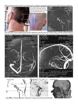
- 1. Proposed minor intervention was cutting only a window-opening inside the sub-arachnoid brain-membrane. The little half circle resulted from the first operation, just the skin was folded aside, and that sufficed largely. During 2nd operation more happened then a fenestration between sub-aranoid membrane and cisterna magna. ?? Sinus Transversus? The surgery hole in the skull is at the top of the large incision. The incision down into the neck was not needed for this. For what reason was that needed? V. Sinus Rectus and V. Transversalis (l) hit? Foreign body material inside head. Natural lacking of V. Occiptalis. Indication of altered anatomic situation, Foreign body material left behind.
- 2. Craniotomy opening is on the backside of the skull. Michel Suture Clip 16x3 mm would sag down in 6 weeks. L R C3 The sub-arachnoid space in Leptomeningen is + 1 mm. Clip is placed mainly past the vertebral arch. used unused Object left behind right under the skull, where officially no surgery took place.
- 3. What happened to the intervertebral disc C2-C3? Hole ? ?
- 4. Dimensions of the clip vary. The clip at the AMC sagittal scan differs in position with the other scan-results. The inter-vertebral disc C2-C3 is poorly visible. With other contrast settings the metal clip is disappearing almost completely. Another aspect (drilling hole?) becomes visible on C3, which other vertebras don’t show. spike Although the coup is at C4 (below the metal artifact), the scan-image measures a width of 1mm space inside the leptomeninges as online also is mentioned to be the regular width by the Oxford dictionary. The clip has a width of 3mm. This in itself is already a kind of health-risk in damaging pressure at the spinal cord.
- 5. Significant narrowing of the Spinal Canal at C3 (damage). The cervical object(s) cause a huge distortion. (In those days I also suffered from a possible lung-health-issue, what is gone now in 10 years time by itself.) Blood (ferro)-traces on MRI from just under the skin go untill deep into the neck (black line around). Seen those traces are not just skin-deep but inside the whole backside of the neck towards the spinal cord, this indicates surgery. (Shape square box in a molar of the lower jaw (r) is visible with contour shape as well.) Besides the fact that surgery in the head starting from the neck would mean passing the skull basis, and performing the fenestration from the cisterna magna towards the sub-arachnoid wall of the cyst, this also would be in essence is surgery in an opposite reversed direction, as it is also opposite of the official report too going from inside the cyst towards the cisterna magna. The white arrow suggests the sagging down route, but MRI shows more body-foreign materials present in the neck. C2 C3 Cistena Magna
- 6. So right after the 1st surgery Ct-manipulation was committed meaning premaditated in preparation of the 2nd operation, where after that 2nd operation concerning the oral cavity the MRI and CT images finally match. Therefor it is not sure if the later CT scans of the AMC november and december 2000 are not manipulated. 2 1 Also no foreign material present in the oral cavity, the neck or inside the cyst in the head just before the 2nd surgical treatment. Premeditated Ct-scan manipulation directly after 1st surgical treatment.
- 7. Forensic investigation about what had be done inside the neck also has been obstructed by third parties after. There are more traces of proof, but for this abrigded presentation already motivating conspiracy of silence is. The scan images not to be made as in a strict rotation of 90° around a vertical axis and upright standing for. In rotation-investigation it proved out that the rotation isn’t 90 degrees counter-clockwise as X-ray would show after a rotation from standing with the back to the wall and standing with the left shoulder to the wall. So what is there to conceal by committing image-manipulations regarding the situation regarding C2 - C3? Above: Double nametags as sign of reuse of image information. Here: evident manipulation trace of copying through negatives: Brand information running black and mirrored on one (resulting) single negative. Rotation is 60 degrees around horizontal axis. (Vertical axis + 30° )
- 8. So this might be an alternative drainage for compensating the missing V. Sinus Rectus en left Vene transversalis being hit during that 2nd operation? Note there is no vene occipitalis naturally, that vene can not have been hit causing a thrombosis of the left vene transversalis as one doctor falsely suggested. Besides cutting nearby the continuum sinuum (crosspoint of venes sagittalis and transversalis) is not at the location of the planned fenestration. Contrastvloeistof ‘vlek’Drainage Indication
- 9. Investigating the situation before 2nd surgery showed an intact left and right vene transversalis being present.
- 10. Then finally some enlargements of 3DVENE scan images showing damage occurred during 2nd surgery. DiaSana VEN_3D_PCA AXIAAL
- 13. COMPARING DiaSana VEN_3D_PCA WITH MRI Vessel_Scout_MIP_SAG
- 14. Also after my stroke 2016 counteracting continued in hiding image information, preventing discovery from. Re-evaluating the actual performances during 2nd operation they have been far more than just cutting a small opening between the sub-arachnoid cyst and the cistern magna. One doctor remarked that the AMC did do remarkable very little investigation to find out why the cyst started to grow before surgery upon. Another doctor also said if they were in good intentions secretly, then they also could have told me more after I did found out some facts already. The case became complex because of counteractions also by third-parties. But if there is no serious matter to hide why there was much praxis of an overall conspiracy of silence. In my layman-opinion the fist-wide cyst gave the space to do such an experiment not needed for treating the cyst, because as reported for the sub-arachnoid cyst only a fenestration towards the cistern magna was needed. And also leaving an unused Michel Clip behind is also strange when checking performed surgery before closing the patient. Only on the digital scan that clip was visible, the reports even did not mention the use of it. In the interest of noble practicing physician and honest healthcare-consumer. Siegfried van Hoek.