SlideShare a Scribd company logo
1 of 11
Download to read offline
ANALYSIS OF THE REPORT OF THE 2nd
SURGICAL TREATMENT 30 okt 2000
1sr
remark: no research has been done after the arise of the cyst. Treatment: going downwards a little piece of
meninges (cyst wall) would be cut out. However, there is cut sideward and there is no report which vein had
been hit accidentally on purpose. With concealment of acts I have been abandoned after and I also became
occupational disabled by this. Various deeds have been done which are not mentioned in the surgery report.
Premeditated unlawful conduct is provable. Below a tight translation of the original (on many parts lacking)
operation report is made, with a translation in layman language with my additional comment is underneath:
IN MEDICAL LANGUAGE
Operation report: total anesthesia, prone
position. Fixation of the head in the Mayfield
clamp 45° rotated to the left into a slight flexi.
Registration procedure in conjunction with the
use of neuro-navigation.
Continued with shaving, iodize, and covering
sterile. Hockey stick-shaped incision over the
left cerebellar hemisphere. Preparing the piece
of skin ready. Diathermic incision, pushing off
periosteum and muscle.
We now get the drilled hole of the endoscopic
fenestration à vue, where trough the dural
cover of the arachnoid cyst is shining through.
From out of this drilling hole a bone piece of 3
x 3 cm becomes milled out. After that piece of
bone has been removed, the dural cover of the
cyst becomes incised and liquor is spurting
out with remarkable high pressure. The
opening is cut in further, and the dura gets
hung up.
In the deep, we get the sight upon the cortex
of the left cerebellar hemisphere and more
median the Foramen of Magendi, as well as
the brain trunk.
1:Skin 2:Periost 3:Bone 4:Tough Brain membrane
5:Arachnoidea 6:Soft Brain membrane (layer 5+6 remaining)
Starting from the dura of the occipital bone
there seems a kind of falx coming from,
proceeding into the thin arachnoid wall, which
is drawn tightly over the cortex of the left
cerebellar hemisphere.
It is imaginable, that from here working like a
kind of valve and liquor entrapment from the
Cistern Magna is here.
IN NORMAL LANGUAGE (comment italicized)
Operation report: complete anaesthetization, lying in
prone position. The head has been fixed, and slanting
rotated, so the spot to operate is at the highest point in
sight. Registration procedure in combination with the
use of a computer-neuro-navigator with MRI-scan
information. (focused precision)
Continued with shaving, sterilizing and covering it
sterile. Hockey stick-shaped incision over the head on
the position of the left cerebellar hemisphere (running
through deep down into the neck at the height of C4!)
Skin peace prepared for incision. An electrical cutting
incision is made after and the periosteum and muscle
then were pushed off after. We now get the drilling
hole (remains of the former treatment) in sight, made
for the endoscopic window opening, where underneath
the dural cover the arachnoid cyst is shining through.
Starting from this drilling hole a bone piece is been
milled out sized 3 x 3 cm. Is that hole to the left of the
circle shaped scar? After this piece of bone has been
removed, the brain-membrane, the dural cover of the
cyst) is to be incised. With remarkable pressure brain
liquid is squirting out after. The opening is cut in
further, and the tough brain-membrane becomes hung
up like a tent sideward and upward. Into the deep we
get sight upon the cerebral cortex of the left small
brains side, and more to the middle the Foramen of
Magendi (, which is draining spinal liquor from the
fourth ventricle towards the Cisterna Magna, and
where towards notably from the cyst a whole had to be
made through Pia Mater and the Sub-Arachnoid
membrane), as well as the Brain-trunk.
So there is a visible quiet a lot with the plain eye!
The bone hole itself is a few centimeters away from the
midline, because the cyst is situated completely at the
left half. Starting From the occipital membrane a
separation membrane is to be seen, which is going
further into the arachnoid wall of the cyst and is also
drawn tightly over the left little brain side. THAT IS
THE FALX CEREBELLI, which is situated aside from
the cyst just beyond the middle at the right half. That
Falx namely is also a sidewall of the cyst. The cyst
also runs a little further on of the other brain half, by
which also the Confluens Sinuum has shifted in
position, just like the Falx Cerebelli (to be seen on MRI
scan). It is imaginable that an entrapment of liquor as a
kind of valve from the Cisterna Magna to the cyst is
active (sub-arachnoid space drains brain fluid away).
s
The majority of cysts in the head are often beyond the inner ear, the liquid is leaking/sweating in and
those kinds of cysts do not take space invasively. My cyst presumable started to grow after a heavy fall
on the back side of my head (encapsulated calcified intra-Dural-bleeding, which according to Dr. Teuns
started to retain moisture after an up following calcium deposit around an initial dried up dural bleeding.
The membrane over the cerebellar cortex is
now to be removed as good as possible with a
pair scissors and diathermy. While cutting into
that kind of falx, a strong vein-bleeding occurs
from a venous sinus that apparently is inside.
In coagulation with bipolar diathermy the
opening becomes only bigger, and we are
losing quiet some blood in a short time. The
only thing we can do is to bandage with
placing patties. We now place a LAILA
spatula on the patty, and likewise we become
able to get the hemostasis under control.
Thereupon the opening after removing the
bone piece gets enlarged to an opening of 4 x
4 cm with the highspeed mill. Next the dura
gets opened further to solve this problem.
Now the “falx”/membrane is cut in distal
further, so we can fold it back towards the
location of the bleeding in the sinus. Carefully
we now try to glue the tissue folded back into
the direction of the bleeding in the sinus with
Tissucol. When removing the spatula, a strong
loss of blood however occurs again after all.
The membrane (the layer underneath) over the cerebral
cortex becomes to be removed as good as possible
with a pair of scissors and electric cutting. The
membrane covering the brain surface is now to be
removed as good as possible with electric cutting and
a pair scissors. While cutting into that kind of falx
(cerebelli), a strong vein-bleeding arises from a vein
that is in here apparently. (HERE SIDEWARD HAS
BEEN CUT AND NOT DOWNWARDS AS AGREED,
SO THIS IS NOT A LITTLE ACCIDENT!
Speaking about anatomy, that bleeding cannot be a
surprise, because visibly and notably some veins are
converging in the Confluens Sinuum: de vein Sinus
Rectus and both the veins Sinus Transversalis (L+R),
and the vein Saggitalis Superioris are all coming
together in the Confluens Sinuum. (The insignificant
small Occiptital vein is also running there.) The cyst in
its size gave the opportunity for this. This also is no
issue of surprise, because above this surgery was all
performed under surgical reference, so where is the
surprise? We may speak of an accident under false
pretences, but only a cutting into the lower part of the
membrane scheme was needed DOWNWARDS to
make a window opening without even getting near the
Falx Cerebelli and the Confluens Sinuum and Veins.
With two-sided heating/cauterizing (, diathermy can
also have a cutting effect!!) the opening only gets
wider and quiet some blood is lost in a short time (,
consider that these major veins inside the head are
without clutters and are indirectly connected to the
heart via the V. Jugelaris interna). The only thing
remaining is using bandage with patties. We now place
a LAILA spatula clamp on the patty (, no Michel Clip
was used for this), and in this way we are able to get
the bleeding under control. (After a very huge loss of
blood). Thereupon the skull bone opening gets
enlarged with the highspeed mill to the size of 4 x 4
cm. Next the dura gets opened further so we get more
space to solve this issue. (Near the incision of the skin
groping the surface of the skull a little pit can be felt
further to the left. Note: why it was needed to mill
more towards the middle line of the brain half’s?
Because several veins could be pinched by starting
around the Conlfuens Sinuum? There has been
performed surgery at the side to cut into the falx
cerebelli and to stop the bleeding... instead going
downwards through the middle. The parts to be placed
do not need to be replaced exactly the same way,
because the milled space provided space for.)
The separation wall is now cut in further away from
the center, so it can get folded back into the direction
of the bleeding in the sinus with Tissucol.
When removing the spatula, a strong loss of blood
however occurs again after all. (Compare repairing a
water hose with the pressure still on it. The blood loss,
and the vacuum in the hart chamber with cardiac
arrythmia also caused lowering the blood pressure,
helping it became possible to stop the bleeding.)
The same tactics gets now applied with
pieces of muscle and Tissucol. This time it
works to control the bleeding. After thorough
rinsing and inspection, it is dry. Thereupon a
large part out of the supposed falx is to be cut
(on that spot where no visible vene/venous
sinus seems to be present) and the sides
become coagulated with bipolar diathermy.
A very large phenestration has been made
from the cyst to the cisterna magna. The dura
gets knotted with Vicryl.
The Dura cannot be closed completely
watertight, and consequently the whole dura
in the opening of the skull bone gets covered
with Duragen. Hereupon pieces of bone tissue
are replaced and fixed with Tissucol. Suture
over with periost and muscle and closing skin
layers after.
Post-operative conclusion: microscopic
phenestration of a large cerebellar cyst to the
left towards the cisterna magna.
We are dealing here with conscious mutilation and
experimental activities without patient consent. To me
a complete different plan of treatment was been told.
Green points towards the agreed direction of surgery
and red points at the actual surgery direction deduced
from the actual surgery report and results from scan
investigation.
The size of the cyst provided the space to remove the
left Vein Sinus Transversalis and also sideward and in
opposite direction of the cyst the Falx Cerebelli in
between both brain half’s has been cut in and the V.
Sinus Rectus is an indication from scan investigation.
(Also, there is a supposition on MRI scan of an
artificial vein-deviation and clandestine surgery in the
neck and oral cavity based upon scan-investigation.)
Now in the same way the repair is done with pieces of
muscle and Fibrin-glue. This time we get the bleeding
under control. After thorough rinsing we can inspect
the prepared wound and is closed and dry (the wound
near the Confluens Sinuum has closed, which is an
achievement upon survival...). Thereupon a large piece
is been cut out of the assumed falx (is falx cerebelli)
on that spot where no visible (!) Vein / venous sinus
seems to be present. (The left sinus V. Transversalis (,
which is not running through the Falx Cerebelli, but is
even running horizontally in an opposite direction
away from the Confluens Sinuum sideward along the
skull) is completely removed. For this there was no
cutting needed into the appointed direction.) The sides
become congealed / cauterized with two-sided heating.
A very large window opening has been made from the
cyst to the Cysterna Magna. (REPRESENTATION IS
FALSE, because the Cysterna Magna is situated below
the cyst and not aside the cyst with the Falx Cerebelli
aside.) The brain-membrane now gets closed stitching
with Vycril. The dura, because of shrinkage, however
cannot be closed completely watertight, and therefore
consequently the complete dura (visible) through the
opening of the skull, gets covered with Duragen.
Hereupon the pieces of bone are replaced with little
pieces of bone tissue around into the open groove,
which also gets attached with Tissucol. The
periosteum (the 1st
cover of the skull) and the muscle
become knotted, and hereafter the skin. Post-operative
finding: microscopic window-opening (with a pair of
scissors!) from the cyst located intra-dural near the left
little-brain towards the Cisterna Magna has succeeded.
(Microscopic? Just above we read a very large
window-opening into the Falx Cerebelli has been cut
with cutting further where we think not to see a vein.)
The left V. SinusTransversalis is missing. That vein is
cut off on three spots in a different direction then the
actual appointed direction. The later explanation from
a Doctor that the left V. Transversalis ‘disappeared’,
was by a Thrombosis caused by the Vene Occiptalis
being hit fails, because this insignificant vein is even
intact (arrow). So: Unregistered activities took place,
which above all weren’t meant specific in my interest
of healthcare and the treatment of the pathology.
X
X
X
My additional comment : cutting sideward in the Falx at the right half was not needed to make a
connection between the cyst situated on the left half and the causal situated brain fluid circulation in the
Cysterna Magna right below. Even with basic medical knowledge of anatomy (, next to the fact that such
big veins are visible through the (1mm) enclosing membrane) they had real sight till the brain trunk and
notably surgery was performed under neurosurgical reference with scans made before with contrast fluid,
SO: there cannot be an issue of surprise in surgery. Even smaller veins are visible through the upper
membranes. Quoting this two phrases: ,,While cutting into that kind of falx, a strong vein-bleeding occurs
from a venous sinus that apparently is inside” -and- ,,Thereupon a large part out of the supposed falx is
to be cut (where no visible vein / venous sinus seems to be present) and the sides become coagulated with
bipolar diathermy”. The first quote is mentioned that hitting this vein(s) would be a surprise, they do call
the wall as a kind of Falx, and with anatomic knowledge a big vein is running through; the second quote
implicitly may point to see the vein, because they are cutting further where in the Falx, where they think
not to see a vein. The V. Sinus Rectus is enclosed in the top of the Falx Cerebelli, but The Vein Sinus
Transversus is running ‘horizontal away’ along the left side wall of the skull in an opposite direction.
The assistant -surgeon spoke about a slipping while cutting, and thus he was not telling about a surprise
but about an accident and then later when they brought me back to consciousness, I became completely
unwell, where after they had to bring me back again completely under narcosis. After, later that evening I
have witnessed the transfer of IC-nursing and heard even more information which lead to the suspicion of
other activities then was agreed, but this all was not reported at all at the generalist. In any case the
medical intervention took much longer, while I went under narcosis around 09.30 hrs. and I came back to
consciousness around 18.10. Was there an issue of a succeeded experiment of vein-deviation wherefor the
left Vene Sinus Tranvesalis had to be removed and also the Vein Sinus Rectus had to be cut off? Why
there was placed a (damaging) clamping ring surrounding the Spinal cord in the Myleum canal?
The incision of the second operation runs through till halfway the neck and for me this was the easiest to
find, because for the first operation this was totally not needed at all. Factual above this, the very same
operation would have been performed by hand, by which only the dimensions would get somewhat
bigger. Privat payed forensic investigation partly was sabotaged: manipulated X-ray scans showed an
unused Michel Clip 14x3 mm2 that would have been left behind, which in six weeks’ time would have
been sagged down from the Cyst through the Foramen Magna into the Leptomeninges (1 mm space with
Trabecula as obstacles) around the Spinal Cord, but the use of that Clip also was not reported. On the RX
that clip is even reaching beyond the Vertebral Arch, whereby that clip also would become situated lying
outside the Leptomeninges, which is also impossible. On the MRI we can see inorganic material wat was
left behind inside the head, but also a narrowing ring around the Myleum Canal. Was it in their wish, that
I would die by their actions by/with that narrowing ring for instance, where after the case would have
been put down with a so-called clip that would have been sagged down that far as a little medical error?
Or was the unused Michel-clip really placed in the head to sag down for mortal damaging next to the
presence of that ring? Many doctors after (outsiders) appeared to conspire in concealment of facts with
false reports as well as forgery / hiding (covering) of scan image information (crime too). And this all is
not done by accident or for no reason: what is there to hide, what is not allowed to get in the publicity?
Underneath a list of some explanatory terms that were found in the report:
Coagulate to clot
Diathermy Electric cutting / cauterizing closing tissue. Mono-D is to scald living tissue.
Vicryl Material to stitch closing wounds, self-dissolving in a later stage
Duragen Matrix with Glue for closing and regenerating the Dura after a neurosurgical treatment.
Tissucol Fibrin-glue, glues wounds together, stitches and closes them and is moisture regulating.
Layla Spatula Surgical extension clamping as spatula as a kind third hand
Patty not indicated, most probably absorbing bandages
Flexie Rotation
Distal vs Median respectively being away from the middle -versus- towards the middle
Analysis of the report of the second surgical treatment  low res

More Related Content

What's hot

Eyelid anatomy and physiology
Eyelid anatomy and physiologyEyelid anatomy and physiology
Eyelid anatomy and physiology
Najara Thapa
 
Anatomy and physiology of the eyelid
Anatomy and physiology of the eyelidAnatomy and physiology of the eyelid
Anatomy and physiology of the eyelid
Alaa Farsakh
 
Anatomy & physiology of eyelids
Anatomy  & physiology of eyelidsAnatomy  & physiology of eyelids
Anatomy & physiology of eyelids
Samuel Ponraj
 

What's hot (20)

Eyelid anatomy and physiology
Eyelid anatomy and physiologyEyelid anatomy and physiology
Eyelid anatomy and physiology
 
Eyelid recon
Eyelid reconEyelid recon
Eyelid recon
 
The eyelid
The eyelidThe eyelid
The eyelid
 
Anatomy of the eyelids
Anatomy of the eyelidsAnatomy of the eyelids
Anatomy of the eyelids
 
Sella supra sella anatomy
Sella supra sella anatomySella supra sella anatomy
Sella supra sella anatomy
 
Anatomy of Eyelids & Its Clinical Correlations
Anatomy of Eyelids & Its Clinical CorrelationsAnatomy of Eyelids & Its Clinical Correlations
Anatomy of Eyelids & Its Clinical Correlations
 
Anatomy and physiology of the eyelid
Anatomy and physiology of the eyelidAnatomy and physiology of the eyelid
Anatomy and physiology of the eyelid
 
Anatomy of eyelid
Anatomy of eyelidAnatomy of eyelid
Anatomy of eyelid
 
Lids and Adnexa Class 2 Part A - Ectropion and Entropion
Lids and Adnexa Class 2 Part A - Ectropion and EntropionLids and Adnexa Class 2 Part A - Ectropion and Entropion
Lids and Adnexa Class 2 Part A - Ectropion and Entropion
 
Anatomy & physiology of eyelids
Anatomy  & physiology of eyelidsAnatomy  & physiology of eyelids
Anatomy & physiology of eyelids
 
Lacrimal system ppt.
Lacrimal system ppt.Lacrimal system ppt.
Lacrimal system ppt.
 
Anatomy of eyelid and eyelid reconstruction
Anatomy of eyelid and eyelid reconstructionAnatomy of eyelid and eyelid reconstruction
Anatomy of eyelid and eyelid reconstruction
 
ICA anatomy
ICA anatomyICA anatomy
ICA anatomy
 
eyelid
eyelideyelid
eyelid
 
Anatomy of Eyelids
Anatomy of EyelidsAnatomy of Eyelids
Anatomy of Eyelids
 
Anatomy of Eyelid
Anatomy of EyelidAnatomy of Eyelid
Anatomy of Eyelid
 
Anatomy of eyelids
Anatomy of eyelidsAnatomy of eyelids
Anatomy of eyelids
 
External carotid artery, branches and ligation
External carotid artery, branches and ligationExternal carotid artery, branches and ligation
External carotid artery, branches and ligation
 
Anat of eyelid
Anat of eyelidAnat of eyelid
Anat of eyelid
 
Lecture 1 orbit-by Dr. Noura- 2018
Lecture 1 orbit-by Dr.  Noura- 2018Lecture 1 orbit-by Dr.  Noura- 2018
Lecture 1 orbit-by Dr. Noura- 2018
 

Similar to Analysis of the report of the second surgical treatment low res

244221 shoulder-and-pectoral
244221 shoulder-and-pectoral244221 shoulder-and-pectoral
244221 shoulder-and-pectoral
YoAmoNYC
 

Similar to Analysis of the report of the second surgical treatment low res (20)

Cavernous sinus 360°
Cavernous sinus 360°Cavernous sinus 360°
Cavernous sinus 360°
 
V introduction anatomy head and neck for background info
V introduction anatomy head and neck for background infoV introduction anatomy head and neck for background info
V introduction anatomy head and neck for background info
 
Surgical management of vestibular schwannoma by drdhiru456
Surgical management of vestibular schwannoma by drdhiru456Surgical management of vestibular schwannoma by drdhiru456
Surgical management of vestibular schwannoma by drdhiru456
 
13. neck dissection - Meghali
13. neck dissection - Meghali13. neck dissection - Meghali
13. neck dissection - Meghali
 
Blood supply of head and neck
Blood supply of head and neck Blood supply of head and neck
Blood supply of head and neck
 
pterional articulo viejo.pdf
pterional articulo viejo.pdfpterional articulo viejo.pdf
pterional articulo viejo.pdf
 
pterional articulo viejo.pdf
pterional articulo viejo.pdfpterional articulo viejo.pdf
pterional articulo viejo.pdf
 
REZ 360°
REZ 360°REZ 360°
REZ 360°
 
Vestibular schwannoma (acoustic neuroma) surgical anatomy and microsurgeries,...
Vestibular schwannoma (acoustic neuroma) surgical anatomy and microsurgeries,...Vestibular schwannoma (acoustic neuroma) surgical anatomy and microsurgeries,...
Vestibular schwannoma (acoustic neuroma) surgical anatomy and microsurgeries,...
 
Imaging in ent
Imaging in entImaging in ent
Imaging in ent
 
Topographic anatomy of the brain and brain part of the skull
Topographic anatomy of the brain and brain part of the skullTopographic anatomy of the brain and brain part of the skull
Topographic anatomy of the brain and brain part of the skull
 
Pituitary 360°
Pituitary 360°Pituitary 360°
Pituitary 360°
 
3)neck dissection
3)neck dissection3)neck dissection
3)neck dissection
 
Pterional craniotomy
Pterional craniotomyPterional craniotomy
Pterional craniotomy
 
Surgery for atrial fibrillation abhijit presentation
Surgery for atrial fibrillation abhijit presentationSurgery for atrial fibrillation abhijit presentation
Surgery for atrial fibrillation abhijit presentation
 
Seminar on applied anatomy and surgical approaches to shoulder
Seminar on applied anatomy and surgical approaches to shoulderSeminar on applied anatomy and surgical approaches to shoulder
Seminar on applied anatomy and surgical approaches to shoulder
 
Neck Dissection Head Neck oncoSurgery.pptx
Neck Dissection Head Neck oncoSurgery.pptxNeck Dissection Head Neck oncoSurgery.pptx
Neck Dissection Head Neck oncoSurgery.pptx
 
Full story brain herniation imaging Dr Ahmed Esawy
Full story brain herniation imaging Dr Ahmed EsawyFull story brain herniation imaging Dr Ahmed Esawy
Full story brain herniation imaging Dr Ahmed Esawy
 
Shoulder and Pectoral Girdle
Shoulder and Pectoral GirdleShoulder and Pectoral Girdle
Shoulder and Pectoral Girdle
 
244221 shoulder-and-pectoral
244221 shoulder-and-pectoral244221 shoulder-and-pectoral
244221 shoulder-and-pectoral
 

More from siegfried van hoek

More from siegfried van hoek (20)

OVER MINDFULLNESS IN JE LIEFDES-RELATIE Valentijnsdag 14 februari 2020 bij As...
OVER MINDFULLNESS IN JE LIEFDES-RELATIE Valentijnsdag 14 februari 2020 bij As...OVER MINDFULLNESS IN JE LIEFDES-RELATIE Valentijnsdag 14 februari 2020 bij As...
OVER MINDFULLNESS IN JE LIEFDES-RELATIE Valentijnsdag 14 februari 2020 bij As...
 
ECHR procedure na gesaboteerde art 12 SV incl soft antwoord ECHR 2017 low res
ECHR procedure na gesaboteerde art 12 SV incl soft antwoord ECHR 2017 low resECHR procedure na gesaboteerde art 12 SV incl soft antwoord ECHR 2017 low res
ECHR procedure na gesaboteerde art 12 SV incl soft antwoord ECHR 2017 low res
 
Abbriged presentation low res on medical abuse
Abbriged presentation low res on medical abuseAbbriged presentation low res on medical abuse
Abbriged presentation low res on medical abuse
 
Ontbijtsessie 06012020 over de zingeving van pijn bij Assadaaka
Ontbijtsessie 06012020 over de zingeving van pijn bij AssadaakaOntbijtsessie 06012020 over de zingeving van pijn bij Assadaaka
Ontbijtsessie 06012020 over de zingeving van pijn bij Assadaaka
 
Ontbijtsessie de week van de eenzaamheid op 28 09-2929 bij Assadaaka.
Ontbijtsessie de week van de eenzaamheid op 28 09-2929 bij Assadaaka.Ontbijtsessie de week van de eenzaamheid op 28 09-2929 bij Assadaaka.
Ontbijtsessie de week van de eenzaamheid op 28 09-2929 bij Assadaaka.
 
V5 introduction anatomy head and neck low res for background info
V5 introduction anatomy head and neck low res  for background infoV5 introduction anatomy head and neck low res  for background info
V5 introduction anatomy head and neck low res for background info
 
PDF of dia presentation MEDICAL ABUSE CASE
PDF of dia presentation MEDICAL ABUSE CASEPDF of dia presentation MEDICAL ABUSE CASE
PDF of dia presentation MEDICAL ABUSE CASE
 
Notulen presentatie assaadaka amsterdam meldcode huislijk geweld
Notulen presentatie assaadaka amsterdam meldcode huislijk geweldNotulen presentatie assaadaka amsterdam meldcode huislijk geweld
Notulen presentatie assaadaka amsterdam meldcode huislijk geweld
 
1st presentation with selected scanimages
1st presentation with selected scanimages1st presentation with selected scanimages
1st presentation with selected scanimages
 
Voorkomen is beter dan Genezen
Voorkomen is beter dan GenezenVoorkomen is beter dan Genezen
Voorkomen is beter dan Genezen
 
Deadly Medical Practices by Trudy Newman (c) 2003
Deadly Medical Practices by Trudy Newman (c) 2003Deadly Medical Practices by Trudy Newman (c) 2003
Deadly Medical Practices by Trudy Newman (c) 2003
 
Verslag klachtzitting artikel 12 strafvordering
Verslag klachtzitting artikel 12 strafvorderingVerslag klachtzitting artikel 12 strafvordering
Verslag klachtzitting artikel 12 strafvordering
 
Scan selecties met minimaal commentaar
Scan selecties met minimaal commentaarScan selecties met minimaal commentaar
Scan selecties met minimaal commentaar
 
Fraude tekenen van manipulatie van fysieke rx negatieven gezien vanuit het a...
Fraude tekenen van manipulatie van fysieke rx negatieven gezien vanuit  het a...Fraude tekenen van manipulatie van fysieke rx negatieven gezien vanuit  het a...
Fraude tekenen van manipulatie van fysieke rx negatieven gezien vanuit het a...
 
This is how it works: CORRUPTION
This is how it works: CORRUPTIONThis is how it works: CORRUPTION
This is how it works: CORRUPTION
 
Signs of manipulations of physical rx negatives seen from the aspect of produ...
Signs of manipulations of physical rx negatives seen from the aspect of produ...Signs of manipulations of physical rx negatives seen from the aspect of produ...
Signs of manipulations of physical rx negatives seen from the aspect of produ...
 
Declaration in between
Declaration in betweenDeclaration in between
Declaration in between
 
Source added for understanding anatomy
Source added for understanding anatomySource added for understanding anatomy
Source added for understanding anatomy
 
Add olvg 2016 added
Add olvg 2016 addedAdd olvg 2016 added
Add olvg 2016 added
 
Vene Occipitalis is naturally missing sometimes at people
Vene Occipitalis is naturally missing sometimes at peopleVene Occipitalis is naturally missing sometimes at people
Vene Occipitalis is naturally missing sometimes at people
 

Recently uploaded

Russian Call Girls Lucknow Just Call 👉👉7877925207 Top Class Call Girl Service...
Russian Call Girls Lucknow Just Call 👉👉7877925207 Top Class Call Girl Service...Russian Call Girls Lucknow Just Call 👉👉7877925207 Top Class Call Girl Service...
Russian Call Girls Lucknow Just Call 👉👉7877925207 Top Class Call Girl Service...
adilkhan87451
 

Recently uploaded (20)

Most Beautiful Call Girl in Bangalore Contact on Whatsapp
Most Beautiful Call Girl in Bangalore Contact on WhatsappMost Beautiful Call Girl in Bangalore Contact on Whatsapp
Most Beautiful Call Girl in Bangalore Contact on Whatsapp
 
Pondicherry Call Girls Book Now 9630942363 Top Class Pondicherry Escort Servi...
Pondicherry Call Girls Book Now 9630942363 Top Class Pondicherry Escort Servi...Pondicherry Call Girls Book Now 9630942363 Top Class Pondicherry Escort Servi...
Pondicherry Call Girls Book Now 9630942363 Top Class Pondicherry Escort Servi...
 
The Most Attractive Hyderabad Call Girls Kothapet 𖠋 9332606886 𖠋 Will You Mis...
The Most Attractive Hyderabad Call Girls Kothapet 𖠋 9332606886 𖠋 Will You Mis...The Most Attractive Hyderabad Call Girls Kothapet 𖠋 9332606886 𖠋 Will You Mis...
The Most Attractive Hyderabad Call Girls Kothapet 𖠋 9332606886 𖠋 Will You Mis...
 
Model Call Girls In Chennai WhatsApp Booking 7427069034 call girl service 24 ...
Model Call Girls In Chennai WhatsApp Booking 7427069034 call girl service 24 ...Model Call Girls In Chennai WhatsApp Booking 7427069034 call girl service 24 ...
Model Call Girls In Chennai WhatsApp Booking 7427069034 call girl service 24 ...
 
Call Girls Kurnool Just Call 8250077686 Top Class Call Girl Service Available
Call Girls Kurnool Just Call 8250077686 Top Class Call Girl Service AvailableCall Girls Kurnool Just Call 8250077686 Top Class Call Girl Service Available
Call Girls Kurnool Just Call 8250077686 Top Class Call Girl Service Available
 
Call Girls Visakhapatnam Just Call 8250077686 Top Class Call Girl Service Ava...
Call Girls Visakhapatnam Just Call 8250077686 Top Class Call Girl Service Ava...Call Girls Visakhapatnam Just Call 8250077686 Top Class Call Girl Service Ava...
Call Girls Visakhapatnam Just Call 8250077686 Top Class Call Girl Service Ava...
 
Trichy Call Girls Book Now 9630942363 Top Class Trichy Escort Service Available
Trichy Call Girls Book Now 9630942363 Top Class Trichy Escort Service AvailableTrichy Call Girls Book Now 9630942363 Top Class Trichy Escort Service Available
Trichy Call Girls Book Now 9630942363 Top Class Trichy Escort Service Available
 
Mumbai ] (Call Girls) in Mumbai 10k @ I'm VIP Independent Escorts Girls 98333...
Mumbai ] (Call Girls) in Mumbai 10k @ I'm VIP Independent Escorts Girls 98333...Mumbai ] (Call Girls) in Mumbai 10k @ I'm VIP Independent Escorts Girls 98333...
Mumbai ] (Call Girls) in Mumbai 10k @ I'm VIP Independent Escorts Girls 98333...
 
Top Quality Call Girl Service Kalyanpur 6378878445 Available Call Girls Any Time
Top Quality Call Girl Service Kalyanpur 6378878445 Available Call Girls Any TimeTop Quality Call Girl Service Kalyanpur 6378878445 Available Call Girls Any Time
Top Quality Call Girl Service Kalyanpur 6378878445 Available Call Girls Any Time
 
Top Rated Hyderabad Call Girls Chintal ⟟ 9332606886 ⟟ Call Me For Genuine Se...
Top Rated  Hyderabad Call Girls Chintal ⟟ 9332606886 ⟟ Call Me For Genuine Se...Top Rated  Hyderabad Call Girls Chintal ⟟ 9332606886 ⟟ Call Me For Genuine Se...
Top Rated Hyderabad Call Girls Chintal ⟟ 9332606886 ⟟ Call Me For Genuine Se...
 
Call Girls Vadodara Just Call 8617370543 Top Class Call Girl Service Available
Call Girls Vadodara Just Call 8617370543 Top Class Call Girl Service AvailableCall Girls Vadodara Just Call 8617370543 Top Class Call Girl Service Available
Call Girls Vadodara Just Call 8617370543 Top Class Call Girl Service Available
 
Call Girls Vasai Virar Just Call 9630942363 Top Class Call Girl Service Avail...
Call Girls Vasai Virar Just Call 9630942363 Top Class Call Girl Service Avail...Call Girls Vasai Virar Just Call 9630942363 Top Class Call Girl Service Avail...
Call Girls Vasai Virar Just Call 9630942363 Top Class Call Girl Service Avail...
 
Call Girls Ahmedabad Just Call 9630942363 Top Class Call Girl Service Available
Call Girls Ahmedabad Just Call 9630942363 Top Class Call Girl Service AvailableCall Girls Ahmedabad Just Call 9630942363 Top Class Call Girl Service Available
Call Girls Ahmedabad Just Call 9630942363 Top Class Call Girl Service Available
 
Premium Call Girls In Jaipur {8445551418} ❤️VVIP SEEMA Call Girl in Jaipur Ra...
Premium Call Girls In Jaipur {8445551418} ❤️VVIP SEEMA Call Girl in Jaipur Ra...Premium Call Girls In Jaipur {8445551418} ❤️VVIP SEEMA Call Girl in Jaipur Ra...
Premium Call Girls In Jaipur {8445551418} ❤️VVIP SEEMA Call Girl in Jaipur Ra...
 
VIP Hyderabad Call Girls Bahadurpally 7877925207 ₹5000 To 25K With AC Room 💚😋
VIP Hyderabad Call Girls Bahadurpally 7877925207 ₹5000 To 25K With AC Room 💚😋VIP Hyderabad Call Girls Bahadurpally 7877925207 ₹5000 To 25K With AC Room 💚😋
VIP Hyderabad Call Girls Bahadurpally 7877925207 ₹5000 To 25K With AC Room 💚😋
 
Call Girls Gwalior Just Call 8617370543 Top Class Call Girl Service Available
Call Girls Gwalior Just Call 8617370543 Top Class Call Girl Service AvailableCall Girls Gwalior Just Call 8617370543 Top Class Call Girl Service Available
Call Girls Gwalior Just Call 8617370543 Top Class Call Girl Service Available
 
Call Girls Raipur Just Call 9630942363 Top Class Call Girl Service Available
Call Girls Raipur Just Call 9630942363 Top Class Call Girl Service AvailableCall Girls Raipur Just Call 9630942363 Top Class Call Girl Service Available
Call Girls Raipur Just Call 9630942363 Top Class Call Girl Service Available
 
Russian Call Girls Lucknow Just Call 👉👉7877925207 Top Class Call Girl Service...
Russian Call Girls Lucknow Just Call 👉👉7877925207 Top Class Call Girl Service...Russian Call Girls Lucknow Just Call 👉👉7877925207 Top Class Call Girl Service...
Russian Call Girls Lucknow Just Call 👉👉7877925207 Top Class Call Girl Service...
 
Call Girls in Delhi Triveni Complex Escort Service(🔝))/WhatsApp 97111⇛47426
Call Girls in Delhi Triveni Complex Escort Service(🔝))/WhatsApp 97111⇛47426Call Girls in Delhi Triveni Complex Escort Service(🔝))/WhatsApp 97111⇛47426
Call Girls in Delhi Triveni Complex Escort Service(🔝))/WhatsApp 97111⇛47426
 
All Time Service Available Call Girls Marine Drive 📳 9820252231 For 18+ VIP C...
All Time Service Available Call Girls Marine Drive 📳 9820252231 For 18+ VIP C...All Time Service Available Call Girls Marine Drive 📳 9820252231 For 18+ VIP C...
All Time Service Available Call Girls Marine Drive 📳 9820252231 For 18+ VIP C...
 

Analysis of the report of the second surgical treatment low res

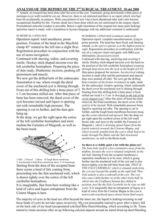
  • 1. ANALYSIS OF THE REPORT OF THE 2nd SURGICAL TREATMENT 30 okt 2000 1sr remark: no research has been done after the arise of the cyst. Treatment: going downwards a little piece of meninges (cyst wall) would be cut out. However, there is cut sideward and there is no report which vein had been hit accidentally on purpose. With concealment of acts I have been abandoned after and I also became occupational disabled by this. Various deeds have been done which are not mentioned in the surgery report. Premeditated unlawful conduct is provable. Below a tight translation of the original (on many parts lacking) operation report is made, with a translation in layman language with my additional comment is underneath: IN MEDICAL LANGUAGE Operation report: total anesthesia, prone position. Fixation of the head in the Mayfield clamp 45° rotated to the left into a slight flexi. Registration procedure in conjunction with the use of neuro-navigation. Continued with shaving, iodize, and covering sterile. Hockey stick-shaped incision over the left cerebellar hemisphere. Preparing the piece of skin ready. Diathermic incision, pushing off periosteum and muscle. We now get the drilled hole of the endoscopic fenestration à vue, where trough the dural cover of the arachnoid cyst is shining through. From out of this drilling hole a bone piece of 3 x 3 cm becomes milled out. After that piece of bone has been removed, the dural cover of the cyst becomes incised and liquor is spurting out with remarkable high pressure. The opening is cut in further, and the dura gets hung up. In the deep, we get the sight upon the cortex of the left cerebellar hemisphere and more median the Foramen of Magendi, as well as the brain trunk. 1:Skin 2:Periost 3:Bone 4:Tough Brain membrane 5:Arachnoidea 6:Soft Brain membrane (layer 5+6 remaining) Starting from the dura of the occipital bone there seems a kind of falx coming from, proceeding into the thin arachnoid wall, which is drawn tightly over the cortex of the left cerebellar hemisphere. It is imaginable, that from here working like a kind of valve and liquor entrapment from the Cistern Magna is here. IN NORMAL LANGUAGE (comment italicized) Operation report: complete anaesthetization, lying in prone position. The head has been fixed, and slanting rotated, so the spot to operate is at the highest point in sight. Registration procedure in combination with the use of a computer-neuro-navigator with MRI-scan information. (focused precision) Continued with shaving, sterilizing and covering it sterile. Hockey stick-shaped incision over the head on the position of the left cerebellar hemisphere (running through deep down into the neck at the height of C4!) Skin peace prepared for incision. An electrical cutting incision is made after and the periosteum and muscle then were pushed off after. We now get the drilling hole (remains of the former treatment) in sight, made for the endoscopic window opening, where underneath the dural cover the arachnoid cyst is shining through. Starting from this drilling hole a bone piece is been milled out sized 3 x 3 cm. Is that hole to the left of the circle shaped scar? After this piece of bone has been removed, the brain-membrane, the dural cover of the cyst) is to be incised. With remarkable pressure brain liquid is squirting out after. The opening is cut in further, and the tough brain-membrane becomes hung up like a tent sideward and upward. Into the deep we get sight upon the cerebral cortex of the left small brains side, and more to the middle the Foramen of Magendi (, which is draining spinal liquor from the fourth ventricle towards the Cisterna Magna, and where towards notably from the cyst a whole had to be made through Pia Mater and the Sub-Arachnoid membrane), as well as the Brain-trunk. So there is a visible quiet a lot with the plain eye! The bone hole itself is a few centimeters away from the midline, because the cyst is situated completely at the left half. Starting From the occipital membrane a separation membrane is to be seen, which is going further into the arachnoid wall of the cyst and is also drawn tightly over the left little brain side. THAT IS THE FALX CEREBELLI, which is situated aside from the cyst just beyond the middle at the right half. That Falx namely is also a sidewall of the cyst. The cyst also runs a little further on of the other brain half, by which also the Confluens Sinuum has shifted in position, just like the Falx Cerebelli (to be seen on MRI scan). It is imaginable that an entrapment of liquor as a kind of valve from the Cisterna Magna to the cyst is active (sub-arachnoid space drains brain fluid away). s The majority of cysts in the head are often beyond the inner ear, the liquid is leaking/sweating in and those kinds of cysts do not take space invasively. My cyst presumable started to grow after a heavy fall on the back side of my head (encapsulated calcified intra-Dural-bleeding, which according to Dr. Teuns started to retain moisture after an up following calcium deposit around an initial dried up dural bleeding.
  • 2. The membrane over the cerebellar cortex is now to be removed as good as possible with a pair scissors and diathermy. While cutting into that kind of falx, a strong vein-bleeding occurs from a venous sinus that apparently is inside. In coagulation with bipolar diathermy the opening becomes only bigger, and we are losing quiet some blood in a short time. The only thing we can do is to bandage with placing patties. We now place a LAILA spatula on the patty, and likewise we become able to get the hemostasis under control. Thereupon the opening after removing the bone piece gets enlarged to an opening of 4 x 4 cm with the highspeed mill. Next the dura gets opened further to solve this problem. Now the “falx”/membrane is cut in distal further, so we can fold it back towards the location of the bleeding in the sinus. Carefully we now try to glue the tissue folded back into the direction of the bleeding in the sinus with Tissucol. When removing the spatula, a strong loss of blood however occurs again after all. The membrane (the layer underneath) over the cerebral cortex becomes to be removed as good as possible with a pair of scissors and electric cutting. The membrane covering the brain surface is now to be removed as good as possible with electric cutting and a pair scissors. While cutting into that kind of falx (cerebelli), a strong vein-bleeding arises from a vein that is in here apparently. (HERE SIDEWARD HAS BEEN CUT AND NOT DOWNWARDS AS AGREED, SO THIS IS NOT A LITTLE ACCIDENT! Speaking about anatomy, that bleeding cannot be a surprise, because visibly and notably some veins are converging in the Confluens Sinuum: de vein Sinus Rectus and both the veins Sinus Transversalis (L+R), and the vein Saggitalis Superioris are all coming together in the Confluens Sinuum. (The insignificant small Occiptital vein is also running there.) The cyst in its size gave the opportunity for this. This also is no issue of surprise, because above this surgery was all performed under surgical reference, so where is the surprise? We may speak of an accident under false pretences, but only a cutting into the lower part of the membrane scheme was needed DOWNWARDS to make a window opening without even getting near the Falx Cerebelli and the Confluens Sinuum and Veins. With two-sided heating/cauterizing (, diathermy can also have a cutting effect!!) the opening only gets wider and quiet some blood is lost in a short time (, consider that these major veins inside the head are without clutters and are indirectly connected to the heart via the V. Jugelaris interna). The only thing remaining is using bandage with patties. We now place a LAILA spatula clamp on the patty (, no Michel Clip was used for this), and in this way we are able to get the bleeding under control. (After a very huge loss of blood). Thereupon the skull bone opening gets enlarged with the highspeed mill to the size of 4 x 4 cm. Next the dura gets opened further so we get more space to solve this issue. (Near the incision of the skin groping the surface of the skull a little pit can be felt further to the left. Note: why it was needed to mill more towards the middle line of the brain half’s? Because several veins could be pinched by starting around the Conlfuens Sinuum? There has been performed surgery at the side to cut into the falx cerebelli and to stop the bleeding... instead going downwards through the middle. The parts to be placed do not need to be replaced exactly the same way, because the milled space provided space for.) The separation wall is now cut in further away from the center, so it can get folded back into the direction of the bleeding in the sinus with Tissucol. When removing the spatula, a strong loss of blood however occurs again after all. (Compare repairing a water hose with the pressure still on it. The blood loss, and the vacuum in the hart chamber with cardiac arrythmia also caused lowering the blood pressure, helping it became possible to stop the bleeding.)
  • 3. The same tactics gets now applied with pieces of muscle and Tissucol. This time it works to control the bleeding. After thorough rinsing and inspection, it is dry. Thereupon a large part out of the supposed falx is to be cut (on that spot where no visible vene/venous sinus seems to be present) and the sides become coagulated with bipolar diathermy. A very large phenestration has been made from the cyst to the cisterna magna. The dura gets knotted with Vicryl. The Dura cannot be closed completely watertight, and consequently the whole dura in the opening of the skull bone gets covered with Duragen. Hereupon pieces of bone tissue are replaced and fixed with Tissucol. Suture over with periost and muscle and closing skin layers after. Post-operative conclusion: microscopic phenestration of a large cerebellar cyst to the left towards the cisterna magna. We are dealing here with conscious mutilation and experimental activities without patient consent. To me a complete different plan of treatment was been told. Green points towards the agreed direction of surgery and red points at the actual surgery direction deduced from the actual surgery report and results from scan investigation. The size of the cyst provided the space to remove the left Vein Sinus Transversalis and also sideward and in opposite direction of the cyst the Falx Cerebelli in between both brain half’s has been cut in and the V. Sinus Rectus is an indication from scan investigation. (Also, there is a supposition on MRI scan of an artificial vein-deviation and clandestine surgery in the neck and oral cavity based upon scan-investigation.) Now in the same way the repair is done with pieces of muscle and Fibrin-glue. This time we get the bleeding under control. After thorough rinsing we can inspect the prepared wound and is closed and dry (the wound near the Confluens Sinuum has closed, which is an achievement upon survival...). Thereupon a large piece is been cut out of the assumed falx (is falx cerebelli) on that spot where no visible (!) Vein / venous sinus seems to be present. (The left sinus V. Transversalis (, which is not running through the Falx Cerebelli, but is even running horizontally in an opposite direction away from the Confluens Sinuum sideward along the skull) is completely removed. For this there was no cutting needed into the appointed direction.) The sides become congealed / cauterized with two-sided heating. A very large window opening has been made from the cyst to the Cysterna Magna. (REPRESENTATION IS FALSE, because the Cysterna Magna is situated below the cyst and not aside the cyst with the Falx Cerebelli aside.) The brain-membrane now gets closed stitching with Vycril. The dura, because of shrinkage, however cannot be closed completely watertight, and therefore consequently the complete dura (visible) through the opening of the skull, gets covered with Duragen. Hereupon the pieces of bone are replaced with little pieces of bone tissue around into the open groove, which also gets attached with Tissucol. The periosteum (the 1st cover of the skull) and the muscle become knotted, and hereafter the skin. Post-operative finding: microscopic window-opening (with a pair of scissors!) from the cyst located intra-dural near the left little-brain towards the Cisterna Magna has succeeded. (Microscopic? Just above we read a very large window-opening into the Falx Cerebelli has been cut with cutting further where we think not to see a vein.) The left V. SinusTransversalis is missing. That vein is cut off on three spots in a different direction then the actual appointed direction. The later explanation from a Doctor that the left V. Transversalis ‘disappeared’, was by a Thrombosis caused by the Vene Occiptalis being hit fails, because this insignificant vein is even intact (arrow). So: Unregistered activities took place, which above all weren’t meant specific in my interest of healthcare and the treatment of the pathology. X X X
  • 4. My additional comment : cutting sideward in the Falx at the right half was not needed to make a connection between the cyst situated on the left half and the causal situated brain fluid circulation in the Cysterna Magna right below. Even with basic medical knowledge of anatomy (, next to the fact that such big veins are visible through the (1mm) enclosing membrane) they had real sight till the brain trunk and notably surgery was performed under neurosurgical reference with scans made before with contrast fluid, SO: there cannot be an issue of surprise in surgery. Even smaller veins are visible through the upper membranes. Quoting this two phrases: ,,While cutting into that kind of falx, a strong vein-bleeding occurs from a venous sinus that apparently is inside” -and- ,,Thereupon a large part out of the supposed falx is to be cut (where no visible vein / venous sinus seems to be present) and the sides become coagulated with bipolar diathermy”. The first quote is mentioned that hitting this vein(s) would be a surprise, they do call the wall as a kind of Falx, and with anatomic knowledge a big vein is running through; the second quote implicitly may point to see the vein, because they are cutting further where in the Falx, where they think not to see a vein. The V. Sinus Rectus is enclosed in the top of the Falx Cerebelli, but The Vein Sinus Transversus is running ‘horizontal away’ along the left side wall of the skull in an opposite direction. The assistant -surgeon spoke about a slipping while cutting, and thus he was not telling about a surprise but about an accident and then later when they brought me back to consciousness, I became completely unwell, where after they had to bring me back again completely under narcosis. After, later that evening I have witnessed the transfer of IC-nursing and heard even more information which lead to the suspicion of other activities then was agreed, but this all was not reported at all at the generalist. In any case the medical intervention took much longer, while I went under narcosis around 09.30 hrs. and I came back to consciousness around 18.10. Was there an issue of a succeeded experiment of vein-deviation wherefor the left Vene Sinus Tranvesalis had to be removed and also the Vein Sinus Rectus had to be cut off? Why there was placed a (damaging) clamping ring surrounding the Spinal cord in the Myleum canal? The incision of the second operation runs through till halfway the neck and for me this was the easiest to find, because for the first operation this was totally not needed at all. Factual above this, the very same operation would have been performed by hand, by which only the dimensions would get somewhat bigger. Privat payed forensic investigation partly was sabotaged: manipulated X-ray scans showed an unused Michel Clip 14x3 mm2 that would have been left behind, which in six weeks’ time would have been sagged down from the Cyst through the Foramen Magna into the Leptomeninges (1 mm space with Trabecula as obstacles) around the Spinal Cord, but the use of that Clip also was not reported. On the RX that clip is even reaching beyond the Vertebral Arch, whereby that clip also would become situated lying outside the Leptomeninges, which is also impossible. On the MRI we can see inorganic material wat was left behind inside the head, but also a narrowing ring around the Myleum Canal. Was it in their wish, that I would die by their actions by/with that narrowing ring for instance, where after the case would have been put down with a so-called clip that would have been sagged down that far as a little medical error? Or was the unused Michel-clip really placed in the head to sag down for mortal damaging next to the presence of that ring? Many doctors after (outsiders) appeared to conspire in concealment of facts with false reports as well as forgery / hiding (covering) of scan image information (crime too). And this all is not done by accident or for no reason: what is there to hide, what is not allowed to get in the publicity? Underneath a list of some explanatory terms that were found in the report: Coagulate to clot Diathermy Electric cutting / cauterizing closing tissue. Mono-D is to scald living tissue. Vicryl Material to stitch closing wounds, self-dissolving in a later stage Duragen Matrix with Glue for closing and regenerating the Dura after a neurosurgical treatment. Tissucol Fibrin-glue, glues wounds together, stitches and closes them and is moisture regulating. Layla Spatula Surgical extension clamping as spatula as a kind third hand Patty not indicated, most probably absorbing bandages Flexie Rotation Distal vs Median respectively being away from the middle -versus- towards the middle