Foramen Magnum Meningioma
•Download as PPTX, PDF•
10 likes•4,051 views

This document discusses foramen magnum meningiomas, a type of brain tumor. It defines the foramen magnum region and describes the structures that pass through it. Foramen magnum meningiomas present with variable neurological symptoms and are challenging to treat due to their proximity to critical structures. Imaging plays an important role in diagnosis and surgical planning. The surgical approach depends on factors such as tumor location and relationship to the vertebral artery. Complications can include lower cranial nerve deficits, cerebrospinal fluid leakage, and vascular injury. Complete resection remains the goal but must be balanced against risk of morbidity.

Recommended
More Related Content
What's hot
What's hot (20)
Similar to Foramen Magnum Meningioma
Similar to Foramen Magnum Meningioma (20)
More from suresh Bishokarma
More from suresh Bishokarma (20)
Recently uploaded
Recently uploaded (20)
Foramen Magnum Meningioma
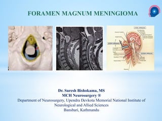
- 1. cka Dr. Suresh Bishokama, MS MCH Neurosurgery ® Department of Neurosurgery, Upendra Devkota Memorial National Institute of Neurological and Allied Sciences Bansbari, Kathmandu FORAMEN MAGNUM MENINGIOMA
- 2. A meningioma is considered to be located into the FM region if its insertion zone is mainly situated into the FM area. FM area is defined by these landmarks 1. Anterior border: lower third of the clivus and upper edge of the body of C2 2. Lateral borders: jugular tubercles and upper aspect of C2 laminas 3. Posterior border: anterior edge of the squamous occipital bone and C2 spinous process STRUCTURE PASSING VIA FM: Limits of the FM 2 PICA, 3 CN XII, 4 vertebral artery V4 segment, 5 C1, 6 dentate ligament, 7: VA3 1. Medulla oblongata 2. Ascending part of spinal accessory n. 3. Vertebral artery
- 3. Foramen magnum meningiomas are challenging tumors, requiring special considerations because of the vicinity of the medulla oblongata, the lower cranial nerves, and the vertebral artery. Meningiomas represent 25 – 30 % of all hospital-based primary intracranial neoplasms. The foramen magnum meningiomas constitutes 1 – 3 % of all cranial meningiomas. FORAMEN MAGNUM MENINGIOMAS
- 4. Variable symptoms. The lesion is often large when discovered because of their slow-growing rate, their indolent development, the difficulty of the diagnosis leading to a long interval since the first symptom, and the wide subarachnoid space at this level. Neck or sub-occipital pain exacerbated by coughing, straining, or sneezing. Motor sensory symptoms develop, usually in one arm, and then in the contralateral leg. (Elsberg phenomenon) Gait disturbances, diplopia, dysphagia, dysarthria, dyspnea, sphincter disturbances, vomiting, nausea are some of the common symptoms. The most common neurological sign is hyperreflexia, followed by weakness of the extremities in all combinations (hemiparesis/plegia, quadriparesis/plegia) and sensory loss. The Babinski sign, a gait disturbances, or lower CN (IX-XI) palsies are present in approximately half of patients with this tumor. CN XI is most commonly affected, presented by the shoulder weakness because of atrophy of sternocleidomastoids and trapezius muscles. Dissociated sensory loss, loss of coordination in the hands, Brown-Sequard syndrome, down- beating nystagmus, and nuchal rigidity and tenderness occur in one quarter to one third of patients. Papilledema, Horner’s syndrome, and dysarthria are less common neurological findings. Because of the lower cranial nerve morbidity associated with surgical resection of these tumors, incidentally discovered tumors in this location can initially be followed. CLINICAL PRESENTATION
- 5. 1. Anteriorly from the inferior third of the clivus to the superior edge of the C2 body. 2. Laterally from the jugular tubercle to the C2 laminae 3. Posteriorly from the anterior border of the occipital squama to the spinal process of C2. Origin of FMM
- 6. The neural elements: 1. Inferior vermis, 2. Cerebellar tonsils, 3. Fourth ventricle, 4. Lower cranial nerves from 9 – 12th, 5. Caudal aspect of the medulla oblongata 6. Rostral aspect of the spinal cord and 7. Upper cervical nerves C1 and C2. Important neurovascular structures The vascular structures: 1.Vertebral arteries, 2.Posterior inferior cerebellar arteries, 3.The meningeal branches of the vertebral artery, 4.The posterior and anterior spinal arteries and 5.The venous plexus.
- 7. Classification of foramen magnum meningiomas (FMM) depending on: 1. Their compartment of development: i. Intradural (most commonly), ii. Extradural (invasive into the bone, nerves and vessels sheaths) iii. Intra-extradural. 2. Their dural insertion: i. Anterior :Insertion on both side of the anterior mdiiline ii. Posterior : Insertion posterior to the dentate ligaments iii. Posterolateral : Insertion between midline and the dentate ligament iv. Anterolateral: 3. Their relation to the VAs: i. Above (the position of the lower cranial nerves cannot be anticipated), ii. Below (the lower cranial nerves are pushed cranially and posteriorly) and iii. On both sides Classification Boulton MR, Cusimano MD, Foramen magnum meningiomas: concepts, classifications and nuance. Neurosurg Focus 14, 2003
- 8. Classification Bruneau M and George B
- 9. The role of neuroimaging is to confirm the clinical diagnosis and to allow the planning of a surgical approach. Magnetic resonance imaging is the modality of choice for defining tumors of the foramen magnum. CT scan: Bony erosions and hyperstosis. Imaging
- 10. With meninigoma below VA, The lower CNs are displaced superiorly while Tumor above VA, the position is unpredictable. Relationship of VA with meningioma
- 11. Conventional angiography is generally useless. There are only two indications for preoperative angiography: 1. If a highly vascularized tumor is suspected and embolization is contemplated 2. To perform a balloon occlusion test in case of VA encasement (extradural or recurrent meningioma and meningioma inserted around the VA). Conventional angiography
- 12. CONSIDERATION 1. The location of the tumor 2. The extent of the tumor (above the foramen magnum) 3. The relation of the tumor with the vertebral artery and with the origin of posterior inferior cerebellar artery 4. Choice of the surgical approach, 5. Extent of bone resection 6. Management of the vertebral artery (VA) and 7. Involvement of lower cranial nerves (CN) SURGICALAPPROACH
- 13. NORMAL RELATIONSHIP OF BRAINSTEM AND TUMOR TO FORAMEN MAGNUM A: Normal relationship of brainstem to foramen magnum. OC = occipital condyle; CMJ = cervicomedullary junction. B-D: As the tumor (T) enlarges, it displaces the brainstem posteriorly and typically to one side, naturally creating a widened surgical corridor. B: A narrow corridor of less than 1 cm between the condyle and cervicomedullary junction. C: Adequate corridor with a 1-2 cm distance. D: Large corridor (> 2 cm) that allows relatively easy access to the anterior foramen magnum. (2
- 14. SURGICAL APPROACHES OF FORAMEN MAGNUM Posterior (for intradural lesions), lateral (intradural lesion situated lateral to and / or in front of the brainstem), anterior (for extradural lesions)
- 15. The midline posterior approach Posterior meningiomas (intra- and extradural extension): posterior to the plane of the dentate ligament and medial to the VA. Postero-lateral approach Intradural process located laterally and/or anteriorly to the neuraxis Extradural lesions developed on the posterior part of the lateral FM wall. Antero-lateral approach Rarely used Meningiomas with extradural extension through the bony structures SURGICALAPPROACHES
- 16. THE FAR-LATERALAPPROACH TO FMMS.
- 17. 1. Temporary: CSF leak Pseudomeningocele Lower cranial nerve deficits (IX–XII) Air embolism Hemiparesis Postop epidural hematoma Wound infection Meningitis Need for tracheostomy/gastrostomy 2. Permanent Lower cranial nerve deficits (IX–XII) hydrocephalus VA injury Tetraplegia Need for tracheostomy/gastrostomy Morbidity with Far lateral surgery
- 18. The transoral approach: did not acquired great acceptance: Risks Cerebrospinal fluid leak and Meningitis, difficult access for tumors with lateral extension and Risk of postoperative craniocervical instability and Velopalatine insufficiency The far-lateral and the extreme-lateral approaches are the most utilized nowadays. In both approaches it is possible to remove partially the occipital condyle but they provide different exposure because of different angles of approaches to the anterior FM. The extreme-lateral approach requires VA transposition for drilling the occipital condyle SURGICALAPPROACHES
- 19. SURGICAL APPROACH TO AN ANTERIOR FORAMEN MAGNUM MENINGOMA A: Suboccipital craniotomy (red) with a narrow corridor does not provide adequate exposure of the tumor for resection. B: Tumor growth naturally widens the surgical corridor, allowing its safe and effective removal via suboccipital craniotomy without drilling of the condyle. C: Transcondylar exposure (blue) widens the corridor by removing the medial condyle (red arrow represents very narrow corridor before excision of the condyle, green arrow represents adequate corridor after this resection). D.Access to much of the tumor has been created
- 20. 1. Anterior location: Difficult 2. Tumor size (bigger lesions are easier to be resected) 3. Tumor invasiveness 4. Extradural extension 5. VA encasement 6. Absence of arachnoidal layer 7. Adherences in recurrent lesions. Prognostic factors
- 21. 1. Yasargil 1976: Morbidity: ~13% ( Others: 45%) 2. Lower CN deficits: Sharp dissection of the arachnoid plane is key to preserving the rootlets of cranial nerves IX, X, XI, and XII. 3. Hemiparesis, tetraparesis, 4. Sensory deficits, 5. Hydrocephalus, 6. Craniospinal fluid leak, meningitis and 7. General complications as pneumonia, and respiratory faiuure. Outcome of surgery
- 22. Thank you NATIONAL INSTITUTE OF NEUROLOGICAL AND ALLIED SCIENCES, BANSBARI, KATHMANDU FORAMEN MAGNUM MENINGIOMA UPENDRA DEVKOTA MEMORIAL NATIONAL INSTITUTE OF NEUROLOGICALAND ALLIED SCIENCES, BANSBARI, KATHMANDU
Editor's Notes
- A: Positioning. The patient is placed in the true lateral decubitus position, with the lesion side up and ipsilateral shoulder rotated slightly anteriorly and inferiorly. The marked inverted hockey stick–shaped incision (dashed line) begins at the mastoid tip and curves medially toward the inion, then caudally at the midline down to the midcervical region. The hatched area underlying the skin incision indicates the site where the initial posterolateral craniectomy will occur. B: Exposure. The skin and superficial/intermediate musculature layers are reflected laterally. A small musculoaponeurotic cuff is left attached to the nuchal line to aid in wound closure. Note the horizontal segment of V3 exposed deep in the suboccipital triangle. C: Craniectomy. A posterolateral, retrocondylar suboccipital craniectomy is performed with the footplate of a high-speed drill. It includes the rim of the foramen magnum and extends laterally to expose the medial edge of the sigmoid sinus. The lateral rim of the foramen magnum, the condylar fossa, the posteromedial aspect of the occipital condyle, and the posterior arch of C-1 (hatched areas) will complete the bone exposure. Note the V3 segment and its association with the sulcus arteriosus of C-1 as it courses superiorly and medially to penetrate the posterior fossa dura. D: Dural opening. A curvilinear incision is created, extending medially from the transverse-sigmoid junction and then caudally at the midline. The dura is reflected laterally and held in place with nylon sutures. The proximal V4 segment is exposed intradurally. Note the spinal component of the accessory nerve (cranial nerve XI) coursing posteriorly and medially to V4, on its way to the jugular foramen (not shown)