Practical anatomy of the git
•
15 likes•953 views

This document provides guidance for a practical anatomy session on the gastrointestinal tract. It outlines 14 spots to identify various structures of the bones, muscles, nerves and vessels of the head and neck. Key structures to be identified include the temporal fossa, mental foramen, mandibular notch, coronoid process, mylohyoid line, lingula, neck of the mandible, parotid gland, parotid duct, masseter muscle, maxillary artery, inferior alveolar nerve and lingual nerve. The relationships and functions of these structures are also reviewed.

Recommended
More Related Content
What's hot
What's hot (20)
Similar to Practical anatomy of the git
Similar to Practical anatomy of the git (20)
Recently uploaded
Recently uploaded (20)
Practical anatomy of the git
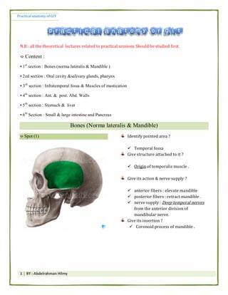
- 1. Practical anatomy of GIT 1 │ BY : Abdelrahman Hilmy N.B : all the theoretical lectures related to practical sessions Should be studied first. ⌔ Content : ▪ 1st section : Bones (norma lateralis & Mandible ) ▪ 2ed section : Oral cavity &salivary glands, pharynx ▪ 3rd section : Infratemporal fossa & Muscles of mastication ▪ 4th section : Ant. & post. Abd. Walls ▪ 5th section : Stomach & liver ▪ 6th Section : Small & large intestine and Pancreas Bones (Norma lateralis & Mandible) ⌔ Spot (1) Identify pointed area ? Temporal fossa Give structure attached to it ? Origin of temporalis muscle . Give its action & nerve supply ? anterior fibers : elevate mandible posterior fibers : retract mandible . nerve supply : Deep temporal nerves from the anterior division of mandibular nerve. Give its insertion ? Coronoid process of mandible .
- 2. Practical anatomy of GIT 2 │ BY : Abdelrahman Hilmy Spot (2) Spot (2) Identify pointed structure ? Mental foramen Give structures passing through ? Mental verve & Vessels N.B mental nerve is on of the 2 terminal brnaches of inferior alveolar neve. Identify pointed structure ? Temporalis muscle . انظجٕد ٗف لهُبْى ٗانه ٍي طؤال ٖا ٔيظبل فبد ٗانه.
- 3. Practical anatomy of GIT 3 │ BY : Abdelrahman Hilmy Spot (4) Spot (5) Identify the pointed structure ? Head of mandible (Condyle) Name joint related & give its type ? Temporo-mandibular joint.( TMJ) Synovial joint Identify the pointed structure ? Mandibular notch Mention nerve related to it ? Nerve to masseter muscle Spot (6) Identify the pointed structure ? Coronoid process Mention Structure attached ? Insertion of temporalis muscle .
- 4. Practical anatomy of GIT 4 │ BY : Abdelrahman Hilmy Spot (7) Spot (8) Identify the pointed structure ? Mylohyoid line Mention str. Attached ? Origin of mylohoid muscle Pterygomandibular lig. Superior Constrictor muscle . Identify the pointed structures 1 & 2 ? 1- Lingula 2- Mandibular foramen Mention str. Attached to No. (1) ? Sphenomandibular lig. Mention str. Passing through No. (2) ? Inferior alveolar nerve & vessels .
- 5. Practical anatomy of GIT 5 │ BY : Abdelrahman Hilmy Spot (9) Spot (10) Spot (11) Identify the pointed structures? Neck of the mandible Mention str. Attached ? Tempromandibular ligament Mention Nerves & Vessels related ? Maxillary artery : medial to neck Auriculotemporal nerve . : medial to neck Identify the pointed structures No. 1 & 2 ? 1- Sublingual fossa 2- submandibular fosaa Mention str. Related ? 1- sunlingual salivary gland 2- submandibular salivary gland . Identify the pointed Str. ? Pterygoid fovea Mention str. Attached ? Insertion of lateral pterygoid muscle
- 6. Practical anatomy of GIT 6 │ BY : Abdelrahman Hilmy Spot (12) Spot (13) Spot (14) Identify the pointed structure ? Zygomatic arch Mention str. Attached ? Temporalis fascia : attachéd to upper border Origin of masseter : from lower border & inner surface . Tempromandibular ligament . Identify area pointed by yellow arrow ? Pterion Identify the pointed structure ? Lateral surface of ramus of mandible Mention str. Attached ? Insertion of masseter muscle Identify the pointed structure ? Angle of mandible Mention str. Attached ? Stylomandibular ligament . Insertion of Medial pterygoid muscle (in inner aspect of the angle )
- 7. Practical anatomy of GIT 7 │ BY : Abdelrahman Hilmy Spot (15) Spot (15) ⌔ Geniohyoid muscle : * Nerve supply : C1 component of hypoglossal N. * Action : Elevate hyoid bone & depress mandible . Identify the pointed structures (red arrow)? Superior temporal line Mention str. Attached ? Temporalis fascia Identify Circled area ? Asterion Identify the pointed structures 1 & 2 ? 1- Superior genial tubercle 2- inferior genial tubercle Mention Str. Attached ? 1- Origin of Genioglossus muscle 2- Origin of geniohyoid muscles
- 8. Practical anatomy of GIT 8 │ BY : Abdelrahman Hilmy Spot (1) Identify the pointed structures? Parotid gland Mention its surfaces ? Lateral (superfacial) surface Anteromedial surface Posteromedial surface Mention Strs. Within it (in order from superfascial to deep) ? Facial nerve Retromandibular vein External carotid artery Mention its blood supply ? External carotid artery and its terminal -----> superficial temporal + maxillary arteries Mention its venous drainage ? Retromandibular vein Mention its capsule ? 1- inner capsule (true capsule): connective-tissue capsule adherent to the gland and sends 2- Outer capsule (false capsule): fromed by the investing layer of deep cervical fascia. Give its Motor nerve supply ? 1- Parasympathetic : Inferior salivary nucleus ⟶ glossopharyngeal nerve ⟶ ⟶⟶ postganglionic fibers are carried by ⟶ auriculo-temporal nerve to supply the gland 2- Sympathetic supply : from plexus surrounding the external carotid artery
- 9. Practical anatomy of GIT 9 │ BY : Abdelrahman Hilmy Mention its lymphatic drainage ? parotid lymph nodes + deep cervical lymph nodes. Mention its sensory nerve supply ? Capsule ⟶ Great auricular nerve . Parynchema ⟶ auriculo-temporal nerve . Mention the structures that leave the gland at its upper end (pole) ? 1- Auriculo-temporal nerve 2- Superfascial temporal artery 3- temporal branches of facial nerve . Mention the structure /s that Enter the gland through its upper end (pole) ? Superfascial temporal vein Mention the structures that enter the gland through its posteromedial surface ? 1- Facial nerve 2- External carotid artery Mention the structures that enter the gland through its anteromedial surface ? 1- Auriculo-temporal nerve 2- Maxillary vein Mention the Strs. That leave the gland at its lower pole (End) ? 1- Cervical branch of facial nerve 2- Anterior & posterior divisions of retromandibular vein . Give its anterior & posterior boundaries ? Anteriorly : masseter muscle (overlies its posterior part ) Posteriorly : sternomastoid muscle (overlies its upper part )
- 10. Practical anatomy of GIT 10 │ BY : Abdelrahman Hilmy Give its superior & inferior boundaries ? Upwards : Zygomatic arch Downwards : angle of mandible . Enumerate the Strs. That leave the gland at its anterior border ? Zygomatic N. Buccal nerve Marginal mandibular N. ⌔ Review the relations of the gland page 26 Spot (2) Identify pointed str. ? Parotid Duct Enumerate Strs. Pierced by it ? (4 Bucc-) 1-Buccal pad of fat 2- Buccopharungeal fascia 3- Buccinator muscle 4-buccal mucosa
- 11. Practical anatomy of GIT 11 │ BY : Abdelrahman Hilmy Mention the site of its opening ? (V-IMP Q) vestibule of the mouth opposite the upper second molar tooth Mention the surface marking of it ? It corresponds to the middle third of a line extending between two points: ① A point midway between the red margin of the upper lip and ala of the nose. ② A point at the lower end of the tragus of the ear. Spot (3) Identify pointed Str. ? Masseter muscle Give its action & nerve supply ? Action : Elevation and protraction of the mandible Nerve supply : anterior division of the mandibular nerve N.B origin & insertion & action & nerve supply of muscles of mastication is important .
- 12. Practical anatomy of GIT 12 │ BY : Abdelrahman Hilmy Spot (4) ⌔انـ ُْبmandibleانـ اػٕف ٌػهؼب يكظٕرحinfratemporal fossaحُ: ؼٕف - artery : maxillary artery فٕق ٗانه انصٕرح ٗف ّػهي يزؼبٔر - 2 muscles : medial & lateral pterygoid - 2 nerves : inferior alveolar nerve & lingual nerve *انـinferior alveolar nerveانـ جٕا داخم ٗثيجمmandibular foramen of mandibleٗثيجم ٔطبػبد يكظٕرح ٗثزجم ٗانه انـ ايب ,, يمطٕعlingual nerveٗثيجمanteriorّني,نهـ ٔرايحtongue. انـ ٗحزجم دٔل ٍانُزفي رحذ ٗانه انؼضهخ ٗف دثٕص حظ ٕن *medial pterygoidانـ ايبlateral pterygoidانـ رحذ ٗثزجم maxillary a. *ٌػهؼب كذا ٖد انصٕر َؼٕف. َزخيم
- 13. Practical anatomy of GIT 13 │ BY : Abdelrahman Hilmy inferior alveolar nerve البون فى داخل بيبقى Lingual nerve Anterior to inferior alveolar n.
- 14. Practical anatomy of GIT 14 │ BY : Abdelrahman Hilmy Spot (5) Identify pointed Str. ? Maxillary a. Mention its origin ? (how & where ?) It arises from External carotid artery within the substance of the parotid gland at the level of neck of mandible . Give its parts ? (3 parts ) 1- First (Mandibular) part : Medial to the neck of the mandible 2- Second (pterygoid ) part : On the lateral surface of the lateral pterygoid muscle 3- third part : Passes between the two heads of the lateral pterygoid to reach the pterygopalatine fossa . ّاي انظزٔف ػبرف يحذع ٌال َّؼزف ٍيضطزي احُب ثض ٗحيكف يغ انظجٕد ٍٔسي كزيزح كزبثخ يحزبج ٌال ٗيج صؼت دا *انظؤال *ٕثزض ٗاحزيبط جشء كم ٍي ٍثزَؼي ٖا َحفظ كذا ثؼذ. Third partSecond partFirst part 1-posterior superior alveolar 2-greater palatine 3-pharyngeal 4- sphenopalatine arteries, 5- artery of the pterygoid canal. 6- infraorbital artery Muscular Branches 1- deep temporal as. 2- masseteric a. 3- buccal a. 4- pterygoid branches for medial and lateral pterygoid muscles MIADA 1- Middle meningeal a. 2- Inferior alveolar a. 3- Accessory meningeal a. 4- Deep auricular a. 5- Anterior tympanic
- 15. Practical anatomy of GIT 15 │ BY : Abdelrahman Hilmy Spot (6) Identify pointed Str. 1 & 2 ? 1- inferior alveolar nerve 2- lingual nerve Mention origin of Str. NO. 1 & 2 ? Both arise from the posterior Division of mandibular nerve . Name the Structure that join Str. No 2 ? Mention its type of fibers ? Chorda tympani nerve It carries parasympathetic & sensory fibers . Mention the area supplied by Str. NO 1 & 2 ? The inferior alveolar nerves Takes the sensation from the lower teeth It gives the mylohyoid nerve wich supply anterior belly of diagastric muscle & mylohyoid muscle . Lingual nerve : takes general sensation from anterior 2 /3 of the tongue . & Chin & lower lip
- 16. Practical anatomy of GIT 16 │ BY : Abdelrahman Hilmy Mention the terminal branches of str. No 1 ? inferior alveolar nerves terminates as ---> incisive & mental branches adjacent to first premolar tooth . Mention the dangerous area of str. No. 2 ? Dangerous area of ligual nerve ---> medial surface of mandible adjacent to last molar tooth . *يذاكز يغ ٗانه ثظزػخ ػهيٓب يزاجغ َٖظز يذاكز ٗانه َٖظز اطئهخ كهٓب ٖدٗثم يؼبَب حيزؼت *ٗصجهي َزف ٔا ٍاكؼ ٗػه حيظبل ػهيٓى لهُب ٗانه ٍاالرُي ٍي ػضهخ ٖا جبة ٕن. Spot (7) Identify pointed Str. ? Submandibular Salivary gland . Mention its parts & how they are devided ? 1- large superficial part. 2- Small deep part. by posterior border of mylohyoid muscle.
- 17. Practical anatomy of GIT 17 │ BY : Abdelrahman Hilmy Mention the site of its superfacial part ? digastric triangle (Submandibular triangle ) Name the Str. That separate it from parotid gland ? stylomandibular ligament Mention the artery related to it ? Facial artery (grooves it) Mention its capsule ? True capsule : connective-tissue capsule adherent to the gland Flase capsule : derived from the investing layer of deep cervical fascia. Mention the site of opening of its duct ? Opens in the floor of mouth on the summit of the sublingual papilla situated at the side of the frenulum of the tongue. Mention the nerve related to its duct ? Lingual nerve (it has a triple relation ship with the duct) Mention its arterial supply ? branches from Facial & lingual arteries. Mention its venous drainage ? Facial & lingual veins. Mention its Lymphatic drainage ? Submandibular and deep cervical lymph nodes
- 18. Practical anatomy of GIT 18 │ BY : Abdelrahman Hilmy Mention its nerve supply ? Sensory: lingual nerve. Motor : ⌔ Sympathetic : plexus around facial & lingual arteries arteries. ⌔ Parasympathetic: postganglionic fibres from the Submandibular ganglion N.B Review relations of each part of the gland . ٗانه انززفبد ّاي يمٕل يظبل ٍيًك انًًيشح انحبجبد رؼزفٕا اَزى يًم ٗٔحيجم ُٗي حيفزح انفبيم كذا ٌال ُْب اكزجٓب ػبيش يغ اَب *related to its medial surfaceْٔكذا * Remember : Spot (8) Identify ? ⌔ Paryngeal recess (Fossa of Rosen Muller ) *دث ٗثيجمانـ ٔرا يحطٕط ٕصtubal elevation Mentio Str. Overlyed by it ? ⌔ Internal carotid artery
- 19. Practical anatomy of GIT 19 │ BY : Abdelrahman Hilmy Spot (9) Identify ? ⌔ Salpingo-pharyngeal fold *انـ ٍي َبسل ٗثيجمtubal elevationانـ ٗػه ٔرايحpharynx. Mentio Str. Forming it ? ⌔ salpingo-pharyngeas muscle & mucous membrane covering it .
- 20. Practical anatomy of GIT 20 │ BY : Abdelrahman Hilmy Spot (10) Identify ? ⌔ Pharyngeal opening of the auditory tube Give the site of the opening of other end of this opening ? mention its function ? ⌔ Tympanic cavity of the middle ear ⌔ equalize the pressure on both sides of the ear drum Name the pointed str. No. 1 ? ⌔ Tubal elevation Mention the Strs. Forming it ? 1- cartilaginous end of the auditory tube & Mucosa covering it . 2- Tubal tonsil (lymphoid tissue).
- 21. Practical anatomy of GIT 21 │ BY : Abdelrahman Hilmy Spot (11) 1- Bed of palatine tonsil 2- palatoglossal arch 3- palatopharyngeal arch Mention the Str. Forming No. 2 & 3 ? 1- Palatoglossus arch formed by : palatoglossus muscle & mucosa covering it 2- palatopharyngeus arch formed by : palatopharyngeus & mucosa covering it Any theoretical Q may be asked about palatine tonsil : A - arterial supply : 1- Tonsillar artery (from facial artery). 2- Ascending palatine artery (from facial artery). 3- Lingual artery (from external carotid artery). 4- Ascending pharyngeal artery (from external carotid artery).
- 22. Practical anatomy of GIT 22 │ BY : Abdelrahman Hilmy 2- nerve supply : 1.Glossopharyngeal nerve. 2. Lesser palatine nerve. 3- Venous drainage : ⌔ Paratonsillar vein 4- Lymphatic Drainage : ⌔Deep cervical lymph nodes (mainly the jugulo-digastric nodes). ⌔ See its relations in page 43. Spot (12) Identify ? ⌔ Pyriform fossa Mention its nerve supply ? ⌔internal laryngeal nerve. Give its medial boundary ? ⌔ Aryepiglottic fold of the larynx Give its lateral boundary ? ⌔ Thyrohyoid membrane above and the lamina of the thyroid cartilage below.
- 23. Practical anatomy of GIT 23 │ BY : Abdelrahman Hilmy Spot (13) Identify ? ⌔ Genioglossus muscle Mention its nerve supply ? ⌔ Hypoglossa lverve Give its action ? ⌔ it Protrudes the tongue . & prevent drop of the tongue Gie its origin ? ⌔ Superior genial tubercle of the mandible.
- 24. Practical anatomy of GIT 24 │ BY : Abdelrahman Hilmy Spot (14) Identify ? ⌔ Intrinsic muscles of the tongue Mention its nerve supply ? ⌔ hypoglossal nerve Give their action ? ⌔ they change shape of the tongue 1- transverse muscle: narrowing of tongue 2- longitudinal muscle : Shortening of tongue 3- vertical muscle : Thinning of tongue Spot (15) Identify ? ⌔Tip of the tongue Give its lymphatic driange ? ⌔ Submental lymph nodes Mention its sensory supply ? 1- General S. : lingual nerve 2- Taste sensation : chorda tympani
- 25. Practical anatomy of GIT 25 │ BY : Abdelrahman Hilmy Spot (16) Spot (17) Give its motor supply ? ⌔ All muscles of the tongue are supplied by hypoglossal nerve, except the palatoglossus muscle which is supplied by cranial part of accessory nerve through the pharyngeal plexus. Identify ? ⌔Vallecula Give its sensory innervation ? ⌔ internal laryngeal nerve . Give its boundaries ? ⌔ Median & latewral glossoepiglottic folds Identify the organ ? ⌔ Tongue Give its arterial supply ? 1. Lingual artery. 2. Tonsillar branch of facial artery. 3. Ascending pharyngeal artery. Give its venous drainage ? ⌔Lingual vein ⟶ internal jugular vein
- 26. Practical anatomy of GIT 26 │ BY : Abdelrahman Hilmy Spot (18) Identify ? ⌔ Soft palate Mention its content ? ⌔ Palatine aponeurosis. ⌔ Muscles. ⌔ Nerves. ⌔ Vessels. ⌔ Lymphoid tissue. Enumerate muscles forming it ? which one is intrinsic ? - Tensor palati - Tensor tympani - palatoglossus - palatopharyngeus - musculus uvulae (intrinsic one ) Name its motor innervation ? ⌔ All the muscles are supplied by the cranial part of accessory nerve except the tensor palati muscle which is supplied by the mandibular nerve.
- 27. Practical anatomy of GIT 27 │ BY : Abdelrahman Hilmy Give its sensory innervation ? 1- General sensation : ⌔ Lesser palatine nerve. ⌔ Glossopharyngeal nerve 2- Taste sensation ⟶ lesser palatine nerves. Give its parasympathetic innervation ? or its Secretomotor innervation ? The facial nerve ⟶ greater petrosal nerve ⟶ sphenopalatine ganglion. ⟶ lesser palatine nerves to palatine glands Mention its arterial supply ? Mention 2 arteries supplying it ? Give their origin ? 1. Greater palatine artery ⟶ branch from the maxillary artery (3rd part ). 2. Ascending palatine artery⟶ branch from the facial artery. Mention its venous drianage ? ⌔ pterygoid + pharyngeal plexuses of veins. Mention its Lymphatic Drainage ? ⌔ upper deep cervical & retropharyngeal lymph nodes Spot (20) Identify ? ⌔ Uvula Name muscle forming it ? Give its nerve supply ? ⌔ Musculus uvulae. ⌔ Cranial part of accessory nerve.
- 28. Practical anatomy of GIT 28 │ BY : Abdelrahman Hilmy Spot (21) Identify ? ⌔ Hard palate Name bones forming it ? 1- palatine processes of the maxillae 2- horizontal plates of the palatine bones Name 2 features of its mucosa ? 1- bilateral corrugations on both sides (palatine rugae ) 2- inferior median raphe (palatine raphe) 3- less vascular & firmly attached to underlying periosteum Mention its Sensory innervation ? 1- General sensation: ⌔ Greater palatine nerve ⌔ Nasopalatine nerve 2- Taste sensation ⟶ lesser palatine nerves
- 29. Practical anatomy of GIT 29 │ BY : Abdelrahman Hilmy ● Don’t forget : ▪ Hard = Greater palatine N. + Nasopalatine N. ▪ Soft = Lesser palatine N. + Glossopharyngeal . Give its lymphatic drainage ? ⌔ submandibular lymph nodes. Arterial supply & venous Drianage ⟶ the same as soft palate . Spot (22) Spot (23) Identify ? ⌔ Posterior 1/3 of tongue Or ( pharyngeal part ) Mention its nerve supply ? ⌔ General sensation & Taste sensation : By Glossopharyngeal nerve Name a characteristic feature of its dorsal surface ? ⌔ Lingual tonsils Identify ? ⌔ Anterior 2/3 of tongue Or ( Oral part of the tongue ) Mention its nerve supply ? ⌔ General sensation : By Lingual nerve ⌔ Taste sensation : by Chorda tympani By Glossopharyngeal nerve Name a characteristic feature of its dorsal surface ? ⌔ Lingual papillae (Fungiform & filiform & Vallate)
- 30. Practical anatomy of GIT 30 │ BY : Abdelrahman Hilmy Name the Features of its under surface ? ⌔ Attached to floor of mouth by frenulum linguae ⌔ on each side (from medil to lateral ) : lingual artery & lingual nerve & deep lingual vein Stomach & liver Spot (1) Identify the pointed str. ? Liver , anterior surface Mention the str. Attached to pointed area ? Give its content ? Falciform ligament Ligamentum teres (round ligament of the liver ) Mention its arterial supply ? 1- hepatic artery : from celiac trunk 2- portal vein : formed by union of splenic vein & superior mesenteric vein . Both of them devides into rt & lt branches
- 31. Practical anatomy of GIT 31 │ BY : Abdelrahman Hilmy Mention its lymphatic drainage ? portal lymph nodes then into the coeliac lymph nodes Except bare area of the liver : subphrenic lymph nodes, or Posterior mediastinal lymph nodes. Mention its venous drainage ? Right & middle & left hepatic veins ------> IVC Enumerate its peritoneal ligaments (connections )? 1- Falciform ligament. 2- Upper layer of coronary ligament. 3- Lower layer of coronary ligament. 4- Right triangular ligament. 5- Left triangular ligament. 6- Lesser omentum. Mention its Embryonic ligaments ? Give the origin of each one ? 1- Ligamentum teres ⟶ obliterated left umblical vein 2- Ligamentum venosum⟶ obliterated ductus venosus Enumerate its bare areas ? 1- Bare area 2- Groove for IVC 3- Porta hepatic 4- Fossa of gall bladder 5- Fissures for ligamentum teres and for ligamentum venosum *انـ طؤال ٗف طٕاءligaments or bare areas. ّكه يغ ثض ٍارُي يطهت ٍيًك
- 32. Practical anatomy of GIT 32 │ BY : Abdelrahman Hilmy Spot (2) Identify ? Bare area of the liver Mention its boundaries ? ⌔ base : groove for IVC ⌔ apex : right triangular ligament ⌔ sides : the two layers of coronary ligament. Mention its realtion ? 1-Diaphragm 2- Supra renal gland Mention its lymphatic drainage ? subphrenic lymph nodes or Posterior mediastinal lymph nodes
- 33. Practical anatomy of GIT 33 │ BY : Abdelrahman Hilmy Spot (3) Identify ? Caudate lobe of the liver Mention its boundaries ? Right side : groove for the IVC Left side : fissure for the ligamentum venosum Superiorly : ligamentum venosum Inferiorly : porta hepatis Or Mention the str. Superior / inferior to it ? or mention str. Related to its side ? Name its processes ? Caudate process : projects to the right Papillary process : projects to the left . Name the part that form superior boundary of epiploic foramen ? Caudate process Mention its posterior relations ? 1- Diaphragm 2- Desevding thoracic aorta 3- T12
- 34. Practical anatomy of GIT 34 │ BY : Abdelrahman Hilmy Spot (4) Identify ? Porta hepatis Name the Str. That passing through it ? a. Hepatic ducts: anterior in position. b. Hepatic artery: intermediate in position. c. Portal vein: posterior in position. d. Lymphatics & sympathetic nerves Mention str. Attached to it ? Lesser omentum. Mention its boundaries ? Anteriorly : quadrate lobe Posteriorly : caudate lobe and process
- 35. Practical anatomy of GIT 35 │ BY : Abdelrahman Hilmy Spot (5) Identify ? Fissure for ligamentum venosum Name Str. Related ? give its embryological origin ? ligamentum venosum >> obliterated ductus venosus . Spot (6)
- 36. Practical anatomy of GIT 36 │ BY : Abdelrahman Hilmy Identify ? Quadrate lobe of the liver Mention its boundaries ? Anteriorly : inferior border of liver Posteriorly : porta hepatis Right side : gall bladder fossa Left side : fissure for ligamentum teres Give its relations ? (TPL) Transverse colon (anteriorly) Pylorus& 1st part of duodenum (middle ) Lesser omentum (posteriorly). The order is imp. as the Q may be : give the relations from upward to downward or the reverse . Spot (7) Identify Str. No 1 & 2 & 3 ? 1- ligamentum teres 2- gall bladder 3- gastric impression
- 37. Practical anatomy of GIT 37 │ BY : Abdelrahman Hilmy Give the embryological origin of Str. No 1 ? Obliterated left umblical vein Mention the parts of Str. No 2 ? Fundus + Body + Neck Mention the Blood supply of Str. No. 2 ? Arterial supply : cystic artery from right branch of hepatic artery Venous drainage : cystic vein which drains to the right branch of portal vein. Mention the surface anatomy of Str. No. 2 ? (V-IMp) Fundus of gall bladder: tip of Right 9th costal cartilage انـ صٕرح ٖد *impressionsٔيظبل حزخ ٖا ٗف انذثٕص يحظ ٍيًكidentify. ػبو طؤال ٖا ِٔيؼب
- 38. Practical anatomy of GIT 38 │ BY : Abdelrahman Hilmy Spot (8) Identify ? Portal vein Mention its origin ? (how & where ?) It is formed by union of the superior mesenteric and splenic veins behind the neck of pancreas and in front of IVC Name 2 of its tributaries ? 1- Superior mesenteric vein. 2- Splenic vein 3- Right gastric vein. 4- Left gastric vein. 5- Paraumbilical vein (in the left branch). 6- Cystic vein (in the right branch).
- 39. Practical anatomy of GIT 39 │ BY : Abdelrahman Hilmy Spot (9) Spot (10) Identify the pointed structure? Lesser curvature of stomach Mention str. Attached to it ? Lesser omentum Mention str. Related to it ? Right & left gastric vessels . Identify the pointed structure? Greater curvature of stomach Mention str. Attached to it ? Greater omentum Gastrophrenic ligament Gastrosplenic ligament Mention str. Related to it ? Right & left gastroepiploic vessels .
- 40. Practical anatomy of GIT 40 │ BY : Abdelrahman Hilmy Spot (11) Mention its nerve supply ? 1- Sympathetic: from celiac plexus around celiac trunk. 2- Parasympathetic: from anterior and posterior gastric nerves. (from vagi) Mention peritoneal space related to this surface ? * Greater sac Spot (12) Identify the organ ? Stomach , anterior surface Name its parts ? Fundus Body Pylorus : antrum & canal & sphincter Mention str. Related to pointed area ? 1- liver. 2- Anterior abdominal wall. 3- Diaphragm. Mention its lymphatic D. ? The gastric lymph vessels into the celiac lymph nodes Identify the pointed Strs. No 1 & 2 ? 1- Cariac orifice 2- pyloric sphincter Mention Give their surface anatomy ? 1-Cardiac orifice: is 1 inch to the left of the median plane at level of T.11 2-Pyloric orifice: is 1/2 inch to the right of the median plane at level of L.1
- 41. Practical anatomy of GIT 41 │ BY : Abdelrahman Hilmy V- imp : Structures forming stomach bed : (relations of posterior surface of stomach) 4 horizontal structures : - Body of pancreas - Splenic artery - Transverse colon -Transvesre mesocolon يجبػز انظؤال ٗيج يبيب *mention 4 str forming stomach bedانـ ٗف انذثٕص يحظ ٔاposterior surface of stomachٍػ ٔيظبل ان َؼزف ٌػهؼب كٕيض ػهيُب انؼيُخ َظجظ حبجخ اْى انزيالػُشـanteriorيبنـposterior surface. *ٔيظبل جزخ ٖا ٗف دثٕص يحظidentify pointed partانـ اجشاء ٗػه يظبل ٍٔيًكpylorusْٗ ٗانه: antrum & canal & Sphincter 4 vertical Str. : - left kidney -left suprarenal gland - left crus od diaphragm - spleen
- 42. Practical anatomy of GIT 42 │ BY : Abdelrahman Hilmy Spot (13) Spot (14) Identify the pointed part ? ⌔ Posterior posterior surface of stomach Mention 4 Str. Forming its bed ? ( See above ) Name its peritoneal covering ? ⌔ its covered by peritoneum of lesser sac N.B if you asked about peritoneal covering of anterior surface the answer will be : peritoneal of greater sac . Mention peritoneal space related to this surface ? Lesser sac (Omental bursa) Identify the pointed part ? ⌔ Fundus of the stomach Mention its peritoneal connections ? 1- Gastro-phrenic ligament. 2- Gastro-splenic ligament . Name its arterial supply ? Origin ? ⌔ Short Gastric arteries ⌔ From : splenic artery .
- 43. Practical anatomy of GIT 43 │ BY : Abdelrahman Hilmy Spot (15) Mention its peritoneal covering ? ⌔ Its completely covered by peritoneum Mention 2 strutures open into it ? give the site of communication ? ⌔ Terminal ileum & appendix ⌔ they open into it at its postero-medial aspect Mention its venous drainage ? ⌔ superior mesenteric vein then into portal vein Name the specimen? ⌔ Ileo-cecal junction & appendix Name the pointed part ? give its arterial supply ? ⌔ Caecum ⌔ anterior & posterior cecal arteries ⟶ ileocolic ⟶ SMA Mention Nerves related posteriorly to it ? ⌔ Femoral nerve ⌔ lateral cutaneous nerve of the thigh. Mention muscles related posteriorly to it ? ⌔ Iiacus muscle ⌔ Poas major muscle Give its anterior relation ? ⌔ anterior abdominal wall & greater omentum & and coils of small intestine. ⌔آَب االٔل انؼيُخ ٍي ٔرزبكذ حبجخ اْىileo cecalيغ stomach .
- 44. Practical anatomy of GIT 44 │ BY : Abdelrahman Hilmy Spot (16) Enumerate its variant positions ? what is the most frequent one ? 1. Retrocecal (65%) ⟶ Most frequent one . 2. Pelvic (30%) 3. Subcecal (3%) 4. Pre or post- ileal (2%) Mark the surface anatomy of its base ? ( McBurney’s point ) ⌔ is represented by a point at the junction of lateral 1/3rd and medial 2/3rd of a line connecting anterior superior iliac spine (ASIS) and the umbilicus. (Spino-umblical line) Mention its sensory innervation ? Give the site of its refered pain ? ⌔ sympathetic fibers from 10 th thoracic spinal cord segment . ⌔ umbilical region Identify (yellow arrow) ? ⌔ Vermiform appendix Name its Mesentry ? Give its content ? ⌔ Mesoappendix ⌔ appendicular artery in its free border Name its arterial supply ? Origin ? ⌔ appendicular artery from the posterior caecal artery from ileocolic artery ⟶ SMA Mention its venous drainage ? ⌔ superior mesenteric Vein Mention its Frequent site ? ⌔ Retrocecal in postion
- 45. Practical anatomy of GIT 45 │ BY : Abdelrahman Hilmy Spot (17) Mention 3 of its chareteristic features that distinguish it from small intestine ? (Leading Q.) 1- taenia coli 2- sacculations 3- Appendices epiploicae Mention muscles posterior to it ? 1- transverses abdominis 2- quadratus lumborum 3- iliacus Mention Nerves posterior to it ? 1- iliohypogastric N. 2- ilioinguinal N. 3- lateral cutaneous of the thigh . The viscera posterior to it >>> right kidney . & ant. Relation : Coils of small intestine + greater omentum Identify ? ⌔ Ascneding colon Mention its peritoneal Covering ? ⌔ it is covered by peritoneum from anterior & its sides only . Name its arterial supply ? Origin ? 1- Ileocolic artery 2- Right Colic artery * From superior mesenteric artery. Where it terminates and how ? * it ends just below the liver, as the right colic (hepatic) flexure.
- 46. Practical anatomy of GIT 46 │ BY : Abdelrahman Hilmy Spot (18) Spot (19) Identify pointed Str. ? ⌔ Ileum Name the attachment of root of its mesentery ? & its length ? ⌔ from duodeno-jejunal flexure to iliocecal junction. ⌔ 6inches Mention its arterial supply ? ⌔ ileal branches of SMA. Identify ? ⌔ Terminal ileum Mention its arterial supply ? ⌔ Ileal branches oF SMA
- 47. Practical anatomy of GIT 47 │ BY : Abdelrahman Hilmy Spot (20) Identify ? ⌔ Jejunum Mention its arterial supply ? ⌔ jejunal arteries of superior mesenteric arteries . Name 2 features of its mesentery ? 1- low content of fat 2- presence of windows . ⌔ Remember this : Small intestine Large intestine Few & simple arterial arcades Numerous and complex arterial arcades Long vasa recta Short vasa recta Numerous Plica Circularis Few Plica Circularis No Peyer’s patches Numerous Peyer’s patches Wide lumen & thick wall Narrow lumen & thin wall
- 48. Practical anatomy of GIT 48 │ BY : Abdelrahman Hilmy Spot (21) Identify (yellow arrow ) ? ⌔ Mesentery of small intestine Name 3 Str. Crossed by its root ? 1. 3rd part of duodenum 2. Abdominal aorta and right gonadal vessels. 3. IVC. 4. Right psoas major. 5. Right ureter. 6. Right genitor-femoral nerve The Q. may be specific : Mention (Nerves & muscle & vessles ) crossed by its root ? Name the attachment of its root ? &Give the length of its attached & free borders ? ⌔ from duodeno-jejunal flexure to iliocecal junction. ⌔ Root : 6inches & free border : 6 meter Enumerate 3 Str. of its contetns ? 1- Superior mesenteric artery. 2- Superior mesenteric vein. 3- Coils of the small intestine 4- Extraperitoneal tissue and fat. 5- Sympathetic nerve fibers 6- Mesenteric LN
- 49. Practical anatomy of GIT 49 │ BY : Abdelrahman Hilmy Spot (22) Identify ? ⌔ Uncinate process of pancreas Name the Strs. Anterior to it ? ⌔ Superior mesenteric artery & Vein Name the Str. Posterior to it ? ⌔ abdominal aorta . Give its arterial supply ? ⌔ Superior, inferior pancreatico-duodenal arteries Mention its lymphatic Drainage ? ⌔ superior mesenteric lymph nodes
- 50. Practical anatomy of GIT 50 │ BY : Abdelrahman Hilmy Spot (23) Identify ? ⌔ Head of pancreas Mention its arterial supply ? Give the orgin ? ⌔ Superior, inferior pancreatico-duodenal arteries 1- Superior pancreatico-duodenal artery : from gastroduodenal >> hepatic a. 2- inferior pancreatico-duodenal arterys: from SMA Mention its lymphatic Draingae ? ⌔ upper part : coeliac lymph nodes ⌔ lower part : superior mesenteric lymph nodes N.B : you must review the relations of each part of pancreas . the Q may be : Mention vessels related meial to it ? ⌔ Superior, inferior pancreatico-duodenal arteries Mention its posterior relation ? ⌔IVC, renal veins and common bile duct. ● the anterior relation is >> transverse colon
- 51. Practical anatomy of GIT 51 │ BY : Abdelrahman Hilmy Spot (24) Identify ? ⌔ Neck of the pancreas Mention its posterior relation ? ⌔ formation of portal vein from splenic and superior mesenteric veins. *انـ ّاي ٕثزض ٗاحزيبط طبل ٕٔن دا انظؤال ٌػهؼب يخصٕؽ جبيجٓب ٗحيجم ٖد انؼيُخ جبة ٕن ْٕanterior relationٗحزجمduodenal junction-gastro Spot (25) .الـ بعلمneckالـ منSMAلو وكمان تحتها من بيطلع اللى الـ قاصدbodyالـ حنب الدبوس حاطط حيبقى اكيدduodenum الـ قاصد ولو طوول عbodyوطبيعة النص فى الدبوس حيحط لغبطة مفهاش برضو بتبين بعده اللى السؤاليعنى. هللا شاء ان
- 52. Practical anatomy of GIT 52 │ BY : Abdelrahman Hilmy Identify ? ⌔ Body of the pancreas Mention its surfaces ? & Borders ? ⌔ Surfaces : anterior, posterior and inferior ⌔ Borders : anterior, superior and inferior Mention the Strs. Attached to its anterior border ? ⌔ transverse mesocolon ⌔ greater omentum. Mentios Str. Related to its upper border ? Give a characteristic feature of it ? ⌔ splenic artery & it has a very tourtous course . Mention 2 branches of the artery running on its upper border ? 1- left gastro-epiploic artery . 2- short gastric artreis 3- arteria pancreatica magna . Mention the vessels related to its posterior surface ? 1- Aorta and origin of sup. mesenteric artery 2- Splenic and left renal vein Mention the muscles related to its posterior surface ? 1- Left psoas major 2- Left crus of diaphragm Mention the Viscera related to its posterior surface ? 1- Left kidney 2- Left supra renal gland Mention the nervous str . related to its posterior surface ? ⌔ Left sympathetic chain ● Relations of inferior surface (Rare but may come): duodeno-jejunal flexure & loops of jejunum and end of transverse colon (from right to left). ● Relations of Anterior surface : stomach, separated from it by the lesser sac.
- 53. Practical anatomy of GIT 53 │ BY : Abdelrahman Hilmy Spot (26) Identify ? ⌔ Tail of the pancreas Mentio Str. Related to it ? ⌔ Hilum of spleen . Name the peritoneal fold through which it runs ? IMP ⌔ lieno-renal ligament Spot (27)
- 54. Practical anatomy of GIT 54 │ BY : Abdelrahman Hilmy Identify the organ ? ⌔ Duodenum Mention its arterial supply ? Origin ? 1. Supra-duodenal artery: from the hepatic artery proper . 2. Superior pancreatico-duodenal artery: from gastro-duodenal . 3. Inferior pancreatico-duodenal artery: from superior mesenteric artery. Mention its beginning ? ⌔ It begins at the pylorus, 1/2 an inch to the right of the median plane at the level of L1 (transpyloric plane). Mention its termination ? ⌔ at the dudeno-jejunal flexure , one inch to the left of median plane . What is the most mobile part of this organ ? Why ? ⌔ the first part , because it is covered by peritoneum anteriorly and posteriorly. Spot (28) Identify pointed part ? ⌔ First part of the Dudenum . Give its vertebral level ? ⌔ at the level of L1 (transpyloric plane). Mention its length ? ⌔ 2 inches Give its peritoneal covering ? ⌔ its 1st inch is covered by peritoneum anteriorly and posteriorly.
- 55. Practical anatomy of GIT 55 │ BY : Abdelrahman Hilmy N.B review the relations of each part of the duodnum first . Mention the Str. Superior to it ? ⌔ Epiploic foramen . Mention its anterior relations ? 1- quadrate lobe of the liver 2- gallbladder. Mention its posterior relations ? 1- neck of the pancreas 2- portal vein 3- bile duct 4- gastro-duodenal artery. Name the artery posterior to it ? Give its origin ? Branches? ⌔ gastro-duodenal artery ⌔ it arises from Hapatic artery . ⌔ Branches : 1- Right gastroepiploic a. 2- Superior pancreatico-duodenal a. Name the Duct posterior to it ? how its formed ? give its termination ? ⌔ Common bile duct ⌔ it is formed by the union of custic duct of gall bladder & common hepatic duct ⌔ it terminates by the union with common pancreatic duct to form Ampulla of vater wich opens into major duodenal papilla . ⌔انـ رثطُب احُب ٖد انظجخد ٗف ُٗػ ثجؼضٓب االجشاء كم َٔزثظ كٕيض ٖانُظز َجًغ اَُب ثض انفكزح ٖد االطئهخ كم حيظبل يغ ْٕ طجؼب duodenumانـ ثًحبضزحarterial supplyانـ ثًحبضزحbiliary system.
- 56. Practical anatomy of GIT 56 │ BY : Abdelrahman Hilmy Spot (29) Identify pointed part ? ⌔ Seconf part of the duodenum Give its vertebral level ? ⌔ descends vertically from the level of L1 to the level of L3 Mention its length ? ⌔ 3 inches Mention its peritoneal covering ? ⌔ only covered by peritoneum anteriorly . Mention its anterior relations ? ⌔ right lobe of the liver & gall bladder & transverse colon & coils of jejunum. Mentios its posterior relation ? ⌔ hilum of the right kidney Mention its medial relation ? ⌔ head of pancreas & bile duct & pancreatico –duodenal arteries Mention vessels related medial to it ? ⌔ Superior & inferior pancreatico –duodenal arteries
- 57. Practical anatomy of GIT 57 │ BY : Abdelrahman Hilmy Spot (30) Identify pointed part ? ⌔ third part of the Duodenum Give its vertebral level ? ⌔ lies horizontally opposite to the level of L3. Mention its length ? ⌔ 4 inches Mentions the vessels posterior & anterior to it ? IMP ⌔ Anterior : superior mesenteric vessels ⌔ Posterior : 1-right gonadal vessels 2-IVC 3- aorta and inferior mesenteric artery. Mention its anterior relation ? ⌔ superior mesenteric vessels in the root of the mesentery . ⌔ coils of small intestine.
- 58. Practical anatomy of GIT 58 │ BY : Abdelrahman Hilmy Abdominal wall muscles Spot (1) Identify ? ⌔ rectus abdominis muscle Give its action ? ⌔ Flexes the trunk ⌔raises the intra-abdominal pressure. Give its nerve supply ? ⌔ lower five intercostal verves & subcostal nerve (intercostal nerves 7-11 and subcostal nerve) Give its origin & insertion ? Mention the contents of its sheath ? ⌔ Superior & inferior epigastric vessels ⌔ Lower 5 intercostal nerves & Subcostal nerve ⌓ Important : You have to know the Structures forming the anterior & posterior walls of each part of the sheath (see the Book page 53 & 54 ) For Example : Name the Str. That form the anterior wall of the middle part of its sheath ? َّا ٔارد َٖظز طؤال ٖا *. ػٕيخ حُزؼت يؼهغ ثبنُب فُبخذ ٗيج
- 59. Practical anatomy of GIT 59 │ BY : Abdelrahman Hilmy *انـ ثميذMuscles of anterior abdominal wallركشٔا ثض ػهيٓى َٖظز طؤال ٖا يظبنُب , ثزبػزٓى انفيجزس ِارجب ٍي ثؼزفٓى . ٍٔاالكؼ ٗصجه انُزف ٗػه اكزز ⌔ Nerve supply of Transversus abdominis & external oblique & internal oblique : ▪ Intercostal nerves 7-11, subcostal N. ▪ iliohypogastric N. ▪ ilioinguinal nerve ⌔ Action : ▪ Flexes and laterally bends the trunk . 2- External abdominal oblique : fibers runs downward & forwards & medially. ( ّجيج ٗف ِايذ حطظ )ٔاحذ
- 60. Practical anatomy of GIT 60 │ BY : Abdelrahman Hilmy 2- internal abdominal oblique : fibers directed upwards, forwards and medially 3 -Transversus abdominis muscle : fibers runs transversely.
- 61. Practical anatomy of GIT 61 │ BY : Abdelrahman Hilmy Spot (2) Identify ? ⌔ Poas major muscle Mentio its nerve supply ? ⌔ Lumbar plexus via anterior rami of L1-2-3 nerves. Give its action ? ⌔ Flexion of the thigh. ⌔Lateral flexion of the trunk. Give its insertion ? ⌔ By a strong tendon to lesser trochanter of femur Mention Nerves that appears from its lateral border ? 1- iliohypogastric N. 2- ilioinguinal N. 3- lateral cutaneuos nerve of the thigh 4- Femoral nerve
- 62. Practical anatomy of GIT 62 │ BY : Abdelrahman Hilmy Mention Nerve that emerges from its Medial border? 1- Obturator nerve 2- Lumbosacral trunk Mention Nerves that emerges from its anterior surface ? give its root value ? its terminal branches ? area supplied by them ? ( IMP Q ) ⌔ genitofemoral nerve ⌔ Root value : L1 & L2 (Ventral rami) ⌔ Terminal brnches : 1- genital branch (enters inguinal canal) : supply cremasteric muscle 2- femoral branch : supply the skin over the femoral triangle. انـ صٕرح ٖٔد *genitofemoral nerveانـ ٍي طبنغ ٗثيجمanterior surface of poas majorفٕق ٗانه ٍي طؤال ٖا ّػهي يظبل ٍيًك . دٔل
- 63. Practical anatomy of GIT 63 │ BY : Abdelrahman Hilmy *(ؽ دٔل ٍي ػضهخ كم ثزبع ٍٔاالكؼ ٗصجهي انُزف َؼزف26انـ ٗػه ركشد اَب )poas majorٌػهؼبmaterialكزيزح اطئهخ ػهيٓب يظبل ٍيًك حهٕح . كذا فٕق ػًهُب يب ٖس ثبنُزفبد يزثطٓب ٍيًك اَخ اال ثبالضبفخ.ٗاحزيبط ػهيٓى ثصٕا يجٕا ٍيًك ٍانزبَيي ٌا ييًُؼغ ثض Vessels & Nerves of Abdomen Femoral nerve : thick & lateral to poas major (IMP)
- 64. Practical anatomy of GIT 64 │ BY : Abdelrahman Hilmy Identify ? ⌔ Femoral nerve Give its Root value ? IMP ⌔ posterior division of the L2,3,4 anterior primary rami How it enters the thigh ? ⌔ behind the inguinal ligament. Muscle supplied by it ? ⌔ Iliacus Spot (2) Identify ? ⌔ Inferior vena cava . Mention its begining ? (or Mention its formation) ? ⌔ It is formed by union of two common iliac veins at level of 5th lumber vertebra Mention its termination ? ⌔ it terminates into right atrium Give the areas drained by it ? ⌔ It Drains the blood from the whole body below the diaphragm
- 65. Practical anatomy of GIT 65 │ BY : Abdelrahman Hilmy Mention its single Tributaries ? (IMP Q ) ⌔ Right gonadal vein ⌔ Right suprarenal vein Mention 2 of its paired Tributaries ? 1- Two common iliac veins 2- Two pairs of lumbar veins: - 3rd, 4th 3- Two renal veins (Rt. & Lt.). 4- Two inferior phrenic veins 5- Two hepatic veins. Spot (3) Identify ? ⌔ Abdominal aorta How it enters the thorax ? ⌔ through aortic hiatus of the Diaphragm at the level of T12. Mention its termination ? ⌔ It ends by dividing into 2 common iliac arteries opposite the 4th lumbar vertebra.
- 66. Practical anatomy of GIT 66 │ BY : Abdelrahman Hilmy Mention 2 of its single branches ? Give their vertebral level ? (IMP Q ) 1- coeliac trunk : upper border of L1 2- superior mesenteric artery : lower border of L1 3- inferior mesenteric artery : L3 4- median sacral artery : L4 Mention 2 of its paired branches ? Give their vertebral level ? 1- inferior phrenic arteries : upper border of L1 2- middle suprarenal arteries : lower border of L1 3- renal arteries : L2 4- gonadal arteries : L3 *ٔانـ ٗاالٔرط ريالػُشIVCٗانه انُزفبد ٗػه اكزز ٔركشٔا ٗاحُيبط ػهيٓب ثصٕاrelated. Single Branches of Abdominal aorta & their vertebral level
- 67. Practical anatomy of GIT 67 │ BY : Abdelrahman Hilmy Spot (4) Identify ? ⌔ Celiac trunk Give the vertebral level of its origin ? ⌔ upper border of L1 Mention the Str. On each side of it ? 1- Right & left coeliac ganglia 2- Right & left Crura of diaphragm Give 2 of its Branches ? 1- Left gastric artery 2- Splenic artery 3- Hepatic artery Mention the parts of the gut supplied by it ? (IMP) ⌔ it supplies Foregut : 1- Esophagus 2- Stomach 3- 1st & ½ of second part od duodenum 4- liver & pancreas & gall bladder
- 68. Practical anatomy of GIT 68 │ BY : Abdelrahman Hilmy Spot (5) Identify ? ⌔ Superior mesenteric artery Give the vertebral level of its origin ? ⌔ Lower border of L1 Mention the Strs. Crossed by it before it enters the mesentery ? 1- uncinate process of the pancreas 2- third part of the duodenum Give 2 of its Branches ? 1- Inferior pancreatico-duodenal artery 2- Middle colic artery 3- Right colic artery 4- Ileo-colic artery 5- Jejunal arteries 6- Ileal arteries N.B the first 4 branches mentioned above arise from left side of SMA & and the last 2 arise from right side . Mention its termination ? ⌔ end at the ileum 2 feet proximal to the caecum.
- 69. Practical anatomy of GIT 69 │ BY : Abdelrahman Hilmy How ? ⌔ its terminal trunk anastomose with ileal branches of ileo-colic artery . Mention parts of the gut supplied by it ? (IMP) ⌔ it supplies Midgut : From the lower ½ of second part of the duodenum till the junction between right 2/3 and left 1/3 of transverse colon . Spot (6) Identify ? ⌔ inferior mesenteric artery . Mention its origin ? ⌔ It arises from the front of the aorta opposite the third lumber vertebra. Mention its termination ? ⌔ it terminates as the superior rectal artery at the pelvic brim . Mention 2 of its branches ? 1- Left colic artery 2- Sigmoid arteries Mention parts of the gut supplied by it ? ⌔ it supplies the Hindgut : 1- left 1/3 of transverse colon 2- descending colon 3- sigmoid colon & rectum 4- upper ½ of anal canal.
- 70. Practical anatomy of GIT 70 │ BY : Abdelrahman Hilmy Rapid Spots : Identify ? 1- Esophageal notch 2- Renal imoression Plus , Any General Q. about the liver we have mentioned above Spot (2) Identify ? 1-Epiglottis 2- piriform fossa
- 71. Practical anatomy of GIT 71 │ BY : Abdelrahman Hilmy ⌔ Rmember : ▪ Sensory innervation of the pharytnx : ⌔ Mucous membrane of the nasopharynx is supplied by ⟶ Maxillary nerve. ⌔ Mucous membrane of the oropharynx is supplied by ⟶ Glossopharyngeal nerve. ⌔ Mucous membrane of the laryngopharynx is supplied by ⟶ internal laryngeal branch of the vagus nerve ▪ Arterial supply of the pharynx : 1- Ascending pharyngeal artery 2- Ascending palatine artery 3- Facial and lingual arteries ▪ Motor nerve supply of pharyngeal muscles : ⌔ Through the pharyngeal plexus (by cranial part of accessory nerve) except the stylopharyngeas muscle which is supplied by the glossopharyngeal nerve Q : Mention the STr. Forming its wall ? ① Mucosa. ② Inner fibrous coat called ⟶ The pharyngobasilar fascia. ③ Muscle layer ⟶ consisting of the following muscles : (3 constrictors +3 pharyngeus ) ④ Outer fibrous layer called ⟶ the buccopharyngeal fascia
- 72. Practical anatomy of GIT 72 │ BY : Abdelrahman Hilmy Q2 : Enumerate Non-Constrictor muscle of the pharynx ? ⌔ Stylopharyngeus ⌔ Palatopharyngeus ⌔ Salpingopharyngeus *ٖا ٗف انذثٕص يحظ ٍيًكانظ يغ فٕق يمهزٓبع اَب رًبو ٖد انحبجذ ٍػ ٔيظبل جشءجٕربد. ⌔ Conjoint tendon is formed by the lower arched fibers of both internal oblique and transversus abdominis muscles ▪ it narrows the inguinal canal thus prevents passage of the intestine through the inguinal canal. انـ ٗاَبرٕي ثزاكزيكبل خهصُب هلل انحًذ كذا *GITكذا طزيؼخ لزايخ حزخ ٔا ٖانُظز ٗف حبجخ يُظجغ ثض يبريذع يُضًُغ ٌػهؼبيمذر لذ ٗػه ٖانُظز ٗف انًًٓخ انحبجبد انى حبٔد اَب انظزٔف,, ٌا ثبنزٕفيك^_^ هللا ػبء