
Recommended
More Related Content
What's hot
What's hot (20)
Similar to Thyroidectomy
Similar to Thyroidectomy (20)
Recently uploaded
Recently uploaded (20)
Thyroidectomy
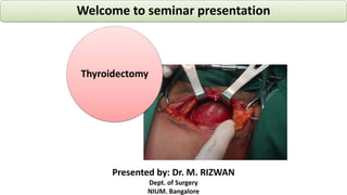
- 1. Thyroidectomy Welcome to seminar presentation Presented by: Dr. M. RIZWAN Dept. of Surgery NIUM. Bangalore
- 2. Learning outcomes Anatomy of the thyroid gland Relations of the Thyroid gland Thyroidectomy & its types Preoperative investigation Details of Procedure Post-operative management Possible complications
- 3. S. Anatomy of thyroid gland
- 4. S. Anatomy of the gland Butterfly shaped Brownish & red Ant.Traingle of neck Measure-5x3x1.5 cm Weight- 20-25 g 2 lobes, central connecting isthmus and pyramidal lobe. Isthmus extends: 2nd - 4th tracheal cartilage. Extension (behind) C5-T1 vertebra U.extent- oblique ridge. L.extent of- 6th T. cartilage
- 5. Capsules of the thyroid gland • True Fibrous capsule • Condensed connective tissue • Encloses the gland • False fascial capsule • Derived from the pretrachial fascia. • Thickend laterally forming the lat. Ligament of berry which fixes the gland to the cricoid cartilage. • Surgical importance……???? It has 2 Capsules:
- 6. Arterial supply to the gland The thyroid is supplied with arterial blood from Superior thyroid artery, a branch of the external carotid artery. Inferior thyroid artery; a branch of the thyrocervical trunk. Thyroid ima artery: subclavian artery/ arch of aorta. Accessssory thyroid artery
- 7. Venous Drainage • Superior thyroid veins, emerges from the apex of each lateral lobe, draining in the internal jugular vein. • Middle thyroid veins, emerges from the lower part of each lat. Lobe. • Inferior thyroid veins, emerges from the isthmus & lower part of the lat. Lobe. draining via the plexus thyreoidea impar in the left brachiocephalic vein. Venous blood drained via:
- 8. Surgical anatomy – cont’d • External laryngeal nerve: • Branch of SLN runs close to superior thyroid artery. • Related to upper pole, supply to CTM • Injury produces voice weakness • Recurrent laryngeal nerve: • Branch of vagus nerve • Related to lower pole of gland • It runs upwards in the tracheo esophageal groove, on posteromedial surface of thyroid • Hooks- AOA -on L side. SCA-on R. side • Injury produces vocal cord paralysis. Important nerves in relation to thyroid
- 10. Lymphatic drainage The lymphatic of the gland drain into: Prelaryngeal L.Ns, infront of cricothyroid memb. Pretracheal L.Ns, infront of trachea. Paratracheal L.Ns, alongside the trachea. Upper & lower deep cervical L.Ns, along the I.J.V . Brachiocephalic L.Ns.
- 11. Relations of the gland Relations of the isthmus. •Relations of the lateral lobes.
- 12. 1. Relations of the isthmus. The isthmus has 2 surfaces (Ant. & Post.) & 2 borders (Upper & lower) Anterior Posterior Upper Lower 1. Skin& superficial fascia. 2. Ant. Jugularveins. 3. Deep fascia. 4. Sternohyoid & sternothyroid muscles. 1. Trachea(2nd, 3rd &4th rings) 1. Related to the sup. Anastomotic a. (between rt & lft sup. Thyroid aa) 2. Thepyramidal lobe may project upwards from the isthmus. 1. Giverise to the Rt &Lt Inf. Thyroid veins. 2. Thethyroid ima a. enters the lower border. 3. An Inf. Anasomotic a. runs along the lower border of theisthmus.
- 13. Anterior 1. Skin& superficial fascia. 2. Ant. Jugularveins. 3. Deepfascia. 4. Sternohyoid & sternothyroid muscles.
- 14. Lower 1. Giverise to the Rt &Lt Inf. Thyroid veins. 2. Thethyroid imaa. enters the lower border. 3. An Inf. Anasomotic a. runs along the lower border of theisthmus. Upper 1. Related to the sup. Anastomotic a. (between rt & lft sup. Thyroid aa) 2. Thepyramidal lobe may project upwards from the isthmus.
- 15. 2. Relations of the lateral lobes. each lobe has 3surfaces: Posterior Medial Anterolateral 1. 2 Parathyroidglands ( sup. & inf. )embedded in the post.Surface. 1. 2arteries: A- Common Carotid a.( inside the carotidsheath) B-Inf. Thyroid a. before it enters the gland. 1. Its upper part relatedto: A- Layrnx(thyroid & cricoid cartilages & cricothyroidm.) B- Pharynx (Inf. Constrictor m.) C-External laryngealn. 2. Its lower part is relatedto: A- trachea B-oesophagus C-Recurrent laryngeal n. in between 1. Skin,superficial fascia & deep fascia. 2. Its upper part is crossedbysup. Belly of omohyoid. 3. Its middle part is covered by sternohyoid & sternothyroidm. 4. Its lower part is overlappedby the ant. Border of sternomastoid.
- 16. Posterior 1. 2 Parathyroid glands ( sup. & inf. )embedded in the post.Surface. 1. 2arteries: A- Common Carotid a.( inside the carotidsheath) B-Inf. Thyroid a. before it enters the gland.
- 17. Medial 1. Its upper part relatedto: A- Layrnx(thyroid & cricoid cartilages & cricothyroidm.) B- Pharynx(Inf.Constrictor m.) C-External laryngeal n. 2. Its lower part is related to: A- trachea B-oesophagus C-Recurrent laryngeal n. in between
- 18. Anterolateral 1. Skin,superficial fascia & deep fascia. 2. Its upper part is crossedbysup. Belly of omohyoid. 3. Its middle part is coveredby sternohyoid & sternothyroid m. 4. Its lower part is overlappedby the ant. Border of sternomastoid.
- 19. Diffused simple goitre Diffused colloidal goitre Simple nodular goitre Solitary or discreet thyroid nodules Chronic thyroiditis Primary toxic goitre Toxic nodular goitre Thyroidectomy – Indications Carcinoma Follicular carcinoma Papillary carcinoma
- 21. Lobectomy Removal of either right or left lobe of thyroid gland Indication- growth limited to one lobe and size less than 2 cm. solitary goitre Well differentiated carcinoma
- 22. Hemi-thyroidectomy 1 lobe + Entire isthmus is removed benign diseases of 1 lobe S. nodule Toxic/ nontoxic (adenoma/ colloid goitre) Histological report= malignancy- Completion thyroidectomynancy H . Report within 1 wk= immediate H .report after 1 wk= after 3 wk Re-exploration in 2nd & 3rd wk Not advisable
- 23. Subtotal thyroidectomy Done in- multi-nodular goitre & Toxic goitre OOC- Toxic goitre Majority of both lobes are Removed . In Toxic goitre- Better to leave only a strips of the tissue on post. Surface of both lower pole In non toxic goitre Advisable- to leave 1/3rd of NTG i.e. 4-5 grams of normal thyroid tissue
- 24. Near-total thyroidectomy on the site p. lesion. Complete lobectomy on contra lateral side. Posterior capsules +a slice of thyroid tissue are left to preserve parathyroid gland Indication Large, diffusely infiltrating and well differentiated CA involving isthmus + contralateral lobe Medullary carcinoma Poorly differentiated lesion
- 25. Total thyroidectomy: whole thyroid gland- Removed Done in - Thyroid Gland malignancy Papillary or follicular carcinoma medullary carcinoma most common operation for multi-nodular goitre.
- 26. Hartley dunhill operation Removal of 1 entire lateral lobe+ isthmus +partial /subtotal of opposite lobe. Done in non- toxic MNG.
- 27. Pre-operative investigations Full blood count (CBC) Serum Urea, Electrolytes, Creatinine Thyroid Profile: T3, T4, TSH FNAC
- 28. Pre-operative preparation & imaging Bring Euthyroid /near euthyroid condition. Ultrasound thyroid gland Radio-iodine (99mTc / 131I) scan X-ray neck X-ray chest– (Both AP / lateral) Indirect laryngoscopy
- 29. INFORMED CONSENT FOR THE SURGERY IS ESSENTIAL
- 30. procedure
- 31. Endo tracheal intubation General anaesthesia + ET Intubation
- 32. Thyroidectomy Steps 1–The preliminaries • Position of patient: – Supine position – Neck elevated (15 to 30 degree) – Neck slightly hyperextended – Sand bag under shoulder – Foot end slightly down
- 33. Thyroidectomy Steps 1 – The preliminaries Preparing the part: –The entire front of neck, from jaw line to nipples, is cleaned with Cholorhexidine, surgical spirit and Betadine.
- 34. Thyroidectomy Steps 1 – The preliminaries • Draping: – Sterile sheets are draped above, below and on either sides of neck, keeping only neck portion visible. – Protection of the eyes – Some surgeons prefer to cover this area with self-adhesive sheet to enhance sterility.
- 35. Principle of thyroidectomy Prevent injury to RLN & ELN Preservation of parathyroid gland & its vascularities Ligation of vessels in order of MTV> ITA >STA > STV > ITV Meticulous technique Absolute haemostasis Avoidance of mass ligation Cosmoses
- 36. Fast revision- structure dissected/ splited • Skin • Subcutaneous tissues • superficial fascia • Platysma (a muscle in superficial fascia of neck) • Investing layer of deep cervical fascia • Pre-tracheal layer of deep cervical fascia • Strap muscles of neck (thin flat muscles) From superficial to deep
- 37. Thyroidectomy Steps 2– Incision and raising flaps • Incision: Kocher’s low collar skin incision, 2-3 cm above supra-sternal notch along neck crease is made. Extent - from posterior border of one sterno- mastoid muscle to other. Skin incision is deepened through subcutaneous tissue, superficial fascia and platysma In female incision is made at slightly higher side in comparisons to male
- 38. Thyroidectomy Steps 2– raising flaps Skin flaps elevation: – Two skin flaps are raised; one above and other below – Upper flap: raised cranially up to the notch of thyroid cartilage. – Lower flap: raised caudally up to suprasternal notch – Flaps are held in place with Joll’s retractor – Upper Flaps consists of – skin, sup Fascia & platysma Technique Infiltrate the superficial fascia with 60 ml of 1:400000 dilution of adrenaline Start the dissection for raising the flaps from lateral to medial side Always remain in subplatysmal plane. Prevent undue raising lower flap
- 39. Thyroidectomy steps 3 – Exposing the gland by spliting the straps muscle • Anterior jugular vein are preserve the ( chance of air embolism) • Investing fascia / deep cervical fascia is incised vertically in midline. • { 2 layer of strap muscles Sterno hyoid-(outer one) sterno thyroid muscle} • After the incision to DCF, Sterno hyoid muscles comes in to view, splited in midline Technique of splitting the straps muscle Midline land mark - thyroid notch/ supra-sternal notch/ 2 ant. jugular vein Tissue in Para median line lifted up dissected vertically in midline with monopolar cautary/ knife- to get in to midline raphe. Split the sternohyoid muscle & retracted laterally Sternothyroid is splited in midline simmliarly to expose thyroid gland.
- 40. Insinuate the 2 index fingers in between pre tracheal fascia (false capsules) & true capsules, stretch the two fingers laterally in b/w the two capsules, medial traction is applied to deliver the gland. While insinuating the fingers care should be taken to Identify the MTV, if identified ligate it / coagulate it. Thyroid gland is held with babcock’s forceps and retracted medially to expose Para carotid tunnel. Carotid sheath opened, C. C. Artery & IJV come in to view. ITA is an important land mark ,it runs up, behind and deep to CCA, take a horizontel turn to supply thyroid and Para thyroid gland. Thyroidectomy steps 3 – Exposing the gland & ITA in P. carotid tunnel
- 41. Identifying The RLN & ELN The recurrent laryngeal nerve is located b/w: Beahrs triangle/ loreyes triangle The common carotid artery- laterally The oesophagus/ trachea - medially The inferior thyroid artery- superiorly. RLN Lies deeper, dissection should be started from inferior to ITA The external laryngeal nerve lies Jols triangle Superior thyroid vessel & upper pole of thyroid- laterally Midline of neck- medially Attachment of Strap muscles & IDLCF to hyoid- superiorly
- 42. Identifying Superior & inferior Parathyroid Gland Superior Parathyroid Gland The SPT gland located on posterior aspect at the level of the upper 2/3rd of thyroid Approximately 1 cm above the crossing point of the RLN and ITA but posterior to RLN It is orange yellow in colour The inferior parathyroid glands located between the lower pole of the thyroid and the isthmus most commonly on the posterolateral surface of the lower pole of the thyroid. 1- 2 cm below & medial to crossing point of the RLN and the ITA and anterior to RLN
- 43. Thyroidectomy steps 4 – Dealing with vessels ligation of ITA The branch of ITA supplying to thyroid gland is ligated just medial to branching of ITA but away from the lower pole of the gland & and than divided. If trunk of the ITA is ligated-compromise the blood supply to parathyroid gland. Ligation of STA It is ligated near to superior pole of thyroid as near as possible and divided. STV & ITV /TIA- ligated and divided subsequently
- 44. Separation from ant. Tracheal aspects by dividing The berry’s ligament & Isthmus In lobectomy/hemithyroidectomy- the berrey’s ligament dissected from trachea with electrocautray. for dividing the isthmus, it is held with kochers or tonsil clamp. Divided and sutured with interlocking continuous suture with 3.0 vicryl. In Sub total thyroidectomy- a rim of thyroid tissue overlying the parathyroid gland is preserved by apply a number of artery forceps at the site of resection. In total thyroidectomy operation is continued similar fashion on other lobe. Preserve the excised specimen in Formalin solution for biopsy
- 45. Thyroidectomy Steps 6 – Winding up process • Redivac (suction) drain is inserted in the cavity left by the excised thyroid gland, • Brought out through a separate stab incision at the side of the neck, • Sutured to the skin with 2-0 Silk sutures.
- 46. Strap muscles are approximated and sutured with 2-0 Vicryl. Cut edges of deep cervical fascia are also sutured with 2-0 Vicryl. Again, haemostasis is minutely checked. Joll’s retractor, which was holding the skin-platysma flaps open, is removed. Thyroidectomy steps 7 – Closure
- 47. Thyroidectomy steps 7 – Closure • Platysma and subcutaneous tissues are closed with 2-0 Vicryl interrupted sutures. • Skin closed with 3-0 Nylon, horizontal mattress sutures or subcuticular sutures. • The latter gives a finer scar, but it requires more technical expertise, finesse and time.
- 48. Post-operative management Patient is kept NPO/NBM (Nil Per Oral / Nil By Mouth) on the day of surgery. Supplemental IV fluid usually given on day of surgery; usually between 2.5 to 3 litres. Compatible blood transfusion if there had been excessive blood loss during surgery.
- 49. Post-operative management Oral intake initiated from next day, starting with ‘clear fluids’, going on to ‘free fluids’, then to soft diet and finally to normal diet Analgesics essential in post-operative period; there is invariably severe pain during first night. Antibiotics avoided in clean elective surgeries
- 50. Post-operative management • Daily vital (PTR, BP) chart is maintained. • Rise of temperature after 3rd post- operative day indicates infection. – This may require inspection of suture line. • Careful note is made of daily output from Redivac drain. • Drain removed after 48 hours or when drainage falls to few ml during last 24-hour period, whichever is earlier.
- 51. Post-operative management • Initial dressing changed after 48-72 hours (to inspect for infection of suture line), • Unless there is soakage, when it should be removed earlier. • Dry dressings sufficient every alternate day, if suture line is clean and dry. • Sutures usually removed on 5th post- operative day. – This gives minimum scarring.
- 52. Thyroidectomy –Possible complications Anaesthesia retaliated complications Haemorrhage /Hematoma manifest after 48 hours of surgery This may compress the airway, becoming life-threatening Hypothyroidism in up to 50% of patients after ten years. Laryngeal nerve injury in about 1% of patients Recurrent laryngeal nerve Unilateral damage results in a hoarse voice. Bilateral damage presents as laryngeal obstruction (surgical emergency: an emergency for tracheostomy) Total vocal cord paralysis – aphonia
- 53. Thyroidectomy –Possible complications cont…. Infection - 2% rate.possibly an increased risk with chronic pre-operative steroid use. Stitch granuloma Devascularisation of the parathyroid Hypoparathyroidism temporary (transient) in many patients permanent in about 1-4% of patient Hypocalcemic tetany (due to accidental removal of parathyroid glands