SlideShare a Scribd company logo
1 of 30
Dr/ ABD ALLAH NAZEER. MD.
Radiological imaging of Hirschsprung disease.
Hirschsprung disease is the most common cause of neonatal colonic
obstruction (15-20%). It is commonly characterized by a short segment of
colonic aganglionosis affecting term neonates, especially boys.
Epidemiology
Hirschsprung disease affects approximately 1:5000-8000 live births. In
short segment disease, there is a significant predilection for males (M:F
of ~3.5:1), which reduces with increasing length of involvement.
Interestingly, it is almost never seen in premature infants.
Clinical presentation
The condition typically presents in term neonates with failure to pass
meconium in the first 1-2 days after birth, although later presentation is
also common. Overall ~75% of cases present within six weeks of birth, and
over 90% of cases present within the first five years of life. A very small
number may present in the adult population.
In cases of delayed presentation with anorectal constipation, manometry
may be useful in distinguishing short/ultra short segment Hirschsprung
disease from other causes.
A definitive diagnosis requires a full thickness rectal biopsy.
Pathology
Hirschsprung disease is characterized by aganglionosis (absence of ganglion cells) in
the distal colon and rectum. It is thought to either occur from a failure of neuroblasts
in neural crest cells to migrate into bowel segments or degeneration of already
migrated neuroblasts. It affects cells both in the myenteric and submucosal plexuses.
Hence, functional obstruction develops as a result of a spasm in the denervated colon.
It can be anatomically divided into four types according to the length of the
aganglionic segment:
short segment disease: ~75% *
rectal and distal sigmoid colonic involvement only
long segment: ~15%
typically extends to splenic flexure / transverse colon
total colonic aganglionosis: ~7.5% (range 2-13%)
also known as Zuezler-Wilson syndrome
occasional extension of aganglionosis into the small bowel
ultrashort segment disease
3-4 cm of internal anal sphincter only
controversial entity
see notes on percentages
It is postulated that hypoganglionosis (reduced number of ganglion cells) handles
intestinal pseudo-obstruction.
Associations
Although Hirschsprung is an isolated abnormality in 70% of
cases, there are some well-documented associations, including:
Down syndrome: in ~10% of Hirschsprung cases
neurocristopathy syndromes
Waardenburg-Shah syndrome
Haddad syndrome
MEN IIa
neuroblastoma
other non-neurocristopathy syndromes
Aarskog syndrome
Bardet-Biedl syndrome
Fryns syndrome
Pallister-Hall syndrome
Smith-Lemli-Opitz syndrome
Hirschsprung Disease Symptoms:
Eighty percent of children with Hirschsprung disease show symptoms in
the first 6 weeks of life. Infants suffering from the disease usually become
symptomatic during the first 24 to 48 hours of life. However, children with
only a short segment of intestine that lacks normal nerve cells may not
show symptoms for several months or even years. While individuals
experience a range of symptoms, the following are the most common:
Not having a bowel movement in the first 48 hours of life
Gradual marked swelling of the abdomen
Gradual onset of vomiting
Fever
Children who do not have early symptoms may present with the
following:
Sepsis (overwhelming infection)
Constipation that worsens over time
Small, watery stool
Loss of appetite
Delayed growth
Hirschsprung disease diagnosis:
Careful physical examination is required, and physical findings are dependent upon the
age at presentation and the severity of the condition. Establishing the diagnosis also
includes undergoing a number of diagnostic studies. These include the following:
Abdominal X-ray. This may indicate a bowel blockage. This study only allows physicians
to suspect the diagnosis, not definitively diagnose it.
Contrast enema. This is a procedure performed to examine the large intestine (colon)
for abnormalities. A contrast agent is given into the rectum in order to coat the inside of
organs so that they will show up on an X-ray. This is the most valuable radiologic study
for establishing the diagnosis.
An X-ray of the abdomen will show a narrowed colon, obstruction and dilated
(exceptionally enlarged) intestine above the obstruction.
Rectal biopsy. This procedure will establish the diagnosis of Hirschsprung disease.
A sample of the cells in the rectum is taken and then looked at under a microscope.
Confirmation of Hirschsprung is based on the absence of ganglion cells and the presence
of nonmyelinated nerves in the biopsy segment.
In infants, a suction rectal biopsy can be done at the bedside. Since there are no sensory
nerves at the site of biopsy, this is not painful. When a suction biopsy is inconclusive,
surgical biopsy is performed under general anesthesia in the operating room.
Anorectal manometry. This determines whether normal reflexes involving the rectum
and the anus are present. Used only in older children, the test can be performed at the
bedside.
Radiography
Radiographs of the neonatal abdomen with
Hirschsprung disease (HD) may show multiple
loops of dilated small bowel with air-fluid levels
that can usually be determined to be a distal
bowel obstruction. An empty rectum is a common
finding. The classic image is a dilated proximal
colon with the aganglionic cone narrowing
towards the distal gut. A cutoff sign in the
rectosigmoid region with an absence of air
distally is a useful finding in Hirschsprung-
associated enterocolitis (HAEC).
Hirschsprung disease. Frontal abdominal radiograph showing marked dilatation of the small bowel with no gas in the rectum.
Hirschsprung disease. Frontal abdominal radiograph showing marked dilatation of the bowel
with no gas in the rectum. In the sitting position, air-fluid levels in the large bowel are seen.
Hirschsprung disease. Lateral abdominal radiograph shows a very
enlarged, stool-filled sigmoid. No air or stool content is seen in the rectum.
Hirschsprung disease. Barium enema technique shows slow contrast-material infusion.
Hirschsprung disease. Lateral view from a barium enema examination
depicting the reduced diameter of the rectum and sigmoid.
Hirschsprung disease. Barium enema showing reduced caliber of the
rectum, followed by a transition zone to an enlarged-caliber sigmoid.
Hirschsprung disease. Barium enema showing reduced caliber of the
rectum, followed by a transition zone to an enlarged-caliber sigmoid.
The transition zone is in the mid-descending colon.
Hirschsprung disease. Barium enema showing reduced caliber and length of
the large bowel, with no clear transition zone (total colonic aganglionosis).
Hirschsprung Disease.
Hirschsprung disease in an infant. Frontal radiograph demonstrates the diameter of the
rectum (arrows) to be smaller than the diameter of the sigmoid colon, an abnormal
rectosigmoid ratio. Note the saw tooth appearance of the abnormal contracted segment.
Hirschsprung Films A, B and C are plain abdominal x-rays. Films D, E and F are x-rays with a barium enema.
Hirschsprung Disease.
Short narrowed segment indicated between the yellow dotted lines; TZ = transition zone
yellow arrows indicate the small bowel (jejunal) pattern to the descending colon
Hirschsprung disease in a newborn with
abdominal distention and failure to pass
meconium. A, Radiograph shows dilatation of
multiple loops of bowel, consistent with a
distal obstruction. B, Early filling lateral view
from contrast enema shows abnormal
rectosigmoid ratio with
rectum (arrowheads) much more narrow than
sigmoid colon (arrows). C, Frontal view from
contrast enema shows rectum to be narrow in
caliber and corkscrew in appearance due to
spasm. The sigmoid colon (S) is dilated
compared to the rectum (arrows).
Hirschsprung Disease. A. A characteristic transition zone (arrows) is seen between the dilated, feces-filled colon above and the relatively
narrowed rectum below. B. The rectum in this newborn infant is smaller than the sigmoid and descending colon, but a well-defined transition
zone is not present. C. A contrast enema in another infant shows spasm and irregularity of the aganglionic segment (arrow). D. A different
infant with a shorter aganglionic segment (arrows) showing a mild corrugated pattern and a well-defined transition zone.
Total Colonic Aganglionosis—Long Segment Hirschsprung Disease .
Total colonic Hirschsprung disease. The entire colon is small in caliber
(arrowheads). The histologic transition zone was in the distal ileum.
Although CT is considered a common imaging modality for excluding other
diseases such as colorectal cancer, which also causes chronic constipation in
adults, to our knowledge, there have been no reports regarding the CT findings
of adult HD and adult HG in the radiology literature. CT depicted the correct
transition zone in nine patients with HG or HD, and the depicted transition zones
were consistent with the histopathologic results. The transition zone was located
in the rectum or rectosigmoid junction in all of the patients with HD. In addition,
the transition zone in the patients with HG corresponded to the histopathologic
results when it was considered to be in the segment with the least number of
ganglion cells, as HG was distributed throughout the entire colon in four patients.
These findings show that HG exhibits compensatory contraction like HD, even
though, to our knowledge, this fact has not been mentioned in previous reports.
There was a discrepancy between the transition zone site seen at histopathologic
analysis and that seen at CT in one patient. The transition zone was in the distal
transverse colon at histopathologic analysis but in the proximal descending colon
at CT. This discrepancy was probably due to the redundancy of transverse and/or
descending colon. A redundant colon depicted at transverse CT can make accurate
localization of the transition zone difficult. Apparently, there seemed to be a short
distance between the two transition zone sites. Also, the histopathologic type of
combined HD and HG might have played a role in the discrepancy in transition
zone sites.
(a) Anteroposterior double-contrast barium enema radiograph (overhead view) shows mildly
narrowed rectum and rectosigmoid junction (arrows) with dilated sigmoid colon (SC). AC =
ascending colon, DC = descending colon. (b) Resected whole-colon specimen shows dilated
sigmoid colon and non dilated ascending colon, transverse colon (TC), and descending colon.
HD with aganglionic segment of upper part of rectum in 19-year-old man. (a, b) Double-contrast barium enema radiographs (overhead and
anteroposterior spot views) show a markedly dilated upper part of rectum and rectosigmoid junction with fecaloma (arrows). AC = ascending
colon, DC = descending colon. Other colonic segments have normal diameters. (c, d) Contrast material–enhanced transverse CT scans show markedly
dilated feces-filled proximal upper part of rectum and the rectosigmoid junction (arrows in c) with a transition zone and a distal narrowed rectum
(arrows in d). The distal rectum also appears to have a thickened wall. The transition zone ratio is measured as the axial diameter at the most dilated
proximal colonic segment (arrows in c) divided by the axial diameter at the narrowest distal part of the rectum (arrows in d).
Double-contrast barium enema radiographs (a, anteroposterior; b, oblique anteroposterior views) obtained in
64-year-old woman who has HD with aganglionic segment of rectosigmoid junction show markedly dilated
distal sigmoid colon and rectosigmoid junction (arrowheads) and a narrowed rectum (arrows).
3a: Contrast-enhanced transverse CT scans (a obtained at lower level than b) obtained in 31-year-old woman with
HG of entire colon show markedly dilated feces-filled ascending colon (AC) and transverse colon (arrowheads)
compared with a nondilated descending colon (DC) with a transition zone of the proximal descending colon (*).
Thank You.

More Related Content

What's hot

Radiology ----Classical Signs in GIT Dr. Muhammad Bin Zulfiqar
Radiology ----Classical Signs in GIT Dr. Muhammad Bin ZulfiqarRadiology ----Classical Signs in GIT Dr. Muhammad Bin Zulfiqar
Radiology ----Classical Signs in GIT Dr. Muhammad Bin ZulfiqarDr. Muhammad Bin Zulfiqar
 
Presentation1.pptx, radiological signs in thoracic radiology.
Presentation1.pptx, radiological signs in thoracic radiology.Presentation1.pptx, radiological signs in thoracic radiology.
Presentation1.pptx, radiological signs in thoracic radiology.Abdellah Nazeer
 
Radiographic manifestations of pulmonary tuberculosis
Radiographic manifestations of pulmonary tuberculosisRadiographic manifestations of pulmonary tuberculosis
Radiographic manifestations of pulmonary tuberculosisDev Lakhera
 
Abdominal x-ray interpretation ppt
Abdominal x-ray interpretation pptAbdominal x-ray interpretation ppt
Abdominal x-ray interpretation pptNaba Kumar Barman
 
Presentation1, radiological imaging of hypertrophic pyloric stenosis.
Presentation1, radiological imaging of hypertrophic pyloric stenosis.Presentation1, radiological imaging of hypertrophic pyloric stenosis.
Presentation1, radiological imaging of hypertrophic pyloric stenosis.Abdellah Nazeer
 
IMAGING IN ABDOMINAL TUBERCULOSIS
IMAGING IN ABDOMINAL TUBERCULOSISIMAGING IN ABDOMINAL TUBERCULOSIS
IMAGING IN ABDOMINAL TUBERCULOSISNavni Garg
 
Acute appendicitis - Ultrasound first
Acute appendicitis  - Ultrasound firstAcute appendicitis  - Ultrasound first
Acute appendicitis - Ultrasound firstSamir Haffar
 
Pneumobilia vs portal vein gas
Pneumobilia vs portal vein gasPneumobilia vs portal vein gas
Pneumobilia vs portal vein gasairwave12
 
The Radiology of Malrotation
The Radiology of MalrotationThe Radiology of Malrotation
The Radiology of Malrotationtboulden
 
Anorectal malformations
Anorectal malformationsAnorectal malformations
Anorectal malformationsDr. Mohit Goel
 
Ultrasound of thyroid nodules
Ultrasound of thyroid nodulesUltrasound of thyroid nodules
Ultrasound of thyroid nodulesSamir Haffar
 
HEPATOCELLULAR CARCINOMA RADIOLOGY
HEPATOCELLULAR CARCINOMA RADIOLOGYHEPATOCELLULAR CARCINOMA RADIOLOGY
HEPATOCELLULAR CARCINOMA RADIOLOGYRMLIMS
 
Imaging of Bowel Obstruction
Imaging of Bowel ObstructionImaging of Bowel Obstruction
Imaging of Bowel ObstructionRathachai Kaewlai
 
Presentation1.pptx, radiological imaging of large bowel diseases
Presentation1.pptx, radiological imaging of large bowel diseasesPresentation1.pptx, radiological imaging of large bowel diseases
Presentation1.pptx, radiological imaging of large bowel diseasesAbdellah Nazeer
 
Presentation1.pptx, radiological imaging of divertiular disease and diverticu...
Presentation1.pptx, radiological imaging of divertiular disease and diverticu...Presentation1.pptx, radiological imaging of divertiular disease and diverticu...
Presentation1.pptx, radiological imaging of divertiular disease and diverticu...Abdellah Nazeer
 

What's hot (20)

Radiology ----Classical Signs in GIT Dr. Muhammad Bin Zulfiqar
Radiology ----Classical Signs in GIT Dr. Muhammad Bin ZulfiqarRadiology ----Classical Signs in GIT Dr. Muhammad Bin Zulfiqar
Radiology ----Classical Signs in GIT Dr. Muhammad Bin Zulfiqar
 
Presentation1.pptx, radiological signs in thoracic radiology.
Presentation1.pptx, radiological signs in thoracic radiology.Presentation1.pptx, radiological signs in thoracic radiology.
Presentation1.pptx, radiological signs in thoracic radiology.
 
Radiographic manifestations of pulmonary tuberculosis
Radiographic manifestations of pulmonary tuberculosisRadiographic manifestations of pulmonary tuberculosis
Radiographic manifestations of pulmonary tuberculosis
 
Abdominal x-ray interpretation ppt
Abdominal x-ray interpretation pptAbdominal x-ray interpretation ppt
Abdominal x-ray interpretation ppt
 
Presentation1, radiological imaging of hypertrophic pyloric stenosis.
Presentation1, radiological imaging of hypertrophic pyloric stenosis.Presentation1, radiological imaging of hypertrophic pyloric stenosis.
Presentation1, radiological imaging of hypertrophic pyloric stenosis.
 
Abdominal X ray
Abdominal X rayAbdominal X ray
Abdominal X ray
 
Holoprosencephaly
HoloprosencephalyHoloprosencephaly
Holoprosencephaly
 
IMAGING IN ABDOMINAL TUBERCULOSIS
IMAGING IN ABDOMINAL TUBERCULOSISIMAGING IN ABDOMINAL TUBERCULOSIS
IMAGING IN ABDOMINAL TUBERCULOSIS
 
Acute appendicitis - Ultrasound first
Acute appendicitis  - Ultrasound firstAcute appendicitis  - Ultrasound first
Acute appendicitis - Ultrasound first
 
Pneumobilia vs portal vein gas
Pneumobilia vs portal vein gasPneumobilia vs portal vein gas
Pneumobilia vs portal vein gas
 
The Radiology of Malrotation
The Radiology of MalrotationThe Radiology of Malrotation
The Radiology of Malrotation
 
Anorectal malformations
Anorectal malformationsAnorectal malformations
Anorectal malformations
 
Ultrasound of thyroid nodules
Ultrasound of thyroid nodulesUltrasound of thyroid nodules
Ultrasound of thyroid nodules
 
Radiology signs
Radiology signsRadiology signs
Radiology signs
 
HEPATOCELLULAR CARCINOMA RADIOLOGY
HEPATOCELLULAR CARCINOMA RADIOLOGYHEPATOCELLULAR CARCINOMA RADIOLOGY
HEPATOCELLULAR CARCINOMA RADIOLOGY
 
Biliary atresia
Biliary atresiaBiliary atresia
Biliary atresia
 
Imaging of Bowel Obstruction
Imaging of Bowel ObstructionImaging of Bowel Obstruction
Imaging of Bowel Obstruction
 
Presentation1.pptx, radiological imaging of large bowel diseases
Presentation1.pptx, radiological imaging of large bowel diseasesPresentation1.pptx, radiological imaging of large bowel diseases
Presentation1.pptx, radiological imaging of large bowel diseases
 
Presentation1.pptx, radiological imaging of divertiular disease and diverticu...
Presentation1.pptx, radiological imaging of divertiular disease and diverticu...Presentation1.pptx, radiological imaging of divertiular disease and diverticu...
Presentation1.pptx, radiological imaging of divertiular disease and diverticu...
 
BASICS of CT Head
BASICS of CT HeadBASICS of CT Head
BASICS of CT Head
 

Viewers also liked

Hirschprung disease by dr manzoor
Hirschprung disease by dr manzoorHirschprung disease by dr manzoor
Hirschprung disease by dr manzoorMohammad Manzoor
 
Hirschsprung's disease
Hirschsprung's diseaseHirschsprung's disease
Hirschsprung's diseaseRam Kumar
 
Hirschsprung Disease
Hirschsprung DiseaseHirschsprung Disease
Hirschsprung DiseaseKing_maged
 
Hirschsprung
HirschsprungHirschsprung
HirschsprungRajiv Lal
 
Presentation1, radiological imaging of ear microcia.
Presentation1, radiological imaging of ear microcia.Presentation1, radiological imaging of ear microcia.
Presentation1, radiological imaging of ear microcia.Abdellah Nazeer
 
Presentation1.pptx, pediatric radiology.
Presentation1.pptx, pediatric radiology.Presentation1.pptx, pediatric radiology.
Presentation1.pptx, pediatric radiology.Abdellah Nazeer
 
Hirchsprang's disease
Hirchsprang's diseaseHirchsprang's disease
Hirchsprang's diseaseDrBenHarris
 
Presentation1, radiological imaging of scimitar syndrome
Presentation1, radiological imaging of scimitar syndromePresentation1, radiological imaging of scimitar syndrome
Presentation1, radiological imaging of scimitar syndromeAbdellah Nazeer
 
Hirschsprung's disease
Hirschsprung's disease Hirschsprung's disease
Hirschsprung's disease Sameh Shehata
 
Presentation1, radiological imaging of wilson disease.
Presentation1, radiological imaging of wilson disease.Presentation1, radiological imaging of wilson disease.
Presentation1, radiological imaging of wilson disease.Abdellah Nazeer
 
Presentation1.pptx, radiological imaging of obstructive jaundice.
Presentation1.pptx, radiological imaging of obstructive jaundice.Presentation1.pptx, radiological imaging of obstructive jaundice.
Presentation1.pptx, radiological imaging of obstructive jaundice.Abdellah Nazeer
 
Hirschprung's disease
Hirschprung's diseaseHirschprung's disease
Hirschprung's diseaseNova Ci Necis
 
Presentation1, radiological imaging of macleod syndrome.
Presentation1, radiological imaging of macleod syndrome.Presentation1, radiological imaging of macleod syndrome.
Presentation1, radiological imaging of macleod syndrome.Abdellah Nazeer
 
Presentation1 radiological imaging of tarsal tunnel syndrome.
Presentation1 radiological imaging of tarsal tunnel syndrome.Presentation1 radiological imaging of tarsal tunnel syndrome.
Presentation1 radiological imaging of tarsal tunnel syndrome.Abdellah Nazeer
 
Presentation1, radiological imaging of leigh disease.
Presentation1, radiological imaging of leigh disease.Presentation1, radiological imaging of leigh disease.
Presentation1, radiological imaging of leigh disease.Abdellah Nazeer
 
Hirschprung disease
Hirschprung diseaseHirschprung disease
Hirschprung diseaseSidra Naeem
 
Bệnh hirschsprung
Bệnh hirschsprungBệnh hirschsprung
Bệnh hirschsprungVo Nhat
 

Viewers also liked (20)

Hirschprung disease by dr manzoor
Hirschprung disease by dr manzoorHirschprung disease by dr manzoor
Hirschprung disease by dr manzoor
 
Hirschsprung's disease
Hirschsprung's diseaseHirschsprung's disease
Hirschsprung's disease
 
Hirschsprung Disease
Hirschsprung DiseaseHirschsprung Disease
Hirschsprung Disease
 
Hirschsprung
HirschsprungHirschsprung
Hirschsprung
 
Presentation1, radiological imaging of ear microcia.
Presentation1, radiological imaging of ear microcia.Presentation1, radiological imaging of ear microcia.
Presentation1, radiological imaging of ear microcia.
 
Hirschsprung disease
Hirschsprung diseaseHirschsprung disease
Hirschsprung disease
 
Presentation1.pptx, pediatric radiology.
Presentation1.pptx, pediatric radiology.Presentation1.pptx, pediatric radiology.
Presentation1.pptx, pediatric radiology.
 
Hirchsprang's disease
Hirchsprang's diseaseHirchsprang's disease
Hirchsprang's disease
 
Hirdschsprug disease
Hirdschsprug disease Hirdschsprug disease
Hirdschsprug disease
 
Presentation1, radiological imaging of scimitar syndrome
Presentation1, radiological imaging of scimitar syndromePresentation1, radiological imaging of scimitar syndrome
Presentation1, radiological imaging of scimitar syndrome
 
Hirschsprung's disease
Hirschsprung's disease Hirschsprung's disease
Hirschsprung's disease
 
Presentation1, radiological imaging of wilson disease.
Presentation1, radiological imaging of wilson disease.Presentation1, radiological imaging of wilson disease.
Presentation1, radiological imaging of wilson disease.
 
Presentation1.pptx, radiological imaging of obstructive jaundice.
Presentation1.pptx, radiological imaging of obstructive jaundice.Presentation1.pptx, radiological imaging of obstructive jaundice.
Presentation1.pptx, radiological imaging of obstructive jaundice.
 
Hirschsprungs disease
Hirschsprungs disease  Hirschsprungs disease
Hirschsprungs disease
 
Hirschprung's disease
Hirschprung's diseaseHirschprung's disease
Hirschprung's disease
 
Presentation1, radiological imaging of macleod syndrome.
Presentation1, radiological imaging of macleod syndrome.Presentation1, radiological imaging of macleod syndrome.
Presentation1, radiological imaging of macleod syndrome.
 
Presentation1 radiological imaging of tarsal tunnel syndrome.
Presentation1 radiological imaging of tarsal tunnel syndrome.Presentation1 radiological imaging of tarsal tunnel syndrome.
Presentation1 radiological imaging of tarsal tunnel syndrome.
 
Presentation1, radiological imaging of leigh disease.
Presentation1, radiological imaging of leigh disease.Presentation1, radiological imaging of leigh disease.
Presentation1, radiological imaging of leigh disease.
 
Hirschprung disease
Hirschprung diseaseHirschprung disease
Hirschprung disease
 
Bệnh hirschsprung
Bệnh hirschsprungBệnh hirschsprung
Bệnh hirschsprung
 

Similar to Presentation1, radiological imaging of hirshsprung disease.

Hirschsprung's disease.pptx
Hirschsprung's  disease.pptxHirschsprung's  disease.pptx
Hirschsprung's disease.pptxjannatsupti
 
Intussusception in children
Intussusception in childrenIntussusception in children
Intussusception in childrenYahea Zakarei
 
Hirschsprung Disease - Approach & Management
Hirschsprung Disease - Approach & ManagementHirschsprung Disease - Approach & Management
Hirschsprung Disease - Approach & ManagementVikas V
 
Presentation1, Ultrasound of the bowel loops and the lymph nodes..pptx
Presentation1, Ultrasound of the bowel loops and the lymph nodes..pptxPresentation1, Ultrasound of the bowel loops and the lymph nodes..pptx
Presentation1, Ultrasound of the bowel loops and the lymph nodes..pptxAbdellah Nazeer
 
Presentation1, ultrasound of the bowel loops and the lymph nodes.
Presentation1, ultrasound of the bowel loops and the lymph nodes.Presentation1, ultrasound of the bowel loops and the lymph nodes.
Presentation1, ultrasound of the bowel loops and the lymph nodes.AbdullahNazeerYassin
 
Presentation1, ultrasound of the bowel loops and the lymph nodes.
Presentation1, ultrasound of the bowel loops and the lymph nodes.Presentation1, ultrasound of the bowel loops and the lymph nodes.
Presentation1, ultrasound of the bowel loops and the lymph nodes.AbdullahNazeerYassin
 
Emergent pediatric us what every radiologist should know
Emergent pediatric us what every radiologist should knowEmergent pediatric us what every radiologist should know
Emergent pediatric us what every radiologist should knowYasser Asiri
 
Radiology and contrast studies in newborns [autosaved]
Radiology and contrast studies in newborns [autosaved]Radiology and contrast studies in newborns [autosaved]
Radiology and contrast studies in newborns [autosaved]faisalUsman20
 
Presentation2, radiological imaging of diaphagmatic hernia.
Presentation2, radiological imaging of diaphagmatic hernia.Presentation2, radiological imaging of diaphagmatic hernia.
Presentation2, radiological imaging of diaphagmatic hernia.Abdellah Nazeer
 
Presentation1, radiological imaging of barium studies.
Presentation1, radiological imaging of barium studies.Presentation1, radiological imaging of barium studies.
Presentation1, radiological imaging of barium studies.Abdellah Nazeer
 
Pediatric High and Low intestinal Obstruction.pptx
Pediatric High and Low intestinal Obstruction.pptxPediatric High and Low intestinal Obstruction.pptx
Pediatric High and Low intestinal Obstruction.pptxDr Muhammad Tahir Javed
 
Hirschsprung’s disease.pptx
Hirschsprung’s disease.pptx Hirschsprung’s disease.pptx
Hirschsprung’s disease.pptx Meena Reddy
 
Intussusception in Adults-Submucosal Lipoma at Transverse colon-A rare cause
 Intussusception in Adults-Submucosal Lipoma at Transverse colon-A rare cause Intussusception in Adults-Submucosal Lipoma at Transverse colon-A rare cause
Intussusception in Adults-Submucosal Lipoma at Transverse colon-A rare causeDr.Santosh Atreya
 
Diagnostic modalities for Gastric diseases.pptx
Diagnostic modalities for Gastric diseases.pptxDiagnostic modalities for Gastric diseases.pptx
Diagnostic modalities for Gastric diseases.pptxPushpa Lal Bhadel
 
L’ecografia nell’ambulatorio delle Malattie Infiammatorie Intestinali
L’ecografia nell’ambulatorio delle Malattie Infiammatorie Intestinali				L’ecografia nell’ambulatorio delle Malattie Infiammatorie Intestinali
L’ecografia nell’ambulatorio delle Malattie Infiammatorie Intestinali ASMaD
 
Hirschsprung's Disease and management.ppt
Hirschsprung's Disease and management.pptHirschsprung's Disease and management.ppt
Hirschsprung's Disease and management.pptesterhs
 
role of Imaging in female infertility
role of Imaging in  female infertilityrole of Imaging in  female infertility
role of Imaging in female infertilitycharusmita chaudhary
 

Similar to Presentation1, radiological imaging of hirshsprung disease. (20)

Hirschsprung's disease.pptx
Hirschsprung's  disease.pptxHirschsprung's  disease.pptx
Hirschsprung's disease.pptx
 
Intussusception in children
Intussusception in childrenIntussusception in children
Intussusception in children
 
D034023026
D034023026D034023026
D034023026
 
Intestinal obstruction
Intestinal obstructionIntestinal obstruction
Intestinal obstruction
 
INTUSSUCEPTION.pptx
INTUSSUCEPTION.pptxINTUSSUCEPTION.pptx
INTUSSUCEPTION.pptx
 
Hirschsprung Disease - Approach & Management
Hirschsprung Disease - Approach & ManagementHirschsprung Disease - Approach & Management
Hirschsprung Disease - Approach & Management
 
Presentation1, Ultrasound of the bowel loops and the lymph nodes..pptx
Presentation1, Ultrasound of the bowel loops and the lymph nodes..pptxPresentation1, Ultrasound of the bowel loops and the lymph nodes..pptx
Presentation1, Ultrasound of the bowel loops and the lymph nodes..pptx
 
Presentation1, ultrasound of the bowel loops and the lymph nodes.
Presentation1, ultrasound of the bowel loops and the lymph nodes.Presentation1, ultrasound of the bowel loops and the lymph nodes.
Presentation1, ultrasound of the bowel loops and the lymph nodes.
 
Presentation1, ultrasound of the bowel loops and the lymph nodes.
Presentation1, ultrasound of the bowel loops and the lymph nodes.Presentation1, ultrasound of the bowel loops and the lymph nodes.
Presentation1, ultrasound of the bowel loops and the lymph nodes.
 
Emergent pediatric us what every radiologist should know
Emergent pediatric us what every radiologist should knowEmergent pediatric us what every radiologist should know
Emergent pediatric us what every radiologist should know
 
Radiology and contrast studies in newborns [autosaved]
Radiology and contrast studies in newborns [autosaved]Radiology and contrast studies in newborns [autosaved]
Radiology and contrast studies in newborns [autosaved]
 
Presentation2, radiological imaging of diaphagmatic hernia.
Presentation2, radiological imaging of diaphagmatic hernia.Presentation2, radiological imaging of diaphagmatic hernia.
Presentation2, radiological imaging of diaphagmatic hernia.
 
Presentation1, radiological imaging of barium studies.
Presentation1, radiological imaging of barium studies.Presentation1, radiological imaging of barium studies.
Presentation1, radiological imaging of barium studies.
 
Pediatric High and Low intestinal Obstruction.pptx
Pediatric High and Low intestinal Obstruction.pptxPediatric High and Low intestinal Obstruction.pptx
Pediatric High and Low intestinal Obstruction.pptx
 
Hirschsprung’s disease.pptx
Hirschsprung’s disease.pptx Hirschsprung’s disease.pptx
Hirschsprung’s disease.pptx
 
Intussusception in Adults-Submucosal Lipoma at Transverse colon-A rare cause
 Intussusception in Adults-Submucosal Lipoma at Transverse colon-A rare cause Intussusception in Adults-Submucosal Lipoma at Transverse colon-A rare cause
Intussusception in Adults-Submucosal Lipoma at Transverse colon-A rare cause
 
Diagnostic modalities for Gastric diseases.pptx
Diagnostic modalities for Gastric diseases.pptxDiagnostic modalities for Gastric diseases.pptx
Diagnostic modalities for Gastric diseases.pptx
 
L’ecografia nell’ambulatorio delle Malattie Infiammatorie Intestinali
L’ecografia nell’ambulatorio delle Malattie Infiammatorie Intestinali				L’ecografia nell’ambulatorio delle Malattie Infiammatorie Intestinali
L’ecografia nell’ambulatorio delle Malattie Infiammatorie Intestinali
 
Hirschsprung's Disease and management.ppt
Hirschsprung's Disease and management.pptHirschsprung's Disease and management.ppt
Hirschsprung's Disease and management.ppt
 
role of Imaging in female infertility
role of Imaging in  female infertilityrole of Imaging in  female infertility
role of Imaging in female infertility
 

More from Abdellah Nazeer

Muculoskeletal Pediatic Imaging..pptx
Muculoskeletal Pediatic Imaging..pptxMuculoskeletal Pediatic Imaging..pptx
Muculoskeletal Pediatic Imaging..pptxAbdellah Nazeer
 
Presentation1 Short cases MD..pptx
Presentation1 Short cases MD..pptxPresentation1 Short cases MD..pptx
Presentation1 Short cases MD..pptxAbdellah Nazeer
 
Presentation1, MD MCQ Cases..pptx
Presentation1, MD MCQ Cases..pptxPresentation1, MD MCQ Cases..pptx
Presentation1, MD MCQ Cases..pptxAbdellah Nazeer
 
Presentation1, Short Cases Quiz..pptx
Presentation1, Short Cases Quiz..pptxPresentation1, Short Cases Quiz..pptx
Presentation1, Short Cases Quiz..pptxAbdellah Nazeer
 
Presentation1, radiological imaging of lateral hindfoot impingement.
Presentation1, radiological imaging of lateral hindfoot impingement.Presentation1, radiological imaging of lateral hindfoot impingement.
Presentation1, radiological imaging of lateral hindfoot impingement.Abdellah Nazeer
 
Presentation2, radiological anatomy of the liver and spleen.
Presentation2, radiological anatomy of the liver and spleen.Presentation2, radiological anatomy of the liver and spleen.
Presentation2, radiological anatomy of the liver and spleen.Abdellah Nazeer
 
Presentation1, artifacts and pitfalls of the wrist and elbow joints.
Presentation1, artifacts and pitfalls of the wrist and elbow joints.Presentation1, artifacts and pitfalls of the wrist and elbow joints.
Presentation1, artifacts and pitfalls of the wrist and elbow joints.Abdellah Nazeer
 
Presentation1, artifact and pitfalls of the knee, hip and ankle joints.
Presentation1, artifact and pitfalls of the knee, hip and ankle joints.Presentation1, artifact and pitfalls of the knee, hip and ankle joints.
Presentation1, artifact and pitfalls of the knee, hip and ankle joints.Abdellah Nazeer
 
Presentation1, radiological imaging of artifact and pitfalls in shoulder join...
Presentation1, radiological imaging of artifact and pitfalls in shoulder join...Presentation1, radiological imaging of artifact and pitfalls in shoulder join...
Presentation1, radiological imaging of artifact and pitfalls in shoulder join...Abdellah Nazeer
 
Presentation1, radiological imaging of internal abdominal hernia.
Presentation1, radiological imaging of internal abdominal hernia.Presentation1, radiological imaging of internal abdominal hernia.
Presentation1, radiological imaging of internal abdominal hernia.Abdellah Nazeer
 
Presentation11, radiological imaging of ovarian torsion.
Presentation11, radiological imaging of ovarian torsion.Presentation11, radiological imaging of ovarian torsion.
Presentation11, radiological imaging of ovarian torsion.Abdellah Nazeer
 
Presentation1, musculoskeletal anatomy.
Presentation1, musculoskeletal anatomy.Presentation1, musculoskeletal anatomy.
Presentation1, musculoskeletal anatomy.Abdellah Nazeer
 
Presentation1, new mri techniques in the diagnosis and monitoring of multiple...
Presentation1, new mri techniques in the diagnosis and monitoring of multiple...Presentation1, new mri techniques in the diagnosis and monitoring of multiple...
Presentation1, new mri techniques in the diagnosis and monitoring of multiple...Abdellah Nazeer
 
Presentation1, radiological application of diffusion weighted mri in neck mas...
Presentation1, radiological application of diffusion weighted mri in neck mas...Presentation1, radiological application of diffusion weighted mri in neck mas...
Presentation1, radiological application of diffusion weighted mri in neck mas...Abdellah Nazeer
 
Presentation1, radiological application of diffusion weighted images in breas...
Presentation1, radiological application of diffusion weighted images in breas...Presentation1, radiological application of diffusion weighted images in breas...
Presentation1, radiological application of diffusion weighted images in breas...Abdellah Nazeer
 
Presentation1, radiological application of diffusion weighted images in abdom...
Presentation1, radiological application of diffusion weighted images in abdom...Presentation1, radiological application of diffusion weighted images in abdom...
Presentation1, radiological application of diffusion weighted images in abdom...Abdellah Nazeer
 
Presentation1, radiological application of diffusion weighted imges in neuror...
Presentation1, radiological application of diffusion weighted imges in neuror...Presentation1, radiological application of diffusion weighted imges in neuror...
Presentation1, radiological application of diffusion weighted imges in neuror...Abdellah Nazeer
 
Presentation1, mr physics.
Presentation1, mr physics.Presentation1, mr physics.
Presentation1, mr physics.Abdellah Nazeer
 
Presentation1. ct physics.
Presentation1. ct physics.Presentation1. ct physics.
Presentation1. ct physics.Abdellah Nazeer
 
Presentation1, ultrasound physics.
Presentation1, ultrasound physics.Presentation1, ultrasound physics.
Presentation1, ultrasound physics.Abdellah Nazeer
 

More from Abdellah Nazeer (20)

Muculoskeletal Pediatic Imaging..pptx
Muculoskeletal Pediatic Imaging..pptxMuculoskeletal Pediatic Imaging..pptx
Muculoskeletal Pediatic Imaging..pptx
 
Presentation1 Short cases MD..pptx
Presentation1 Short cases MD..pptxPresentation1 Short cases MD..pptx
Presentation1 Short cases MD..pptx
 
Presentation1, MD MCQ Cases..pptx
Presentation1, MD MCQ Cases..pptxPresentation1, MD MCQ Cases..pptx
Presentation1, MD MCQ Cases..pptx
 
Presentation1, Short Cases Quiz..pptx
Presentation1, Short Cases Quiz..pptxPresentation1, Short Cases Quiz..pptx
Presentation1, Short Cases Quiz..pptx
 
Presentation1, radiological imaging of lateral hindfoot impingement.
Presentation1, radiological imaging of lateral hindfoot impingement.Presentation1, radiological imaging of lateral hindfoot impingement.
Presentation1, radiological imaging of lateral hindfoot impingement.
 
Presentation2, radiological anatomy of the liver and spleen.
Presentation2, radiological anatomy of the liver and spleen.Presentation2, radiological anatomy of the liver and spleen.
Presentation2, radiological anatomy of the liver and spleen.
 
Presentation1, artifacts and pitfalls of the wrist and elbow joints.
Presentation1, artifacts and pitfalls of the wrist and elbow joints.Presentation1, artifacts and pitfalls of the wrist and elbow joints.
Presentation1, artifacts and pitfalls of the wrist and elbow joints.
 
Presentation1, artifact and pitfalls of the knee, hip and ankle joints.
Presentation1, artifact and pitfalls of the knee, hip and ankle joints.Presentation1, artifact and pitfalls of the knee, hip and ankle joints.
Presentation1, artifact and pitfalls of the knee, hip and ankle joints.
 
Presentation1, radiological imaging of artifact and pitfalls in shoulder join...
Presentation1, radiological imaging of artifact and pitfalls in shoulder join...Presentation1, radiological imaging of artifact and pitfalls in shoulder join...
Presentation1, radiological imaging of artifact and pitfalls in shoulder join...
 
Presentation1, radiological imaging of internal abdominal hernia.
Presentation1, radiological imaging of internal abdominal hernia.Presentation1, radiological imaging of internal abdominal hernia.
Presentation1, radiological imaging of internal abdominal hernia.
 
Presentation11, radiological imaging of ovarian torsion.
Presentation11, radiological imaging of ovarian torsion.Presentation11, radiological imaging of ovarian torsion.
Presentation11, radiological imaging of ovarian torsion.
 
Presentation1, musculoskeletal anatomy.
Presentation1, musculoskeletal anatomy.Presentation1, musculoskeletal anatomy.
Presentation1, musculoskeletal anatomy.
 
Presentation1, new mri techniques in the diagnosis and monitoring of multiple...
Presentation1, new mri techniques in the diagnosis and monitoring of multiple...Presentation1, new mri techniques in the diagnosis and monitoring of multiple...
Presentation1, new mri techniques in the diagnosis and monitoring of multiple...
 
Presentation1, radiological application of diffusion weighted mri in neck mas...
Presentation1, radiological application of diffusion weighted mri in neck mas...Presentation1, radiological application of diffusion weighted mri in neck mas...
Presentation1, radiological application of diffusion weighted mri in neck mas...
 
Presentation1, radiological application of diffusion weighted images in breas...
Presentation1, radiological application of diffusion weighted images in breas...Presentation1, radiological application of diffusion weighted images in breas...
Presentation1, radiological application of diffusion weighted images in breas...
 
Presentation1, radiological application of diffusion weighted images in abdom...
Presentation1, radiological application of diffusion weighted images in abdom...Presentation1, radiological application of diffusion weighted images in abdom...
Presentation1, radiological application of diffusion weighted images in abdom...
 
Presentation1, radiological application of diffusion weighted imges in neuror...
Presentation1, radiological application of diffusion weighted imges in neuror...Presentation1, radiological application of diffusion weighted imges in neuror...
Presentation1, radiological application of diffusion weighted imges in neuror...
 
Presentation1, mr physics.
Presentation1, mr physics.Presentation1, mr physics.
Presentation1, mr physics.
 
Presentation1. ct physics.
Presentation1. ct physics.Presentation1. ct physics.
Presentation1. ct physics.
 
Presentation1, ultrasound physics.
Presentation1, ultrasound physics.Presentation1, ultrasound physics.
Presentation1, ultrasound physics.
 

Recently uploaded

Physicochemical properties (descriptors) in QSAR.pdf
Physicochemical properties (descriptors) in QSAR.pdfPhysicochemical properties (descriptors) in QSAR.pdf
Physicochemical properties (descriptors) in QSAR.pdfRAJ K. MAURYA
 
Dr. A Sumathi - LINEARITY CONCEPT OF SIGNIFICANCE.pdf
Dr. A Sumathi - LINEARITY CONCEPT OF SIGNIFICANCE.pdfDr. A Sumathi - LINEARITY CONCEPT OF SIGNIFICANCE.pdf
Dr. A Sumathi - LINEARITY CONCEPT OF SIGNIFICANCE.pdfSumathi Arumugam
 
Female Call Girls Tonk Just Call Dipal 🥰8250077686🥰 Top Class Call Girl Serv...
Female Call Girls Tonk  Just Call Dipal 🥰8250077686🥰 Top Class Call Girl Serv...Female Call Girls Tonk  Just Call Dipal 🥰8250077686🥰 Top Class Call Girl Serv...
Female Call Girls Tonk Just Call Dipal 🥰8250077686🥰 Top Class Call Girl Serv...Dipal Arora
 
Female Call Girls Jodhpur Just Call Dipal 🥰8250077686🥰 Top Class Call Girl Se...
Female Call Girls Jodhpur Just Call Dipal 🥰8250077686🥰 Top Class Call Girl Se...Female Call Girls Jodhpur Just Call Dipal 🥰8250077686🥰 Top Class Call Girl Se...
Female Call Girls Jodhpur Just Call Dipal 🥰8250077686🥰 Top Class Call Girl Se...Dipal Arora
 
Premium Call Girls Jammu 🧿 7427069034 🧿 High Class Call Girl Service Available
Premium Call Girls Jammu 🧿 7427069034 🧿 High Class Call Girl Service AvailablePremium Call Girls Jammu 🧿 7427069034 🧿 High Class Call Girl Service Available
Premium Call Girls Jammu 🧿 7427069034 🧿 High Class Call Girl Service Availablechaddageeta79
 
Female Call Girls Sri Ganganagar Just Call Dipal 🥰8250077686🥰 Top Class Call ...
Female Call Girls Sri Ganganagar Just Call Dipal 🥰8250077686🥰 Top Class Call ...Female Call Girls Sri Ganganagar Just Call Dipal 🥰8250077686🥰 Top Class Call ...
Female Call Girls Sri Ganganagar Just Call Dipal 🥰8250077686🥰 Top Class Call ...Dipal Arora
 
💞Call Girls Agra Just Call 🍑👄9084454195 🍑👄 Top Class Call Girl Service Agra A...
💞Call Girls Agra Just Call 🍑👄9084454195 🍑👄 Top Class Call Girl Service Agra A...💞Call Girls Agra Just Call 🍑👄9084454195 🍑👄 Top Class Call Girl Service Agra A...
💞Call Girls Agra Just Call 🍑👄9084454195 🍑👄 Top Class Call Girl Service Agra A...Inaayaeventcompany
 
Difference Between Skeletal Smooth and Cardiac Muscles
Difference Between Skeletal Smooth and Cardiac MusclesDifference Between Skeletal Smooth and Cardiac Muscles
Difference Between Skeletal Smooth and Cardiac MusclesMedicoseAcademics
 
See it and Catch it! Recognizing the Thought Traps that Negatively Impact How...
See it and Catch it! Recognizing the Thought Traps that Negatively Impact How...See it and Catch it! Recognizing the Thought Traps that Negatively Impact How...
See it and Catch it! Recognizing the Thought Traps that Negatively Impact How...bkling
 
7 steps How to prevent Thalassemia : Dr Sharda Jain & Vandana Gupta
7 steps How to prevent Thalassemia : Dr Sharda Jain & Vandana Gupta7 steps How to prevent Thalassemia : Dr Sharda Jain & Vandana Gupta
7 steps How to prevent Thalassemia : Dr Sharda Jain & Vandana GuptaLifecare Centre
 
Shazia Iqbal 2024 - Bioorganic Chemistry.pdf
Shazia Iqbal 2024 - Bioorganic Chemistry.pdfShazia Iqbal 2024 - Bioorganic Chemistry.pdf
Shazia Iqbal 2024 - Bioorganic Chemistry.pdfTrustlife
 
👉 Gulbarga Call Girls Service Just Call 🍑👄7427069034 🍑👄 Top Class Call Girl S...
👉 Gulbarga Call Girls Service Just Call 🍑👄7427069034 🍑👄 Top Class Call Girl S...👉 Gulbarga Call Girls Service Just Call 🍑👄7427069034 🍑👄 Top Class Call Girl S...
👉 Gulbarga Call Girls Service Just Call 🍑👄7427069034 🍑👄 Top Class Call Girl S...chaddageeta79
 
ABO Blood grouping in-compatibility in pregnancy
ABO Blood grouping in-compatibility in pregnancyABO Blood grouping in-compatibility in pregnancy
ABO Blood grouping in-compatibility in pregnancyMs. Sapna Pal
 
ANATOMY AND PHYSIOLOGY OF REPRODUCTIVE SYSTEM.pptx
ANATOMY AND PHYSIOLOGY OF REPRODUCTIVE SYSTEM.pptxANATOMY AND PHYSIOLOGY OF REPRODUCTIVE SYSTEM.pptx
ANATOMY AND PHYSIOLOGY OF REPRODUCTIVE SYSTEM.pptxSwetaba Besh
 
Female Call Girls Sawai Madhopur Just Call Dipal 🥰8250077686🥰 Top Class Call ...
Female Call Girls Sawai Madhopur Just Call Dipal 🥰8250077686🥰 Top Class Call ...Female Call Girls Sawai Madhopur Just Call Dipal 🥰8250077686🥰 Top Class Call ...
Female Call Girls Sawai Madhopur Just Call Dipal 🥰8250077686🥰 Top Class Call ...Dipal Arora
 
Female Call Girls Sikar Just Call Dipal 🥰8250077686🥰 Top Class Call Girl Serv...
Female Call Girls Sikar Just Call Dipal 🥰8250077686🥰 Top Class Call Girl Serv...Female Call Girls Sikar Just Call Dipal 🥰8250077686🥰 Top Class Call Girl Serv...
Female Call Girls Sikar Just Call Dipal 🥰8250077686🥰 Top Class Call Girl Serv...Dipal Arora
 
Call Girl In Mysore 💯Niamh 📲🔝7427069034🔝Call Girls No💰Advance Cash On Deliver...
Call Girl In Mysore 💯Niamh 📲🔝7427069034🔝Call Girls No💰Advance Cash On Deliver...Call Girl In Mysore 💯Niamh 📲🔝7427069034🔝Call Girls No💰Advance Cash On Deliver...
Call Girl In Mysore 💯Niamh 📲🔝7427069034🔝Call Girls No💰Advance Cash On Deliver...chaddageeta79
 
Cardiac Output, Venous Return, and Their Regulation
Cardiac Output, Venous Return, and Their RegulationCardiac Output, Venous Return, and Their Regulation
Cardiac Output, Venous Return, and Their RegulationMedicoseAcademics
 
TEST BANK For Porth's Essentials of Pathophysiology, 5th Edition by Tommie L ...
TEST BANK For Porth's Essentials of Pathophysiology, 5th Edition by Tommie L ...TEST BANK For Porth's Essentials of Pathophysiology, 5th Edition by Tommie L ...
TEST BANK For Porth's Essentials of Pathophysiology, 5th Edition by Tommie L ...rightmanforbloodline
 
Test bank for critical care nursing a holistic approach 11th edition morton f...
Test bank for critical care nursing a holistic approach 11th edition morton f...Test bank for critical care nursing a holistic approach 11th edition morton f...
Test bank for critical care nursing a holistic approach 11th edition morton f...robinsonayot
 

Recently uploaded (20)

Physicochemical properties (descriptors) in QSAR.pdf
Physicochemical properties (descriptors) in QSAR.pdfPhysicochemical properties (descriptors) in QSAR.pdf
Physicochemical properties (descriptors) in QSAR.pdf
 
Dr. A Sumathi - LINEARITY CONCEPT OF SIGNIFICANCE.pdf
Dr. A Sumathi - LINEARITY CONCEPT OF SIGNIFICANCE.pdfDr. A Sumathi - LINEARITY CONCEPT OF SIGNIFICANCE.pdf
Dr. A Sumathi - LINEARITY CONCEPT OF SIGNIFICANCE.pdf
 
Female Call Girls Tonk Just Call Dipal 🥰8250077686🥰 Top Class Call Girl Serv...
Female Call Girls Tonk  Just Call Dipal 🥰8250077686🥰 Top Class Call Girl Serv...Female Call Girls Tonk  Just Call Dipal 🥰8250077686🥰 Top Class Call Girl Serv...
Female Call Girls Tonk Just Call Dipal 🥰8250077686🥰 Top Class Call Girl Serv...
 
Female Call Girls Jodhpur Just Call Dipal 🥰8250077686🥰 Top Class Call Girl Se...
Female Call Girls Jodhpur Just Call Dipal 🥰8250077686🥰 Top Class Call Girl Se...Female Call Girls Jodhpur Just Call Dipal 🥰8250077686🥰 Top Class Call Girl Se...
Female Call Girls Jodhpur Just Call Dipal 🥰8250077686🥰 Top Class Call Girl Se...
 
Premium Call Girls Jammu 🧿 7427069034 🧿 High Class Call Girl Service Available
Premium Call Girls Jammu 🧿 7427069034 🧿 High Class Call Girl Service AvailablePremium Call Girls Jammu 🧿 7427069034 🧿 High Class Call Girl Service Available
Premium Call Girls Jammu 🧿 7427069034 🧿 High Class Call Girl Service Available
 
Female Call Girls Sri Ganganagar Just Call Dipal 🥰8250077686🥰 Top Class Call ...
Female Call Girls Sri Ganganagar Just Call Dipal 🥰8250077686🥰 Top Class Call ...Female Call Girls Sri Ganganagar Just Call Dipal 🥰8250077686🥰 Top Class Call ...
Female Call Girls Sri Ganganagar Just Call Dipal 🥰8250077686🥰 Top Class Call ...
 
💞Call Girls Agra Just Call 🍑👄9084454195 🍑👄 Top Class Call Girl Service Agra A...
💞Call Girls Agra Just Call 🍑👄9084454195 🍑👄 Top Class Call Girl Service Agra A...💞Call Girls Agra Just Call 🍑👄9084454195 🍑👄 Top Class Call Girl Service Agra A...
💞Call Girls Agra Just Call 🍑👄9084454195 🍑👄 Top Class Call Girl Service Agra A...
 
Difference Between Skeletal Smooth and Cardiac Muscles
Difference Between Skeletal Smooth and Cardiac MusclesDifference Between Skeletal Smooth and Cardiac Muscles
Difference Between Skeletal Smooth and Cardiac Muscles
 
See it and Catch it! Recognizing the Thought Traps that Negatively Impact How...
See it and Catch it! Recognizing the Thought Traps that Negatively Impact How...See it and Catch it! Recognizing the Thought Traps that Negatively Impact How...
See it and Catch it! Recognizing the Thought Traps that Negatively Impact How...
 
7 steps How to prevent Thalassemia : Dr Sharda Jain & Vandana Gupta
7 steps How to prevent Thalassemia : Dr Sharda Jain & Vandana Gupta7 steps How to prevent Thalassemia : Dr Sharda Jain & Vandana Gupta
7 steps How to prevent Thalassemia : Dr Sharda Jain & Vandana Gupta
 
Shazia Iqbal 2024 - Bioorganic Chemistry.pdf
Shazia Iqbal 2024 - Bioorganic Chemistry.pdfShazia Iqbal 2024 - Bioorganic Chemistry.pdf
Shazia Iqbal 2024 - Bioorganic Chemistry.pdf
 
👉 Gulbarga Call Girls Service Just Call 🍑👄7427069034 🍑👄 Top Class Call Girl S...
👉 Gulbarga Call Girls Service Just Call 🍑👄7427069034 🍑👄 Top Class Call Girl S...👉 Gulbarga Call Girls Service Just Call 🍑👄7427069034 🍑👄 Top Class Call Girl S...
👉 Gulbarga Call Girls Service Just Call 🍑👄7427069034 🍑👄 Top Class Call Girl S...
 
ABO Blood grouping in-compatibility in pregnancy
ABO Blood grouping in-compatibility in pregnancyABO Blood grouping in-compatibility in pregnancy
ABO Blood grouping in-compatibility in pregnancy
 
ANATOMY AND PHYSIOLOGY OF REPRODUCTIVE SYSTEM.pptx
ANATOMY AND PHYSIOLOGY OF REPRODUCTIVE SYSTEM.pptxANATOMY AND PHYSIOLOGY OF REPRODUCTIVE SYSTEM.pptx
ANATOMY AND PHYSIOLOGY OF REPRODUCTIVE SYSTEM.pptx
 
Female Call Girls Sawai Madhopur Just Call Dipal 🥰8250077686🥰 Top Class Call ...
Female Call Girls Sawai Madhopur Just Call Dipal 🥰8250077686🥰 Top Class Call ...Female Call Girls Sawai Madhopur Just Call Dipal 🥰8250077686🥰 Top Class Call ...
Female Call Girls Sawai Madhopur Just Call Dipal 🥰8250077686🥰 Top Class Call ...
 
Female Call Girls Sikar Just Call Dipal 🥰8250077686🥰 Top Class Call Girl Serv...
Female Call Girls Sikar Just Call Dipal 🥰8250077686🥰 Top Class Call Girl Serv...Female Call Girls Sikar Just Call Dipal 🥰8250077686🥰 Top Class Call Girl Serv...
Female Call Girls Sikar Just Call Dipal 🥰8250077686🥰 Top Class Call Girl Serv...
 
Call Girl In Mysore 💯Niamh 📲🔝7427069034🔝Call Girls No💰Advance Cash On Deliver...
Call Girl In Mysore 💯Niamh 📲🔝7427069034🔝Call Girls No💰Advance Cash On Deliver...Call Girl In Mysore 💯Niamh 📲🔝7427069034🔝Call Girls No💰Advance Cash On Deliver...
Call Girl In Mysore 💯Niamh 📲🔝7427069034🔝Call Girls No💰Advance Cash On Deliver...
 
Cardiac Output, Venous Return, and Their Regulation
Cardiac Output, Venous Return, and Their RegulationCardiac Output, Venous Return, and Their Regulation
Cardiac Output, Venous Return, and Their Regulation
 
TEST BANK For Porth's Essentials of Pathophysiology, 5th Edition by Tommie L ...
TEST BANK For Porth's Essentials of Pathophysiology, 5th Edition by Tommie L ...TEST BANK For Porth's Essentials of Pathophysiology, 5th Edition by Tommie L ...
TEST BANK For Porth's Essentials of Pathophysiology, 5th Edition by Tommie L ...
 
Test bank for critical care nursing a holistic approach 11th edition morton f...
Test bank for critical care nursing a holistic approach 11th edition morton f...Test bank for critical care nursing a holistic approach 11th edition morton f...
Test bank for critical care nursing a holistic approach 11th edition morton f...
 

Presentation1, radiological imaging of hirshsprung disease.

  • 1. Dr/ ABD ALLAH NAZEER. MD. Radiological imaging of Hirschsprung disease.
  • 2. Hirschsprung disease is the most common cause of neonatal colonic obstruction (15-20%). It is commonly characterized by a short segment of colonic aganglionosis affecting term neonates, especially boys. Epidemiology Hirschsprung disease affects approximately 1:5000-8000 live births. In short segment disease, there is a significant predilection for males (M:F of ~3.5:1), which reduces with increasing length of involvement. Interestingly, it is almost never seen in premature infants. Clinical presentation The condition typically presents in term neonates with failure to pass meconium in the first 1-2 days after birth, although later presentation is also common. Overall ~75% of cases present within six weeks of birth, and over 90% of cases present within the first five years of life. A very small number may present in the adult population. In cases of delayed presentation with anorectal constipation, manometry may be useful in distinguishing short/ultra short segment Hirschsprung disease from other causes. A definitive diagnosis requires a full thickness rectal biopsy.
  • 3. Pathology Hirschsprung disease is characterized by aganglionosis (absence of ganglion cells) in the distal colon and rectum. It is thought to either occur from a failure of neuroblasts in neural crest cells to migrate into bowel segments or degeneration of already migrated neuroblasts. It affects cells both in the myenteric and submucosal plexuses. Hence, functional obstruction develops as a result of a spasm in the denervated colon. It can be anatomically divided into four types according to the length of the aganglionic segment: short segment disease: ~75% * rectal and distal sigmoid colonic involvement only long segment: ~15% typically extends to splenic flexure / transverse colon total colonic aganglionosis: ~7.5% (range 2-13%) also known as Zuezler-Wilson syndrome occasional extension of aganglionosis into the small bowel ultrashort segment disease 3-4 cm of internal anal sphincter only controversial entity see notes on percentages It is postulated that hypoganglionosis (reduced number of ganglion cells) handles intestinal pseudo-obstruction.
  • 4. Associations Although Hirschsprung is an isolated abnormality in 70% of cases, there are some well-documented associations, including: Down syndrome: in ~10% of Hirschsprung cases neurocristopathy syndromes Waardenburg-Shah syndrome Haddad syndrome MEN IIa neuroblastoma other non-neurocristopathy syndromes Aarskog syndrome Bardet-Biedl syndrome Fryns syndrome Pallister-Hall syndrome Smith-Lemli-Opitz syndrome
  • 5. Hirschsprung Disease Symptoms: Eighty percent of children with Hirschsprung disease show symptoms in the first 6 weeks of life. Infants suffering from the disease usually become symptomatic during the first 24 to 48 hours of life. However, children with only a short segment of intestine that lacks normal nerve cells may not show symptoms for several months or even years. While individuals experience a range of symptoms, the following are the most common: Not having a bowel movement in the first 48 hours of life Gradual marked swelling of the abdomen Gradual onset of vomiting Fever Children who do not have early symptoms may present with the following: Sepsis (overwhelming infection) Constipation that worsens over time Small, watery stool Loss of appetite Delayed growth
  • 6. Hirschsprung disease diagnosis: Careful physical examination is required, and physical findings are dependent upon the age at presentation and the severity of the condition. Establishing the diagnosis also includes undergoing a number of diagnostic studies. These include the following: Abdominal X-ray. This may indicate a bowel blockage. This study only allows physicians to suspect the diagnosis, not definitively diagnose it. Contrast enema. This is a procedure performed to examine the large intestine (colon) for abnormalities. A contrast agent is given into the rectum in order to coat the inside of organs so that they will show up on an X-ray. This is the most valuable radiologic study for establishing the diagnosis. An X-ray of the abdomen will show a narrowed colon, obstruction and dilated (exceptionally enlarged) intestine above the obstruction. Rectal biopsy. This procedure will establish the diagnosis of Hirschsprung disease. A sample of the cells in the rectum is taken and then looked at under a microscope. Confirmation of Hirschsprung is based on the absence of ganglion cells and the presence of nonmyelinated nerves in the biopsy segment. In infants, a suction rectal biopsy can be done at the bedside. Since there are no sensory nerves at the site of biopsy, this is not painful. When a suction biopsy is inconclusive, surgical biopsy is performed under general anesthesia in the operating room. Anorectal manometry. This determines whether normal reflexes involving the rectum and the anus are present. Used only in older children, the test can be performed at the bedside.
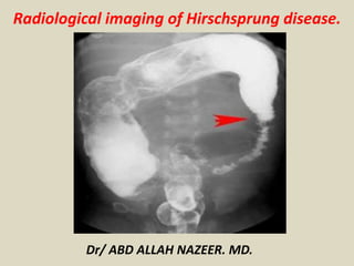
  • 7. Radiography Radiographs of the neonatal abdomen with Hirschsprung disease (HD) may show multiple loops of dilated small bowel with air-fluid levels that can usually be determined to be a distal bowel obstruction. An empty rectum is a common finding. The classic image is a dilated proximal colon with the aganglionic cone narrowing towards the distal gut. A cutoff sign in the rectosigmoid region with an absence of air distally is a useful finding in Hirschsprung- associated enterocolitis (HAEC).
  • 8. Hirschsprung disease. Frontal abdominal radiograph showing marked dilatation of the small bowel with no gas in the rectum.
  • 9. Hirschsprung disease. Frontal abdominal radiograph showing marked dilatation of the bowel with no gas in the rectum. In the sitting position, air-fluid levels in the large bowel are seen.
  • 10. Hirschsprung disease. Lateral abdominal radiograph shows a very enlarged, stool-filled sigmoid. No air or stool content is seen in the rectum.
  • 11. Hirschsprung disease. Barium enema technique shows slow contrast-material infusion.
  • 12. Hirschsprung disease. Lateral view from a barium enema examination depicting the reduced diameter of the rectum and sigmoid.
  • 13. Hirschsprung disease. Barium enema showing reduced caliber of the rectum, followed by a transition zone to an enlarged-caliber sigmoid. Hirschsprung disease. Barium enema showing reduced caliber of the rectum, followed by a transition zone to an enlarged-caliber sigmoid.
  • 14. The transition zone is in the mid-descending colon.
  • 15. Hirschsprung disease. Barium enema showing reduced caliber and length of the large bowel, with no clear transition zone (total colonic aganglionosis).
  • 17. Hirschsprung disease in an infant. Frontal radiograph demonstrates the diameter of the rectum (arrows) to be smaller than the diameter of the sigmoid colon, an abnormal rectosigmoid ratio. Note the saw tooth appearance of the abnormal contracted segment.
  • 18. Hirschsprung Films A, B and C are plain abdominal x-rays. Films D, E and F are x-rays with a barium enema.
  • 20. Short narrowed segment indicated between the yellow dotted lines; TZ = transition zone yellow arrows indicate the small bowel (jejunal) pattern to the descending colon
  • 21. Hirschsprung disease in a newborn with abdominal distention and failure to pass meconium. A, Radiograph shows dilatation of multiple loops of bowel, consistent with a distal obstruction. B, Early filling lateral view from contrast enema shows abnormal rectosigmoid ratio with rectum (arrowheads) much more narrow than sigmoid colon (arrows). C, Frontal view from contrast enema shows rectum to be narrow in caliber and corkscrew in appearance due to spasm. The sigmoid colon (S) is dilated compared to the rectum (arrows).
  • 22. Hirschsprung Disease. A. A characteristic transition zone (arrows) is seen between the dilated, feces-filled colon above and the relatively narrowed rectum below. B. The rectum in this newborn infant is smaller than the sigmoid and descending colon, but a well-defined transition zone is not present. C. A contrast enema in another infant shows spasm and irregularity of the aganglionic segment (arrow). D. A different infant with a shorter aganglionic segment (arrows) showing a mild corrugated pattern and a well-defined transition zone.
  • 23. Total Colonic Aganglionosis—Long Segment Hirschsprung Disease .
  • 24. Total colonic Hirschsprung disease. The entire colon is small in caliber (arrowheads). The histologic transition zone was in the distal ileum.
  • 25. Although CT is considered a common imaging modality for excluding other diseases such as colorectal cancer, which also causes chronic constipation in adults, to our knowledge, there have been no reports regarding the CT findings of adult HD and adult HG in the radiology literature. CT depicted the correct transition zone in nine patients with HG or HD, and the depicted transition zones were consistent with the histopathologic results. The transition zone was located in the rectum or rectosigmoid junction in all of the patients with HD. In addition, the transition zone in the patients with HG corresponded to the histopathologic results when it was considered to be in the segment with the least number of ganglion cells, as HG was distributed throughout the entire colon in four patients. These findings show that HG exhibits compensatory contraction like HD, even though, to our knowledge, this fact has not been mentioned in previous reports. There was a discrepancy between the transition zone site seen at histopathologic analysis and that seen at CT in one patient. The transition zone was in the distal transverse colon at histopathologic analysis but in the proximal descending colon at CT. This discrepancy was probably due to the redundancy of transverse and/or descending colon. A redundant colon depicted at transverse CT can make accurate localization of the transition zone difficult. Apparently, there seemed to be a short distance between the two transition zone sites. Also, the histopathologic type of combined HD and HG might have played a role in the discrepancy in transition zone sites.
  • 26. (a) Anteroposterior double-contrast barium enema radiograph (overhead view) shows mildly narrowed rectum and rectosigmoid junction (arrows) with dilated sigmoid colon (SC). AC = ascending colon, DC = descending colon. (b) Resected whole-colon specimen shows dilated sigmoid colon and non dilated ascending colon, transverse colon (TC), and descending colon.
  • 27. HD with aganglionic segment of upper part of rectum in 19-year-old man. (a, b) Double-contrast barium enema radiographs (overhead and anteroposterior spot views) show a markedly dilated upper part of rectum and rectosigmoid junction with fecaloma (arrows). AC = ascending colon, DC = descending colon. Other colonic segments have normal diameters. (c, d) Contrast material–enhanced transverse CT scans show markedly dilated feces-filled proximal upper part of rectum and the rectosigmoid junction (arrows in c) with a transition zone and a distal narrowed rectum (arrows in d). The distal rectum also appears to have a thickened wall. The transition zone ratio is measured as the axial diameter at the most dilated proximal colonic segment (arrows in c) divided by the axial diameter at the narrowest distal part of the rectum (arrows in d).
  • 28. Double-contrast barium enema radiographs (a, anteroposterior; b, oblique anteroposterior views) obtained in 64-year-old woman who has HD with aganglionic segment of rectosigmoid junction show markedly dilated distal sigmoid colon and rectosigmoid junction (arrowheads) and a narrowed rectum (arrows).
  • 29. 3a: Contrast-enhanced transverse CT scans (a obtained at lower level than b) obtained in 31-year-old woman with HG of entire colon show markedly dilated feces-filled ascending colon (AC) and transverse colon (arrowheads) compared with a nondilated descending colon (DC) with a transition zone of the proximal descending colon (*).