SlideShare a Scribd company logo
1 of 55
Radiological Imaging of Abdominal Internal Hernias.
Dr/ ABD ALLAH NAZEER. MD.
Paraduodenal hernias, although uncommon, have
classically been the most common type of internal
hernia. However, the incidence of postoperative
internal hernias has been increasing recently. The
two most common types, the left and right
paraduodenal hernia involve small bowel
herniating through a congenital opening in the
mesenteries. These internal hernias may result in
closed-loop bowel obstruction.
Clinical presentation
The patient typically presents with symptoms of
small bowel obstruction: abdominal pain, nausea,
vomiting.
Radiographic features
These hernias usually appear as a sac-like cluster of small bowel loops
in an atypical presentation. A closed-loop obstruction may occur within
these loops due to the hernia.
However, it is not unusual for small bowel loops to cluster in an atypical
position in normal patients. Thin patients may be especially challenging
since it may be difficult to follow the course of the collapsed loops of
small bowel.
Because of this, vascular landmarks around a potential internal hernia
"sac" are critical for making a confident diagnosis.
Left paraduodenal hernia
cluster of small bowel loops in the left anterior pararenal space
the cluster of small bowel loops is behind the inferior mesenteric vein
(IMV) and behind the ascending left colic artery
Right paraduodenal hernia
cluster of small bowel loops is inferior to the third portion of the duodenum
the cluster of small bowel loops is behind the superior mesenteric vein
(SMV), the superior mesenteric artery (SMA), and the right colic vein
Left Paraduodenal Hernia. Superior Duodenal Fossa ( pink ovoid zone). A. Axial
view. Through upper Treizt angle (red arrow). B. Coronal view. Behind Inferior
Mesenteric Vein (white arrow). Loops Herniated Position (circles).
Left Paraduodenal Hernia- A. Left saclike mass of herniated small bowel loops
through Landzert fossa. Mass effect on left anterior abdominal wall
(arrowhead). B. Loops situated between the stomach and pancreas.
Displacement of inferior cava vein (red arrow) and stretching and engorgement
of vessels converging to entrance and inside the hernia sac (yellow arrows).
Left Paraduodenal Hernia. A. Hernia sac (open arrow) located between
stomach (with arrow)and pancreatic tail (black arrow). B. MIP coronal view.
Twisting mesenteric vessels in the core of the herniated loops (red arrow).
Paraduodenal hernia: Contrast-enhanced CT scan of the upper abdomen shows
a saclike mass of dilated jejunal loops between the pancreatic head (P) and
stomach. The descending mesocolon (D) and stomach are displaced laterally
Pericecal hernias (PCHs)
In the classic literature PCHs correspond of 13% of all internal hernias. Bowel
loops, most commonly an ileal segment, herniate into the right paracolic gutter
through a congenital or acquired (most commonly by adhesions) unusual defect in
the cecal mesentery. Four different recesses in the pericecal region formed by folds
of the peritoneum have been described: superior and inferior ileocecal recess,
retrocecal recess and paracolic sulci. However, the diagnostic features
and surgical management of the four subtypes do not differ.
Clinical findings
Patients commonly report recurrent episodes of colicky intense right lower
abdominal pain. Chronic incarceration may produce symptoms compatible with
appendiceal disorders, intestinal diseases or intestinal obstruction caused by
adhesions. In PCHs however have been reported a higher incidence of occlusive
symptoms with rapid progression to strangulation and a mortality rate that can
be high as 75%.
CT findings
With CT, a cluster of fixed and dilated small bowel loops with a sac-like
appearance is noted, possibly extending into the right paracolic gutter, lateral to
the cecum and posterior to the ascending colon, which can be displaced anteriorly
or medially
Pericecal Hernia: Contrast-enhanced CT scan of the lower abdomen shows dilated small
bowel loops (S) and a cluster of fluid-filled small bowel loops (arrow). The ascending colon
(A) is displaced anteriorly, and ascites (arrowhead) is seen in the right paracolic gutter
Paracecal Hernia. A. Coronal view. Retrocecal recess (ovoid zone). B. Cadaver
simulating herniation. A,B: Loops Herniated Position (circle). Terminal Ileum (blue
arrow). Cecum (yellow arrow). Flow direction of herniated loops (curved arrows).
Pericecal hernia in an 83-year-old woman with a 1-day history of nausea and
right lower abdominal pain. (a) Axial contrast-enhanced CT image shows
incarcerated intestine with a saclike appearance (arrowheads) that displaces
the ascending colon (arrow) medially. (b) Oblique MPR CT image clearly shows
the hernia orifice (arrows). Laparoscopic surgery showed incarcerated
intestine in a hernia sac lateral to the ascending colon.
Pericecal hernia through the retrocecal recess in an 84-year-old man with colicky
right lower quadrant pain and vomiting of 48 hours duration. He underwent an
appendectomy at 54 years of age. (a) Contrast-enhanced CT scan of the mid-
abdomen shows a cluster of encapsulated small bowel loops (arrowheads) in the
lateral aspect of the right paracolic gutter and behind the ascending colon (A).
Dilated and stretched mesenteric vessels (arrow) are seen within the cluster. (b)
CT scan of the lower abdomen shows beaking and collapsed bowel loops (arrow)
at the retrocecal recess (arrowhead). The ascending colon (A) is displaced
anteriorly. Laparotomy was performed 12 hours after CT.
Transmesenteric (TMHs) hernias:
CT findings
Blachar et al. in 2001 have described the presence of clustered,
compressed small bowel loops in the periphery of the abdominal
cavity and the lack of omental fat between the loops and the
abdominal wall as the most useful CT signs for the detection of a
transmesenteric hernia. The herniated bowels appeared lateral to
the colon (a reversal of the normal anatomic arrangement) with
central, inferior and posterior displacement of the transverse colon
and inferior and medial displacement of the hepatic flexure. Another
study concluded that the only statistically significant CT signs
predictors of a transmesenteric hernia are clustering of small bowel
loops, especially those that are adjacent to the abdominal wall,
mesenteric vessel abnormalities including stretching, crowding and
engorgement, a displacement of the main mesenteric trunk to the
right and signs of small bowel obstruction.
Left Transmesenteric Hernia. Newborn 12 days old. Simple Rx (A) and
intestinal transit (B,C). Left conglomerate (circle in A,C) dilated loops of
jejunum from Treitz angle (arrow in B) due to obstruction by hernia.
Stomach (St) is also dilated. Distal loops are normal caliber (open arrow).
Lesser sac hernias are a type of internal hernia, where
abdominal contents protrude through the foramen of Winslow,
hence they are also known as foramen of Winslow hernia.
Epidemiology
Lesser sac hernias are rare, accounting for <0.1% of abdominal
hernias and 8% of internal hernias.
Pathology
Typically contains small bowel only (~67%) but may also contain
cecum/ascending colon and less commonly transverse colon,
gallbladder, or omentum.
Risk factors
common intestinal mesentery
intra-peritoneal right colon
long small bowel mesentery
large foramen of Winslow
elongated right liver (e.g. Riedel lobe)
Radiographic features
Plain radiograph
gas-filled loops of small bowel in the upper abdomen
CT
mesenteric fat/vessels posterior to portal vein, common
bile duct, hepatic artery and anterior to the inferior vena
cava
mesenteric vessels passing into the lesser sac via the
foramen of Winslow
gas and/or fluid in the lesser sac with bird beak sign
towards the foramen of Winslow
abnormal cecal position
Differential diagnosis
left paraduodenal hernia
Arrows showing gas contained bowel loops obstruction in foramen of winslow hernia
Foramen of winslow hernia: Enteroclysis shows small bowel at right hepatic flexure.
Foramen of Winslow Hernia. Patient without obstruction but with anomalous position of small
bowel loops (circle). They are located under the liver and upper the uncinate pancreas process
(white arrow in A) and stretching of vessels toward the herniated bowel loop (yellow arrows).
Lesser sac hernia through the lesser omentum in a 44-year-old man who presented with acute
abdominal pain. (a) Axial contrast-enhanced CT image shows converging mesenteric vessels and fat
(white arrows) intersecting the distal part of the stomach (black arrows) and the right gastric artery
(arrowhead). Traction and distortion of the distal part of the stomach are also seen. (b) Transparent
volume-rendered CT image shows a cluster of intestine (arrowheads) in the lesser sac, with a beak
shape (arrows) pointing toward the lesser curvature of the stomach (S). Surgery confirmed
herniated intestine in the lesser sac protruding through a defect in the lesser omentum
Hernia through the Broad Ligament
Hernias through a defect of the broad ligament account for only 4%–5% of
all internal hernias. The herniated viscus is the small intestine in more than
90% of cases. The typical patient with this hernia is a middle-aged woman
who has been pregnant and has no history of abdominal surgery. More
than 85% of these hernias have occurred in parous women.
Broad ligament defects are classified as congenital or acquired. Congenital
cases have an embryologic basis due to a developmental peritoneal defect
around the uterus. Acquired defects are due to surgical trauma, pregnancy
and birth trauma, perforations following vaginal manipulation, and prior
pelvic inflammatory disease. A classification for the broad ligament has
been proposed on the basis of the anatomic position of the defect: type 1 =
defect caudal to the round ligament, type 2 = defect above the round
ligament, and type 3 = defect between the round ligament and the
remainder of the broad ligament through the mesoligamentum teres.
The following are the characteristic CT appearances: (a) a cluster of dilated
small bowel loops with air-fluid levels in the pelvic cavity and (b) bowel
loops compressing the rectosigmoid dorsolaterally and the uterus ventrally
Broad Ligament Hernia A,B (Axial). C (Coronal). Herniated and dilated loops
posterior to uterus ( yellow arrows). Bladder (with arrow). Uterus (red arrow)
Broad ligament hernia in a 58-year-old woman with a 1-
day history of intermittent abdominal pain and vomiting
and a history of three normal pregnancies. (a, b) Axial
contrast-enhanced CT images obtained at different levels
show a cluster of small bowel loops in the pouch of
Douglas (arrowheads) and crowding mesenteric
vessels (arrows). The uterus (*) is deviated inferiorly and
to the right. (c) Coronal MPR CT image shows the hernia
orifice (white arrows) below the tubal and ovarian
branches (black arrow) of the left ovarian and uterine
vessels. Enlargement of the distance between the uterus
(*) and the left ovary (arrowhead) is also seen. The
patient’s abdominal pain acutely intensified, and open
surgery was performed. (d) Intraoperative photograph
(anterior view) shows incarceration of the small intestine
through a thumb-tip–sized defect (arrows) in the left
broad ligament of the uterus. Approximately 70 cm of
incarcerated intestine was infarcted and was resected,
and the broad ligament was sectioned.
Hernia through a defect of the right perirectal fossa in a 28-
year-old woman with continuous lower abdominal pain of 34
hours duration. (a, b) Contrast-enhanced CT scans of the pelvis
(b obtained 10 mm below a) show dilated and fluid-filled small
bowel loops (S). A cluster of dilated bowel loops (arrow) is
located to the right of the rectum (R) and behind the uterine
cervix (U). Laparotomy was performed 4 hours after CT.
Transomental hernias (TOHs)
Traditionally, TOHs make up 1-4% of all IHs. The term
“transomental hernia” usually refers to herniation, most
commonly of small bowel loops, cecum and sigmoid colon,
through or into a congenital or acquired abnormal defect
of the greater omentum from 2 to 10 cm in diameter
involving both leaves (four peritoneal layers) and located
in the periphery near the free edge .
CT findings
CT findings are often identical to those of a
transmesenteric hernia, however characteristic features
of TOHs are dilated bowel loops, without a sac-like
appearance, located in the most anterior portion of the
peritoneal cavity with omental vessels that run vertically
around the hernia orifice.
Transomental Hernia. A. Sagittal view. Great Omentum (blue zone). Hernia pathway
( curved arrow). Position of herniated loop (circle) B. Chance of herniation: Cecum
(yellow arrow). Small Bowell (pink arrow) or Sigmoid Colon (with arrow). C. Simulating
herniation of jejunal loop (curved arrow) through great omentum ( blue arrows)
Sigmoid mesocolon hernia: Contrast-Enhanced CT scans of the pelvis show
multiple dilated small bowel loops (S). A dilated inferior mesenteric vein (arrow)
appears as a landmark at the edge of the inferior mesentery. A saclike mass of
incarcerated jejunal loops (arrowhead) is located anterior to the left psoas muscle
Supravesical and pelvic (PIHs) internal hernias
CT findings:
CT findings of broad ligament hernias include a cluster of
dilated small bowel loops herniated in the pelvic cavity
laterally to the uterus with a displacement of the
rectosigmoid dorso-laterally and of the uterus ventrally
and enlargement of the distance between the uterus and
the ovary deviating in opposite directions. Furthermore
mesenteric vessels of herniated loops penetrating the
broad ligament may be seen (33, 88). In supravesical
internal hernia usually CT scans show bowel loops with a
sac-like appearance pass into the space of Retzius and lay
in front of the compressed bladder on the left or right.
These patients can present with bladder irritation and
dysuria, as the bladder is compressed by the small bowel
Internal supravesical hernia in a 67-year-old woman with a two-day
history of lower abdominal pain. Contrast-enhanced axial CT scan
shows intestine loops (white circle) to the left of the urinary bladder.
Internal supravesical hernia in a 74-year-old man with a 1-day history of right lower
abdominal pain. (a) Axial contrast-enhanced CT image shows incarcerated intestine
with a saclike appearance (arrow) that compresses the anterior wall (arrowheads) of
the urinary bladder on the right. (b) Intraoperative photograph (superoanterior view)
shows incarceration of the ileum (blue arrow) into the right supravesical fossa (green
arrows). Approximately 7 cm of intestine was infarcted and was resected. The defect
was approximately 2 cm in diameter. The hernia sac was resected, and the hernia
orifice was closed with sutures. B = urinary bladder, yellow arrow = pubis.
Petersen hernia in a 60-year-old woman with acute
abdominal pain and vomiting. The patient had a history
of laparoscopic distal gastrectomy and antecolic
reconstruction of a Roux-en-Y anastomosis for gastric
cancer performed 11 months earlier. (a) Axial contrast-
enhanced CT image shows a dilated biliopancreatic limb
(*) and mesenteric swirl (arrows) behind the Roux limb
(arrowhead). (b) Coronal MPR CT image shows a cluster
of intestine (★) with engorged mesenteric lymph nodes
located in the left lower quadrant, as well as a dilated
biliopancreatic limb (*). Surgery confirmed herniation of
a long segment of small intestine, including
jejunojejunostomy through a Petersen defect. Arrowhead
= Roux limb, black arrow = jejunojejunostomy, white
arrow = gastrojejunostomy.
Internal hernia with Whirl sign.
Closed loop small bowel obstruction. Contrast enhanced axial CT scan shows a
radial array of distended small bowel loops (B) with stretched and thickened
mesenteric vessels converging to a central point (white arrow). Bowel wall
thickening (arrowhead) and mesenteric edema (asterisk) can also be observed.
Thank You.

More Related Content

What's hot

Presentation1.pptx, radiological imaging of large bowel diseases
Presentation1.pptx, radiological imaging of large bowel diseasesPresentation1.pptx, radiological imaging of large bowel diseases
Presentation1.pptx, radiological imaging of large bowel diseasesAbdellah Nazeer
 
Diagnositc Imaging of the Esophagus
Diagnositc Imaging of the EsophagusDiagnositc Imaging of the Esophagus
Diagnositc Imaging of the EsophagusMohamed M.A. Zaitoun
 
Presentation1.pptx imaging of the peritoneum and mesentry.
Presentation1.pptx imaging of the peritoneum and mesentry.Presentation1.pptx imaging of the peritoneum and mesentry.
Presentation1.pptx imaging of the peritoneum and mesentry.Abdellah Nazeer
 
Presentation2, radiological anatomy of the liver and spleen.
Presentation2, radiological anatomy of the liver and spleen.Presentation2, radiological anatomy of the liver and spleen.
Presentation2, radiological anatomy of the liver and spleen.Abdellah Nazeer
 
Imaging in Skull base
Imaging in Skull baseImaging in Skull base
Imaging in Skull baseRakesh Ca
 
General abdomen ultrasound sonography
General abdomen ultrasound sonographyGeneral abdomen ultrasound sonography
General abdomen ultrasound sonographySafi. Khan
 
Diagnostic Imaging of Cholangiocarcinoma
Diagnostic Imaging of CholangiocarcinomaDiagnostic Imaging of Cholangiocarcinoma
Diagnostic Imaging of CholangiocarcinomaMohamed M.A. Zaitoun
 
Ultrasound imaging of Bowel pathology
Ultrasound imaging of Bowel pathologyUltrasound imaging of Bowel pathology
Ultrasound imaging of Bowel pathologyairwave12
 
Radiological anatomy of liver segments
Radiological anatomy of liver segmentsRadiological anatomy of liver segments
Radiological anatomy of liver segmentsTarun Goyal
 
Imaging of spleen ct and mri
Imaging of spleen ct and mriImaging of spleen ct and mri
Imaging of spleen ct and mriPankaj Kaira
 
Salivary gland imaging radiology ppt
Salivary gland imaging radiology pptSalivary gland imaging radiology ppt
Salivary gland imaging radiology pptDr pradeep Kumar
 
Imaging of Malignant Liver Lesions
Imaging of Malignant Liver LesionsImaging of Malignant Liver Lesions
Imaging of Malignant Liver LesionsSahil Chaudhry
 
Radiological approach to gastric ulcer disease
Radiological approach to gastric ulcer diseaseRadiological approach to gastric ulcer disease
Radiological approach to gastric ulcer diseaseNavneet Ranjan
 
Acute Abdomen-Radiology
Acute Abdomen-RadiologyAcute Abdomen-Radiology
Acute Abdomen-RadiologyParvathy Nair
 
Diagnostic Imaging of Adrenal Glands
Diagnostic Imaging of Adrenal GlandsDiagnostic Imaging of Adrenal Glands
Diagnostic Imaging of Adrenal GlandsMohamed M.A. Zaitoun
 
Presentation1.pptx, ultrasound examination of the urinary bladder and prostate.
Presentation1.pptx, ultrasound examination of the urinary bladder and prostate.Presentation1.pptx, ultrasound examination of the urinary bladder and prostate.
Presentation1.pptx, ultrasound examination of the urinary bladder and prostate.Abdellah Nazeer
 
Biliary tract imaging final...........
Biliary tract imaging  final...........Biliary tract imaging  final...........
Biliary tract imaging final...........Ram Jharpula
 
Presentation1.pptx, radiological imaging of divertiular disease and diverticu...
Presentation1.pptx, radiological imaging of divertiular disease and diverticu...Presentation1.pptx, radiological imaging of divertiular disease and diverticu...
Presentation1.pptx, radiological imaging of divertiular disease and diverticu...Abdellah Nazeer
 

What's hot (20)

Presentation1.pptx, radiological imaging of large bowel diseases
Presentation1.pptx, radiological imaging of large bowel diseasesPresentation1.pptx, radiological imaging of large bowel diseases
Presentation1.pptx, radiological imaging of large bowel diseases
 
Diagnositc Imaging of the Esophagus
Diagnositc Imaging of the EsophagusDiagnositc Imaging of the Esophagus
Diagnositc Imaging of the Esophagus
 
Presentation1.pptx imaging of the peritoneum and mesentry.
Presentation1.pptx imaging of the peritoneum and mesentry.Presentation1.pptx imaging of the peritoneum and mesentry.
Presentation1.pptx imaging of the peritoneum and mesentry.
 
Radiology Spotters
Radiology Spotters Radiology Spotters
Radiology Spotters
 
Presentation2, radiological anatomy of the liver and spleen.
Presentation2, radiological anatomy of the liver and spleen.Presentation2, radiological anatomy of the liver and spleen.
Presentation2, radiological anatomy of the liver and spleen.
 
Imaging in Skull base
Imaging in Skull baseImaging in Skull base
Imaging in Skull base
 
General abdomen ultrasound sonography
General abdomen ultrasound sonographyGeneral abdomen ultrasound sonography
General abdomen ultrasound sonography
 
Diagnostic Imaging of Cholangiocarcinoma
Diagnostic Imaging of CholangiocarcinomaDiagnostic Imaging of Cholangiocarcinoma
Diagnostic Imaging of Cholangiocarcinoma
 
Ultrasound imaging of Bowel pathology
Ultrasound imaging of Bowel pathologyUltrasound imaging of Bowel pathology
Ultrasound imaging of Bowel pathology
 
Radiological anatomy of liver segments
Radiological anatomy of liver segmentsRadiological anatomy of liver segments
Radiological anatomy of liver segments
 
Diagnostic Imaging of the Liver
Diagnostic Imaging of the LiverDiagnostic Imaging of the Liver
Diagnostic Imaging of the Liver
 
Imaging of spleen ct and mri
Imaging of spleen ct and mriImaging of spleen ct and mri
Imaging of spleen ct and mri
 
Salivary gland imaging radiology ppt
Salivary gland imaging radiology pptSalivary gland imaging radiology ppt
Salivary gland imaging radiology ppt
 
Imaging of Malignant Liver Lesions
Imaging of Malignant Liver LesionsImaging of Malignant Liver Lesions
Imaging of Malignant Liver Lesions
 
Radiological approach to gastric ulcer disease
Radiological approach to gastric ulcer diseaseRadiological approach to gastric ulcer disease
Radiological approach to gastric ulcer disease
 
Acute Abdomen-Radiology
Acute Abdomen-RadiologyAcute Abdomen-Radiology
Acute Abdomen-Radiology
 
Diagnostic Imaging of Adrenal Glands
Diagnostic Imaging of Adrenal GlandsDiagnostic Imaging of Adrenal Glands
Diagnostic Imaging of Adrenal Glands
 
Presentation1.pptx, ultrasound examination of the urinary bladder and prostate.
Presentation1.pptx, ultrasound examination of the urinary bladder and prostate.Presentation1.pptx, ultrasound examination of the urinary bladder and prostate.
Presentation1.pptx, ultrasound examination of the urinary bladder and prostate.
 
Biliary tract imaging final...........
Biliary tract imaging  final...........Biliary tract imaging  final...........
Biliary tract imaging final...........
 
Presentation1.pptx, radiological imaging of divertiular disease and diverticu...
Presentation1.pptx, radiological imaging of divertiular disease and diverticu...Presentation1.pptx, radiological imaging of divertiular disease and diverticu...
Presentation1.pptx, radiological imaging of divertiular disease and diverticu...
 

Similar to Presentation1, radiological imaging of internal abdominal hernia.

Hirschsprung's disease.pptx
Hirschsprung's  disease.pptxHirschsprung's  disease.pptx
Hirschsprung's disease.pptxjannatsupti
 
Pancreas ultrasound
Pancreas ultrasoundPancreas ultrasound
Pancreas ultrasoundSafi. Khan
 
Diagnostic modalities for Gastric diseases.pptx
Diagnostic modalities for Gastric diseases.pptxDiagnostic modalities for Gastric diseases.pptx
Diagnostic modalities for Gastric diseases.pptxPushpa Lal Bhadel
 
5th y radiology revision
5th y radiology revision5th y radiology revision
5th y radiology revisionAhmed Samir
 
Intussusception
IntussusceptionIntussusception
Intussusceptionairwave12
 
Abdomen ct patho 3
Abdomen ct  patho 3Abdomen ct  patho 3
Abdomen ct patho 3Sarah Shara
 
Presentation11, radiological imaging of ovarian torsion.
Presentation11, radiological imaging of ovarian torsion.Presentation11, radiological imaging of ovarian torsion.
Presentation11, radiological imaging of ovarian torsion.Abdellah Nazeer
 
Radiographic anatomy of gastrointestinal tract
Radiographic anatomy of gastrointestinal tractRadiographic anatomy of gastrointestinal tract
Radiographic anatomy of gastrointestinal tractairwave12
 
Rapid review of radiology abdomen
Rapid review of radiology abdomenRapid review of radiology abdomen
Rapid review of radiology abdomenDouble M
 
“Little Old Ladies Hernia”: A Case Report and Review of Literature_Crimson Pu...
“Little Old Ladies Hernia”: A Case Report and Review of Literature_Crimson Pu...“Little Old Ladies Hernia”: A Case Report and Review of Literature_Crimson Pu...
“Little Old Ladies Hernia”: A Case Report and Review of Literature_Crimson Pu...CrimsonPublishersAICS
 
Presentation1, Ultrasound of the bowel loops and the lymph nodes..pptx
Presentation1, Ultrasound of the bowel loops and the lymph nodes..pptxPresentation1, Ultrasound of the bowel loops and the lymph nodes..pptx
Presentation1, Ultrasound of the bowel loops and the lymph nodes..pptxAbdellah Nazeer
 
Presentation1, ultrasound of the bowel loops and the lymph nodes.
Presentation1, ultrasound of the bowel loops and the lymph nodes.Presentation1, ultrasound of the bowel loops and the lymph nodes.
Presentation1, ultrasound of the bowel loops and the lymph nodes.AbdullahNazeerYassin
 
Presentation1, ultrasound of the bowel loops and the lymph nodes.
Presentation1, ultrasound of the bowel loops and the lymph nodes.Presentation1, ultrasound of the bowel loops and the lymph nodes.
Presentation1, ultrasound of the bowel loops and the lymph nodes.AbdullahNazeerYassin
 
Acute abdomen
Acute abdomenAcute abdomen
Acute abdomenairwave12
 
Abdomen Gastrointestinal tract.pptx
Abdomen Gastrointestinal tract.pptxAbdomen Gastrointestinal tract.pptx
Abdomen Gastrointestinal tract.pptxPushkarBhure1
 

Similar to Presentation1, radiological imaging of internal abdominal hernia. (20)

Hirschsprung's disease.pptx
Hirschsprung's  disease.pptxHirschsprung's  disease.pptx
Hirschsprung's disease.pptx
 
Pancreas ultrasound
Pancreas ultrasoundPancreas ultrasound
Pancreas ultrasound
 
Diagnostic modalities for Gastric diseases.pptx
Diagnostic modalities for Gastric diseases.pptxDiagnostic modalities for Gastric diseases.pptx
Diagnostic modalities for Gastric diseases.pptx
 
5th y radiology revision
5th y radiology revision5th y radiology revision
5th y radiology revision
 
Learn ultrasonography
Learn ultrasonographyLearn ultrasonography
Learn ultrasonography
 
Intussusception
IntussusceptionIntussusception
Intussusception
 
Abdomen ct patho 3
Abdomen ct  patho 3Abdomen ct  patho 3
Abdomen ct patho 3
 
Sahad gb
Sahad gbSahad gb
Sahad gb
 
Presentation11, radiological imaging of ovarian torsion.
Presentation11, radiological imaging of ovarian torsion.Presentation11, radiological imaging of ovarian torsion.
Presentation11, radiological imaging of ovarian torsion.
 
Radiographic anatomy of gastrointestinal tract
Radiographic anatomy of gastrointestinal tractRadiographic anatomy of gastrointestinal tract
Radiographic anatomy of gastrointestinal tract
 
Spleen[1]
Spleen[1]Spleen[1]
Spleen[1]
 
Abdomen imaging.pptx
Abdomen imaging.pptxAbdomen imaging.pptx
Abdomen imaging.pptx
 
Rapid review of radiology abdomen
Rapid review of radiology abdomenRapid review of radiology abdomen
Rapid review of radiology abdomen
 
Paed.ppt
Paed.pptPaed.ppt
Paed.ppt
 
“Little Old Ladies Hernia”: A Case Report and Review of Literature_Crimson Pu...
“Little Old Ladies Hernia”: A Case Report and Review of Literature_Crimson Pu...“Little Old Ladies Hernia”: A Case Report and Review of Literature_Crimson Pu...
“Little Old Ladies Hernia”: A Case Report and Review of Literature_Crimson Pu...
 
Presentation1, Ultrasound of the bowel loops and the lymph nodes..pptx
Presentation1, Ultrasound of the bowel loops and the lymph nodes..pptxPresentation1, Ultrasound of the bowel loops and the lymph nodes..pptx
Presentation1, Ultrasound of the bowel loops and the lymph nodes..pptx
 
Presentation1, ultrasound of the bowel loops and the lymph nodes.
Presentation1, ultrasound of the bowel loops and the lymph nodes.Presentation1, ultrasound of the bowel loops and the lymph nodes.
Presentation1, ultrasound of the bowel loops and the lymph nodes.
 
Presentation1, ultrasound of the bowel loops and the lymph nodes.
Presentation1, ultrasound of the bowel loops and the lymph nodes.Presentation1, ultrasound of the bowel loops and the lymph nodes.
Presentation1, ultrasound of the bowel loops and the lymph nodes.
 
Acute abdomen
Acute abdomenAcute abdomen
Acute abdomen
 
Abdomen Gastrointestinal tract.pptx
Abdomen Gastrointestinal tract.pptxAbdomen Gastrointestinal tract.pptx
Abdomen Gastrointestinal tract.pptx
 

More from Abdellah Nazeer

Muculoskeletal Pediatic Imaging..pptx
Muculoskeletal Pediatic Imaging..pptxMuculoskeletal Pediatic Imaging..pptx
Muculoskeletal Pediatic Imaging..pptxAbdellah Nazeer
 
Presentation1 Short cases MD..pptx
Presentation1 Short cases MD..pptxPresentation1 Short cases MD..pptx
Presentation1 Short cases MD..pptxAbdellah Nazeer
 
Presentation1, MD MCQ Cases..pptx
Presentation1, MD MCQ Cases..pptxPresentation1, MD MCQ Cases..pptx
Presentation1, MD MCQ Cases..pptxAbdellah Nazeer
 
Presentation1, Short Cases Quiz..pptx
Presentation1, Short Cases Quiz..pptxPresentation1, Short Cases Quiz..pptx
Presentation1, Short Cases Quiz..pptxAbdellah Nazeer
 
Presentation1, radiological imaging of lateral hindfoot impingement.
Presentation1, radiological imaging of lateral hindfoot impingement.Presentation1, radiological imaging of lateral hindfoot impingement.
Presentation1, radiological imaging of lateral hindfoot impingement.Abdellah Nazeer
 
Presentation1, artifacts and pitfalls of the wrist and elbow joints.
Presentation1, artifacts and pitfalls of the wrist and elbow joints.Presentation1, artifacts and pitfalls of the wrist and elbow joints.
Presentation1, artifacts and pitfalls of the wrist and elbow joints.Abdellah Nazeer
 
Presentation1, artifact and pitfalls of the knee, hip and ankle joints.
Presentation1, artifact and pitfalls of the knee, hip and ankle joints.Presentation1, artifact and pitfalls of the knee, hip and ankle joints.
Presentation1, artifact and pitfalls of the knee, hip and ankle joints.Abdellah Nazeer
 
Presentation1, radiological imaging of artifact and pitfalls in shoulder join...
Presentation1, radiological imaging of artifact and pitfalls in shoulder join...Presentation1, radiological imaging of artifact and pitfalls in shoulder join...
Presentation1, radiological imaging of artifact and pitfalls in shoulder join...Abdellah Nazeer
 
Presentation1, musculoskeletal anatomy.
Presentation1, musculoskeletal anatomy.Presentation1, musculoskeletal anatomy.
Presentation1, musculoskeletal anatomy.Abdellah Nazeer
 
Presentation1, new mri techniques in the diagnosis and monitoring of multiple...
Presentation1, new mri techniques in the diagnosis and monitoring of multiple...Presentation1, new mri techniques in the diagnosis and monitoring of multiple...
Presentation1, new mri techniques in the diagnosis and monitoring of multiple...Abdellah Nazeer
 
Presentation1, radiological application of diffusion weighted mri in neck mas...
Presentation1, radiological application of diffusion weighted mri in neck mas...Presentation1, radiological application of diffusion weighted mri in neck mas...
Presentation1, radiological application of diffusion weighted mri in neck mas...Abdellah Nazeer
 
Presentation1, radiological application of diffusion weighted images in breas...
Presentation1, radiological application of diffusion weighted images in breas...Presentation1, radiological application of diffusion weighted images in breas...
Presentation1, radiological application of diffusion weighted images in breas...Abdellah Nazeer
 
Presentation1, radiological application of diffusion weighted images in abdom...
Presentation1, radiological application of diffusion weighted images in abdom...Presentation1, radiological application of diffusion weighted images in abdom...
Presentation1, radiological application of diffusion weighted images in abdom...Abdellah Nazeer
 
Presentation1, radiological application of diffusion weighted imges in neuror...
Presentation1, radiological application of diffusion weighted imges in neuror...Presentation1, radiological application of diffusion weighted imges in neuror...
Presentation1, radiological application of diffusion weighted imges in neuror...Abdellah Nazeer
 
Presentation1, mr physics.
Presentation1, mr physics.Presentation1, mr physics.
Presentation1, mr physics.Abdellah Nazeer
 
Presentation1. ct physics.
Presentation1. ct physics.Presentation1. ct physics.
Presentation1. ct physics.Abdellah Nazeer
 
Presentation1, ultrasound physics.
Presentation1, ultrasound physics.Presentation1, ultrasound physics.
Presentation1, ultrasound physics.Abdellah Nazeer
 
Presentation4, x ray physics
Presentation4, x ray physicsPresentation4, x ray physics
Presentation4, x ray physicsAbdellah Nazeer
 
Presentation1, quiz for the chest.
Presentation1, quiz for the chest.Presentation1, quiz for the chest.
Presentation1, quiz for the chest.Abdellah Nazeer
 

More from Abdellah Nazeer (20)

Muculoskeletal Pediatic Imaging..pptx
Muculoskeletal Pediatic Imaging..pptxMuculoskeletal Pediatic Imaging..pptx
Muculoskeletal Pediatic Imaging..pptx
 
Presentation1 Short cases MD..pptx
Presentation1 Short cases MD..pptxPresentation1 Short cases MD..pptx
Presentation1 Short cases MD..pptx
 
Presentation1, MD MCQ Cases..pptx
Presentation1, MD MCQ Cases..pptxPresentation1, MD MCQ Cases..pptx
Presentation1, MD MCQ Cases..pptx
 
Presentation1, Short Cases Quiz..pptx
Presentation1, Short Cases Quiz..pptxPresentation1, Short Cases Quiz..pptx
Presentation1, Short Cases Quiz..pptx
 
Presentation1, radiological imaging of lateral hindfoot impingement.
Presentation1, radiological imaging of lateral hindfoot impingement.Presentation1, radiological imaging of lateral hindfoot impingement.
Presentation1, radiological imaging of lateral hindfoot impingement.
 
Presentation1, artifacts and pitfalls of the wrist and elbow joints.
Presentation1, artifacts and pitfalls of the wrist and elbow joints.Presentation1, artifacts and pitfalls of the wrist and elbow joints.
Presentation1, artifacts and pitfalls of the wrist and elbow joints.
 
Presentation1, artifact and pitfalls of the knee, hip and ankle joints.
Presentation1, artifact and pitfalls of the knee, hip and ankle joints.Presentation1, artifact and pitfalls of the knee, hip and ankle joints.
Presentation1, artifact and pitfalls of the knee, hip and ankle joints.
 
Presentation1, radiological imaging of artifact and pitfalls in shoulder join...
Presentation1, radiological imaging of artifact and pitfalls in shoulder join...Presentation1, radiological imaging of artifact and pitfalls in shoulder join...
Presentation1, radiological imaging of artifact and pitfalls in shoulder join...
 
Presentation1, musculoskeletal anatomy.
Presentation1, musculoskeletal anatomy.Presentation1, musculoskeletal anatomy.
Presentation1, musculoskeletal anatomy.
 
Presentation1, new mri techniques in the diagnosis and monitoring of multiple...
Presentation1, new mri techniques in the diagnosis and monitoring of multiple...Presentation1, new mri techniques in the diagnosis and monitoring of multiple...
Presentation1, new mri techniques in the diagnosis and monitoring of multiple...
 
Presentation1, radiological application of diffusion weighted mri in neck mas...
Presentation1, radiological application of diffusion weighted mri in neck mas...Presentation1, radiological application of diffusion weighted mri in neck mas...
Presentation1, radiological application of diffusion weighted mri in neck mas...
 
Presentation1, radiological application of diffusion weighted images in breas...
Presentation1, radiological application of diffusion weighted images in breas...Presentation1, radiological application of diffusion weighted images in breas...
Presentation1, radiological application of diffusion weighted images in breas...
 
Presentation1, radiological application of diffusion weighted images in abdom...
Presentation1, radiological application of diffusion weighted images in abdom...Presentation1, radiological application of diffusion weighted images in abdom...
Presentation1, radiological application of diffusion weighted images in abdom...
 
Presentation1, radiological application of diffusion weighted imges in neuror...
Presentation1, radiological application of diffusion weighted imges in neuror...Presentation1, radiological application of diffusion weighted imges in neuror...
Presentation1, radiological application of diffusion weighted imges in neuror...
 
Presentation1, mr physics.
Presentation1, mr physics.Presentation1, mr physics.
Presentation1, mr physics.
 
Presentation1. ct physics.
Presentation1. ct physics.Presentation1. ct physics.
Presentation1. ct physics.
 
Presentation1, ultrasound physics.
Presentation1, ultrasound physics.Presentation1, ultrasound physics.
Presentation1, ultrasound physics.
 
Presentation4, x ray physics
Presentation4, x ray physicsPresentation4, x ray physics
Presentation4, x ray physics
 
Positions
PositionsPositions
Positions
 
Presentation1, quiz for the chest.
Presentation1, quiz for the chest.Presentation1, quiz for the chest.
Presentation1, quiz for the chest.
 

Recently uploaded

💚Reliable Call Girls Chandigarh 💯Niamh 📲🔝8868886958🔝Call Girl In Chandigarh N...
💚Reliable Call Girls Chandigarh 💯Niamh 📲🔝8868886958🔝Call Girl In Chandigarh N...💚Reliable Call Girls Chandigarh 💯Niamh 📲🔝8868886958🔝Call Girl In Chandigarh N...
💚Reliable Call Girls Chandigarh 💯Niamh 📲🔝8868886958🔝Call Girl In Chandigarh N...Sheetaleventcompany
 
Call Girls in Lucknow Just Call 👉👉8630512678 Top Class Call Girl Service Avai...
Call Girls in Lucknow Just Call 👉👉8630512678 Top Class Call Girl Service Avai...Call Girls in Lucknow Just Call 👉👉8630512678 Top Class Call Girl Service Avai...
Call Girls in Lucknow Just Call 👉👉8630512678 Top Class Call Girl Service Avai...soniyagrag336
 
ANATOMY AND PHYSIOLOGY OF RESPIRATORY SYSTEM.pptx
ANATOMY AND PHYSIOLOGY OF RESPIRATORY SYSTEM.pptxANATOMY AND PHYSIOLOGY OF RESPIRATORY SYSTEM.pptx
ANATOMY AND PHYSIOLOGY OF RESPIRATORY SYSTEM.pptxSwetaba Besh
 
Chandigarh Call Girls Service ❤️🍑 9809698092 👄🫦Independent Escort Service Cha...
Chandigarh Call Girls Service ❤️🍑 9809698092 👄🫦Independent Escort Service Cha...Chandigarh Call Girls Service ❤️🍑 9809698092 👄🫦Independent Escort Service Cha...
Chandigarh Call Girls Service ❤️🍑 9809698092 👄🫦Independent Escort Service Cha...Sheetaleventcompany
 
Call Girl In Chandigarh 📞9809698092📞 Just📲 Call Inaaya Chandigarh Call Girls ...
Call Girl In Chandigarh 📞9809698092📞 Just📲 Call Inaaya Chandigarh Call Girls ...Call Girl In Chandigarh 📞9809698092📞 Just📲 Call Inaaya Chandigarh Call Girls ...
Call Girl In Chandigarh 📞9809698092📞 Just📲 Call Inaaya Chandigarh Call Girls ...Sheetaleventcompany
 
Pune Call Girl Service 📞9xx000xx09📞Just Call Divya📲 Call Girl In Pune No💰Adva...
Pune Call Girl Service 📞9xx000xx09📞Just Call Divya📲 Call Girl In Pune No💰Adva...Pune Call Girl Service 📞9xx000xx09📞Just Call Divya📲 Call Girl In Pune No💰Adva...
Pune Call Girl Service 📞9xx000xx09📞Just Call Divya📲 Call Girl In Pune No💰Adva...Sheetaleventcompany
 
Cardiac Output, Venous Return, and Their Regulation
Cardiac Output, Venous Return, and Their RegulationCardiac Output, Venous Return, and Their Regulation
Cardiac Output, Venous Return, and Their RegulationMedicoseAcademics
 
7 steps How to prevent Thalassemia : Dr Sharda Jain & Vandana Gupta
7 steps How to prevent Thalassemia : Dr Sharda Jain & Vandana Gupta7 steps How to prevent Thalassemia : Dr Sharda Jain & Vandana Gupta
7 steps How to prevent Thalassemia : Dr Sharda Jain & Vandana GuptaLifecare Centre
 
Difference Between Skeletal Smooth and Cardiac Muscles
Difference Between Skeletal Smooth and Cardiac MusclesDifference Between Skeletal Smooth and Cardiac Muscles
Difference Between Skeletal Smooth and Cardiac MusclesMedicoseAcademics
 
❤️Call Girl Service In Chandigarh☎️9814379184☎️ Call Girl in Chandigarh☎️ Cha...
❤️Call Girl Service In Chandigarh☎️9814379184☎️ Call Girl in Chandigarh☎️ Cha...❤️Call Girl Service In Chandigarh☎️9814379184☎️ Call Girl in Chandigarh☎️ Cha...
❤️Call Girl Service In Chandigarh☎️9814379184☎️ Call Girl in Chandigarh☎️ Cha...Sheetaleventcompany
 
Dehradun Call Girls Service {8854095900} ❤️VVIP ROCKY Call Girl in Dehradun U...
Dehradun Call Girls Service {8854095900} ❤️VVIP ROCKY Call Girl in Dehradun U...Dehradun Call Girls Service {8854095900} ❤️VVIP ROCKY Call Girl in Dehradun U...
Dehradun Call Girls Service {8854095900} ❤️VVIP ROCKY Call Girl in Dehradun U...Sheetaleventcompany
 
❤️Amritsar Escorts Service☎️9815674956☎️ Call Girl service in Amritsar☎️ Amri...
❤️Amritsar Escorts Service☎️9815674956☎️ Call Girl service in Amritsar☎️ Amri...❤️Amritsar Escorts Service☎️9815674956☎️ Call Girl service in Amritsar☎️ Amri...
❤️Amritsar Escorts Service☎️9815674956☎️ Call Girl service in Amritsar☎️ Amri...Sheetaleventcompany
 
Call Girls Shahdol Just Call 8250077686 Top Class Call Girl Service Available
Call Girls Shahdol Just Call 8250077686 Top Class Call Girl Service AvailableCall Girls Shahdol Just Call 8250077686 Top Class Call Girl Service Available
Call Girls Shahdol Just Call 8250077686 Top Class Call Girl Service AvailableDipal Arora
 
💚Call Girls In Amritsar 💯Anvi 📲🔝8725944379🔝Amritsar Call Girl No💰Advance Cash...
💚Call Girls In Amritsar 💯Anvi 📲🔝8725944379🔝Amritsar Call Girl No💰Advance Cash...💚Call Girls In Amritsar 💯Anvi 📲🔝8725944379🔝Amritsar Call Girl No💰Advance Cash...
💚Call Girls In Amritsar 💯Anvi 📲🔝8725944379🔝Amritsar Call Girl No💰Advance Cash...Sheetaleventcompany
 
Independent Bangalore Call Girls (Adult Only) 💯Call Us 🔝 7304373326 🔝 💃 Escor...
Independent Bangalore Call Girls (Adult Only) 💯Call Us 🔝 7304373326 🔝 💃 Escor...Independent Bangalore Call Girls (Adult Only) 💯Call Us 🔝 7304373326 🔝 💃 Escor...
Independent Bangalore Call Girls (Adult Only) 💯Call Us 🔝 7304373326 🔝 💃 Escor...Sheetaleventcompany
 
Premium Call Girls Nagpur {9xx000xx09} ❤️VVIP POOJA Call Girls in Nagpur Maha...
Premium Call Girls Nagpur {9xx000xx09} ❤️VVIP POOJA Call Girls in Nagpur Maha...Premium Call Girls Nagpur {9xx000xx09} ❤️VVIP POOJA Call Girls in Nagpur Maha...
Premium Call Girls Nagpur {9xx000xx09} ❤️VVIP POOJA Call Girls in Nagpur Maha...Sheetaleventcompany
 
Circulatory Shock, types and stages, compensatory mechanisms
Circulatory Shock, types and stages, compensatory mechanismsCirculatory Shock, types and stages, compensatory mechanisms
Circulatory Shock, types and stages, compensatory mechanismsMedicoseAcademics
 
Cheap Rate Call Girls Bangalore {9179660964} ❤️VVIP BEBO Call Girls in Bangal...
Cheap Rate Call Girls Bangalore {9179660964} ❤️VVIP BEBO Call Girls in Bangal...Cheap Rate Call Girls Bangalore {9179660964} ❤️VVIP BEBO Call Girls in Bangal...
Cheap Rate Call Girls Bangalore {9179660964} ❤️VVIP BEBO Call Girls in Bangal...Sheetaleventcompany
 
tongue disease lecture Dr Assadawy legacy
tongue disease lecture Dr Assadawy legacytongue disease lecture Dr Assadawy legacy
tongue disease lecture Dr Assadawy legacyDrMohamed Assadawy
 
👉 Chennai Sexy Aunty’s WhatsApp Number 👉📞 7427069034 👉📞 Just📲 Call Ruhi Colle...
👉 Chennai Sexy Aunty’s WhatsApp Number 👉📞 7427069034 👉📞 Just📲 Call Ruhi Colle...👉 Chennai Sexy Aunty’s WhatsApp Number 👉📞 7427069034 👉📞 Just📲 Call Ruhi Colle...
👉 Chennai Sexy Aunty’s WhatsApp Number 👉📞 7427069034 👉📞 Just📲 Call Ruhi Colle...rajnisinghkjn
 

Recently uploaded (20)

💚Reliable Call Girls Chandigarh 💯Niamh 📲🔝8868886958🔝Call Girl In Chandigarh N...
💚Reliable Call Girls Chandigarh 💯Niamh 📲🔝8868886958🔝Call Girl In Chandigarh N...💚Reliable Call Girls Chandigarh 💯Niamh 📲🔝8868886958🔝Call Girl In Chandigarh N...
💚Reliable Call Girls Chandigarh 💯Niamh 📲🔝8868886958🔝Call Girl In Chandigarh N...
 
Call Girls in Lucknow Just Call 👉👉8630512678 Top Class Call Girl Service Avai...
Call Girls in Lucknow Just Call 👉👉8630512678 Top Class Call Girl Service Avai...Call Girls in Lucknow Just Call 👉👉8630512678 Top Class Call Girl Service Avai...
Call Girls in Lucknow Just Call 👉👉8630512678 Top Class Call Girl Service Avai...
 
ANATOMY AND PHYSIOLOGY OF RESPIRATORY SYSTEM.pptx
ANATOMY AND PHYSIOLOGY OF RESPIRATORY SYSTEM.pptxANATOMY AND PHYSIOLOGY OF RESPIRATORY SYSTEM.pptx
ANATOMY AND PHYSIOLOGY OF RESPIRATORY SYSTEM.pptx
 
Chandigarh Call Girls Service ❤️🍑 9809698092 👄🫦Independent Escort Service Cha...
Chandigarh Call Girls Service ❤️🍑 9809698092 👄🫦Independent Escort Service Cha...Chandigarh Call Girls Service ❤️🍑 9809698092 👄🫦Independent Escort Service Cha...
Chandigarh Call Girls Service ❤️🍑 9809698092 👄🫦Independent Escort Service Cha...
 
Call Girl In Chandigarh 📞9809698092📞 Just📲 Call Inaaya Chandigarh Call Girls ...
Call Girl In Chandigarh 📞9809698092📞 Just📲 Call Inaaya Chandigarh Call Girls ...Call Girl In Chandigarh 📞9809698092📞 Just📲 Call Inaaya Chandigarh Call Girls ...
Call Girl In Chandigarh 📞9809698092📞 Just📲 Call Inaaya Chandigarh Call Girls ...
 
Pune Call Girl Service 📞9xx000xx09📞Just Call Divya📲 Call Girl In Pune No💰Adva...
Pune Call Girl Service 📞9xx000xx09📞Just Call Divya📲 Call Girl In Pune No💰Adva...Pune Call Girl Service 📞9xx000xx09📞Just Call Divya📲 Call Girl In Pune No💰Adva...
Pune Call Girl Service 📞9xx000xx09📞Just Call Divya📲 Call Girl In Pune No💰Adva...
 
Cardiac Output, Venous Return, and Their Regulation
Cardiac Output, Venous Return, and Their RegulationCardiac Output, Venous Return, and Their Regulation
Cardiac Output, Venous Return, and Their Regulation
 
7 steps How to prevent Thalassemia : Dr Sharda Jain & Vandana Gupta
7 steps How to prevent Thalassemia : Dr Sharda Jain & Vandana Gupta7 steps How to prevent Thalassemia : Dr Sharda Jain & Vandana Gupta
7 steps How to prevent Thalassemia : Dr Sharda Jain & Vandana Gupta
 
Difference Between Skeletal Smooth and Cardiac Muscles
Difference Between Skeletal Smooth and Cardiac MusclesDifference Between Skeletal Smooth and Cardiac Muscles
Difference Between Skeletal Smooth and Cardiac Muscles
 
❤️Call Girl Service In Chandigarh☎️9814379184☎️ Call Girl in Chandigarh☎️ Cha...
❤️Call Girl Service In Chandigarh☎️9814379184☎️ Call Girl in Chandigarh☎️ Cha...❤️Call Girl Service In Chandigarh☎️9814379184☎️ Call Girl in Chandigarh☎️ Cha...
❤️Call Girl Service In Chandigarh☎️9814379184☎️ Call Girl in Chandigarh☎️ Cha...
 
Dehradun Call Girls Service {8854095900} ❤️VVIP ROCKY Call Girl in Dehradun U...
Dehradun Call Girls Service {8854095900} ❤️VVIP ROCKY Call Girl in Dehradun U...Dehradun Call Girls Service {8854095900} ❤️VVIP ROCKY Call Girl in Dehradun U...
Dehradun Call Girls Service {8854095900} ❤️VVIP ROCKY Call Girl in Dehradun U...
 
❤️Amritsar Escorts Service☎️9815674956☎️ Call Girl service in Amritsar☎️ Amri...
❤️Amritsar Escorts Service☎️9815674956☎️ Call Girl service in Amritsar☎️ Amri...❤️Amritsar Escorts Service☎️9815674956☎️ Call Girl service in Amritsar☎️ Amri...
❤️Amritsar Escorts Service☎️9815674956☎️ Call Girl service in Amritsar☎️ Amri...
 
Call Girls Shahdol Just Call 8250077686 Top Class Call Girl Service Available
Call Girls Shahdol Just Call 8250077686 Top Class Call Girl Service AvailableCall Girls Shahdol Just Call 8250077686 Top Class Call Girl Service Available
Call Girls Shahdol Just Call 8250077686 Top Class Call Girl Service Available
 
💚Call Girls In Amritsar 💯Anvi 📲🔝8725944379🔝Amritsar Call Girl No💰Advance Cash...
💚Call Girls In Amritsar 💯Anvi 📲🔝8725944379🔝Amritsar Call Girl No💰Advance Cash...💚Call Girls In Amritsar 💯Anvi 📲🔝8725944379🔝Amritsar Call Girl No💰Advance Cash...
💚Call Girls In Amritsar 💯Anvi 📲🔝8725944379🔝Amritsar Call Girl No💰Advance Cash...
 
Independent Bangalore Call Girls (Adult Only) 💯Call Us 🔝 7304373326 🔝 💃 Escor...
Independent Bangalore Call Girls (Adult Only) 💯Call Us 🔝 7304373326 🔝 💃 Escor...Independent Bangalore Call Girls (Adult Only) 💯Call Us 🔝 7304373326 🔝 💃 Escor...
Independent Bangalore Call Girls (Adult Only) 💯Call Us 🔝 7304373326 🔝 💃 Escor...
 
Premium Call Girls Nagpur {9xx000xx09} ❤️VVIP POOJA Call Girls in Nagpur Maha...
Premium Call Girls Nagpur {9xx000xx09} ❤️VVIP POOJA Call Girls in Nagpur Maha...Premium Call Girls Nagpur {9xx000xx09} ❤️VVIP POOJA Call Girls in Nagpur Maha...
Premium Call Girls Nagpur {9xx000xx09} ❤️VVIP POOJA Call Girls in Nagpur Maha...
 
Circulatory Shock, types and stages, compensatory mechanisms
Circulatory Shock, types and stages, compensatory mechanismsCirculatory Shock, types and stages, compensatory mechanisms
Circulatory Shock, types and stages, compensatory mechanisms
 
Cheap Rate Call Girls Bangalore {9179660964} ❤️VVIP BEBO Call Girls in Bangal...
Cheap Rate Call Girls Bangalore {9179660964} ❤️VVIP BEBO Call Girls in Bangal...Cheap Rate Call Girls Bangalore {9179660964} ❤️VVIP BEBO Call Girls in Bangal...
Cheap Rate Call Girls Bangalore {9179660964} ❤️VVIP BEBO Call Girls in Bangal...
 
tongue disease lecture Dr Assadawy legacy
tongue disease lecture Dr Assadawy legacytongue disease lecture Dr Assadawy legacy
tongue disease lecture Dr Assadawy legacy
 
👉 Chennai Sexy Aunty’s WhatsApp Number 👉📞 7427069034 👉📞 Just📲 Call Ruhi Colle...
👉 Chennai Sexy Aunty’s WhatsApp Number 👉📞 7427069034 👉📞 Just📲 Call Ruhi Colle...👉 Chennai Sexy Aunty’s WhatsApp Number 👉📞 7427069034 👉📞 Just📲 Call Ruhi Colle...
👉 Chennai Sexy Aunty’s WhatsApp Number 👉📞 7427069034 👉📞 Just📲 Call Ruhi Colle...
 

Presentation1, radiological imaging of internal abdominal hernia.

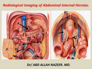
  • 1. Radiological Imaging of Abdominal Internal Hernias. Dr/ ABD ALLAH NAZEER. MD.
  • 2.
  • 3.
  • 4.
  • 5.
  • 6.
  • 7. Paraduodenal hernias, although uncommon, have classically been the most common type of internal hernia. However, the incidence of postoperative internal hernias has been increasing recently. The two most common types, the left and right paraduodenal hernia involve small bowel herniating through a congenital opening in the mesenteries. These internal hernias may result in closed-loop bowel obstruction. Clinical presentation The patient typically presents with symptoms of small bowel obstruction: abdominal pain, nausea, vomiting.
  • 8.
  • 9. Radiographic features These hernias usually appear as a sac-like cluster of small bowel loops in an atypical presentation. A closed-loop obstruction may occur within these loops due to the hernia. However, it is not unusual for small bowel loops to cluster in an atypical position in normal patients. Thin patients may be especially challenging since it may be difficult to follow the course of the collapsed loops of small bowel. Because of this, vascular landmarks around a potential internal hernia "sac" are critical for making a confident diagnosis. Left paraduodenal hernia cluster of small bowel loops in the left anterior pararenal space the cluster of small bowel loops is behind the inferior mesenteric vein (IMV) and behind the ascending left colic artery Right paraduodenal hernia cluster of small bowel loops is inferior to the third portion of the duodenum the cluster of small bowel loops is behind the superior mesenteric vein (SMV), the superior mesenteric artery (SMA), and the right colic vein
  • 10.
  • 11.
  • 12.
  • 13.
  • 14.
  • 15. Left Paraduodenal Hernia. Superior Duodenal Fossa ( pink ovoid zone). A. Axial view. Through upper Treizt angle (red arrow). B. Coronal view. Behind Inferior Mesenteric Vein (white arrow). Loops Herniated Position (circles).
  • 16. Left Paraduodenal Hernia- A. Left saclike mass of herniated small bowel loops through Landzert fossa. Mass effect on left anterior abdominal wall (arrowhead). B. Loops situated between the stomach and pancreas. Displacement of inferior cava vein (red arrow) and stretching and engorgement of vessels converging to entrance and inside the hernia sac (yellow arrows).
  • 17. Left Paraduodenal Hernia. A. Hernia sac (open arrow) located between stomach (with arrow)and pancreatic tail (black arrow). B. MIP coronal view. Twisting mesenteric vessels in the core of the herniated loops (red arrow).
  • 18. Paraduodenal hernia: Contrast-enhanced CT scan of the upper abdomen shows a saclike mass of dilated jejunal loops between the pancreatic head (P) and stomach. The descending mesocolon (D) and stomach are displaced laterally
  • 19. Pericecal hernias (PCHs) In the classic literature PCHs correspond of 13% of all internal hernias. Bowel loops, most commonly an ileal segment, herniate into the right paracolic gutter through a congenital or acquired (most commonly by adhesions) unusual defect in the cecal mesentery. Four different recesses in the pericecal region formed by folds of the peritoneum have been described: superior and inferior ileocecal recess, retrocecal recess and paracolic sulci. However, the diagnostic features and surgical management of the four subtypes do not differ. Clinical findings Patients commonly report recurrent episodes of colicky intense right lower abdominal pain. Chronic incarceration may produce symptoms compatible with appendiceal disorders, intestinal diseases or intestinal obstruction caused by adhesions. In PCHs however have been reported a higher incidence of occlusive symptoms with rapid progression to strangulation and a mortality rate that can be high as 75%. CT findings With CT, a cluster of fixed and dilated small bowel loops with a sac-like appearance is noted, possibly extending into the right paracolic gutter, lateral to the cecum and posterior to the ascending colon, which can be displaced anteriorly or medially
  • 20.
  • 21. Pericecal Hernia: Contrast-enhanced CT scan of the lower abdomen shows dilated small bowel loops (S) and a cluster of fluid-filled small bowel loops (arrow). The ascending colon (A) is displaced anteriorly, and ascites (arrowhead) is seen in the right paracolic gutter
  • 22. Paracecal Hernia. A. Coronal view. Retrocecal recess (ovoid zone). B. Cadaver simulating herniation. A,B: Loops Herniated Position (circle). Terminal Ileum (blue arrow). Cecum (yellow arrow). Flow direction of herniated loops (curved arrows).
  • 23. Pericecal hernia in an 83-year-old woman with a 1-day history of nausea and right lower abdominal pain. (a) Axial contrast-enhanced CT image shows incarcerated intestine with a saclike appearance (arrowheads) that displaces the ascending colon (arrow) medially. (b) Oblique MPR CT image clearly shows the hernia orifice (arrows). Laparoscopic surgery showed incarcerated intestine in a hernia sac lateral to the ascending colon.
  • 24. Pericecal hernia through the retrocecal recess in an 84-year-old man with colicky right lower quadrant pain and vomiting of 48 hours duration. He underwent an appendectomy at 54 years of age. (a) Contrast-enhanced CT scan of the mid- abdomen shows a cluster of encapsulated small bowel loops (arrowheads) in the lateral aspect of the right paracolic gutter and behind the ascending colon (A). Dilated and stretched mesenteric vessels (arrow) are seen within the cluster. (b) CT scan of the lower abdomen shows beaking and collapsed bowel loops (arrow) at the retrocecal recess (arrowhead). The ascending colon (A) is displaced anteriorly. Laparotomy was performed 12 hours after CT.
  • 25. Transmesenteric (TMHs) hernias: CT findings Blachar et al. in 2001 have described the presence of clustered, compressed small bowel loops in the periphery of the abdominal cavity and the lack of omental fat between the loops and the abdominal wall as the most useful CT signs for the detection of a transmesenteric hernia. The herniated bowels appeared lateral to the colon (a reversal of the normal anatomic arrangement) with central, inferior and posterior displacement of the transverse colon and inferior and medial displacement of the hepatic flexure. Another study concluded that the only statistically significant CT signs predictors of a transmesenteric hernia are clustering of small bowel loops, especially those that are adjacent to the abdominal wall, mesenteric vessel abnormalities including stretching, crowding and engorgement, a displacement of the main mesenteric trunk to the right and signs of small bowel obstruction.
  • 26.
  • 27. Left Transmesenteric Hernia. Newborn 12 days old. Simple Rx (A) and intestinal transit (B,C). Left conglomerate (circle in A,C) dilated loops of jejunum from Treitz angle (arrow in B) due to obstruction by hernia. Stomach (St) is also dilated. Distal loops are normal caliber (open arrow).
  • 28.
  • 29.
  • 30.
  • 31.
  • 32. Lesser sac hernias are a type of internal hernia, where abdominal contents protrude through the foramen of Winslow, hence they are also known as foramen of Winslow hernia. Epidemiology Lesser sac hernias are rare, accounting for <0.1% of abdominal hernias and 8% of internal hernias. Pathology Typically contains small bowel only (~67%) but may also contain cecum/ascending colon and less commonly transverse colon, gallbladder, or omentum. Risk factors common intestinal mesentery intra-peritoneal right colon long small bowel mesentery large foramen of Winslow elongated right liver (e.g. Riedel lobe)
  • 33. Radiographic features Plain radiograph gas-filled loops of small bowel in the upper abdomen CT mesenteric fat/vessels posterior to portal vein, common bile duct, hepatic artery and anterior to the inferior vena cava mesenteric vessels passing into the lesser sac via the foramen of Winslow gas and/or fluid in the lesser sac with bird beak sign towards the foramen of Winslow abnormal cecal position Differential diagnosis left paraduodenal hernia
  • 34.
  • 35. Arrows showing gas contained bowel loops obstruction in foramen of winslow hernia
  • 36. Foramen of winslow hernia: Enteroclysis shows small bowel at right hepatic flexure.
  • 37.
  • 38.
  • 39. Foramen of Winslow Hernia. Patient without obstruction but with anomalous position of small bowel loops (circle). They are located under the liver and upper the uncinate pancreas process (white arrow in A) and stretching of vessels toward the herniated bowel loop (yellow arrows).
  • 40. Lesser sac hernia through the lesser omentum in a 44-year-old man who presented with acute abdominal pain. (a) Axial contrast-enhanced CT image shows converging mesenteric vessels and fat (white arrows) intersecting the distal part of the stomach (black arrows) and the right gastric artery (arrowhead). Traction and distortion of the distal part of the stomach are also seen. (b) Transparent volume-rendered CT image shows a cluster of intestine (arrowheads) in the lesser sac, with a beak shape (arrows) pointing toward the lesser curvature of the stomach (S). Surgery confirmed herniated intestine in the lesser sac protruding through a defect in the lesser omentum
  • 41. Hernia through the Broad Ligament Hernias through a defect of the broad ligament account for only 4%–5% of all internal hernias. The herniated viscus is the small intestine in more than 90% of cases. The typical patient with this hernia is a middle-aged woman who has been pregnant and has no history of abdominal surgery. More than 85% of these hernias have occurred in parous women. Broad ligament defects are classified as congenital or acquired. Congenital cases have an embryologic basis due to a developmental peritoneal defect around the uterus. Acquired defects are due to surgical trauma, pregnancy and birth trauma, perforations following vaginal manipulation, and prior pelvic inflammatory disease. A classification for the broad ligament has been proposed on the basis of the anatomic position of the defect: type 1 = defect caudal to the round ligament, type 2 = defect above the round ligament, and type 3 = defect between the round ligament and the remainder of the broad ligament through the mesoligamentum teres. The following are the characteristic CT appearances: (a) a cluster of dilated small bowel loops with air-fluid levels in the pelvic cavity and (b) bowel loops compressing the rectosigmoid dorsolaterally and the uterus ventrally
  • 42.
  • 43. Broad Ligament Hernia A,B (Axial). C (Coronal). Herniated and dilated loops posterior to uterus ( yellow arrows). Bladder (with arrow). Uterus (red arrow)
  • 44. Broad ligament hernia in a 58-year-old woman with a 1- day history of intermittent abdominal pain and vomiting and a history of three normal pregnancies. (a, b) Axial contrast-enhanced CT images obtained at different levels show a cluster of small bowel loops in the pouch of Douglas (arrowheads) and crowding mesenteric vessels (arrows). The uterus (*) is deviated inferiorly and to the right. (c) Coronal MPR CT image shows the hernia orifice (white arrows) below the tubal and ovarian branches (black arrow) of the left ovarian and uterine vessels. Enlargement of the distance between the uterus (*) and the left ovary (arrowhead) is also seen. The patient’s abdominal pain acutely intensified, and open surgery was performed. (d) Intraoperative photograph (anterior view) shows incarceration of the small intestine through a thumb-tip–sized defect (arrows) in the left broad ligament of the uterus. Approximately 70 cm of incarcerated intestine was infarcted and was resected, and the broad ligament was sectioned.
  • 45. Hernia through a defect of the right perirectal fossa in a 28- year-old woman with continuous lower abdominal pain of 34 hours duration. (a, b) Contrast-enhanced CT scans of the pelvis (b obtained 10 mm below a) show dilated and fluid-filled small bowel loops (S). A cluster of dilated bowel loops (arrow) is located to the right of the rectum (R) and behind the uterine cervix (U). Laparotomy was performed 4 hours after CT.
  • 46. Transomental hernias (TOHs) Traditionally, TOHs make up 1-4% of all IHs. The term “transomental hernia” usually refers to herniation, most commonly of small bowel loops, cecum and sigmoid colon, through or into a congenital or acquired abnormal defect of the greater omentum from 2 to 10 cm in diameter involving both leaves (four peritoneal layers) and located in the periphery near the free edge . CT findings CT findings are often identical to those of a transmesenteric hernia, however characteristic features of TOHs are dilated bowel loops, without a sac-like appearance, located in the most anterior portion of the peritoneal cavity with omental vessels that run vertically around the hernia orifice.
  • 47. Transomental Hernia. A. Sagittal view. Great Omentum (blue zone). Hernia pathway ( curved arrow). Position of herniated loop (circle) B. Chance of herniation: Cecum (yellow arrow). Small Bowell (pink arrow) or Sigmoid Colon (with arrow). C. Simulating herniation of jejunal loop (curved arrow) through great omentum ( blue arrows)
  • 48. Sigmoid mesocolon hernia: Contrast-Enhanced CT scans of the pelvis show multiple dilated small bowel loops (S). A dilated inferior mesenteric vein (arrow) appears as a landmark at the edge of the inferior mesentery. A saclike mass of incarcerated jejunal loops (arrowhead) is located anterior to the left psoas muscle
  • 49. Supravesical and pelvic (PIHs) internal hernias CT findings: CT findings of broad ligament hernias include a cluster of dilated small bowel loops herniated in the pelvic cavity laterally to the uterus with a displacement of the rectosigmoid dorso-laterally and of the uterus ventrally and enlargement of the distance between the uterus and the ovary deviating in opposite directions. Furthermore mesenteric vessels of herniated loops penetrating the broad ligament may be seen (33, 88). In supravesical internal hernia usually CT scans show bowel loops with a sac-like appearance pass into the space of Retzius and lay in front of the compressed bladder on the left or right. These patients can present with bladder irritation and dysuria, as the bladder is compressed by the small bowel
  • 50. Internal supravesical hernia in a 67-year-old woman with a two-day history of lower abdominal pain. Contrast-enhanced axial CT scan shows intestine loops (white circle) to the left of the urinary bladder.
  • 51. Internal supravesical hernia in a 74-year-old man with a 1-day history of right lower abdominal pain. (a) Axial contrast-enhanced CT image shows incarcerated intestine with a saclike appearance (arrow) that compresses the anterior wall (arrowheads) of the urinary bladder on the right. (b) Intraoperative photograph (superoanterior view) shows incarceration of the ileum (blue arrow) into the right supravesical fossa (green arrows). Approximately 7 cm of intestine was infarcted and was resected. The defect was approximately 2 cm in diameter. The hernia sac was resected, and the hernia orifice was closed with sutures. B = urinary bladder, yellow arrow = pubis.
  • 52. Petersen hernia in a 60-year-old woman with acute abdominal pain and vomiting. The patient had a history of laparoscopic distal gastrectomy and antecolic reconstruction of a Roux-en-Y anastomosis for gastric cancer performed 11 months earlier. (a) Axial contrast- enhanced CT image shows a dilated biliopancreatic limb (*) and mesenteric swirl (arrows) behind the Roux limb (arrowhead). (b) Coronal MPR CT image shows a cluster of intestine (★) with engorged mesenteric lymph nodes located in the left lower quadrant, as well as a dilated biliopancreatic limb (*). Surgery confirmed herniation of a long segment of small intestine, including jejunojejunostomy through a Petersen defect. Arrowhead = Roux limb, black arrow = jejunojejunostomy, white arrow = gastrojejunostomy.
  • 53. Internal hernia with Whirl sign.
  • 54. Closed loop small bowel obstruction. Contrast enhanced axial CT scan shows a radial array of distended small bowel loops (B) with stretched and thickened mesenteric vessels converging to a central point (white arrow). Bowel wall thickening (arrowhead) and mesenteric edema (asterisk) can also be observed.