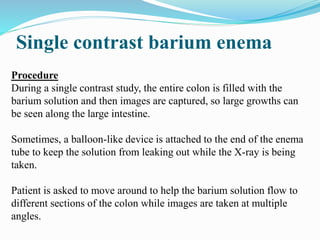
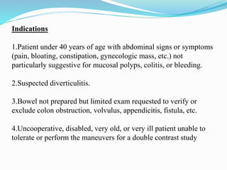
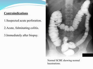
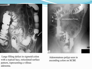
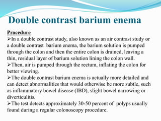
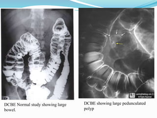
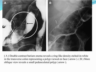
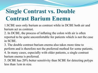
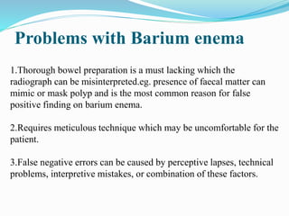
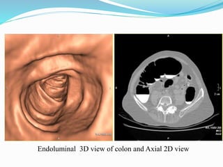
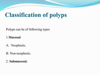
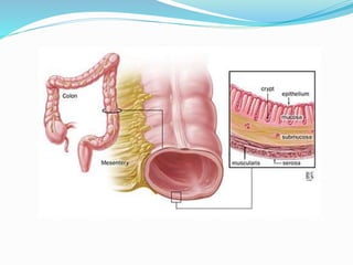
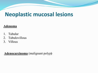
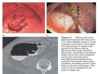
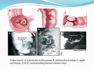
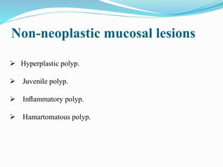
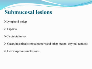
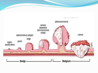
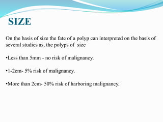
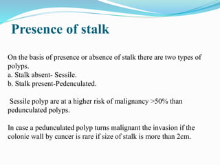
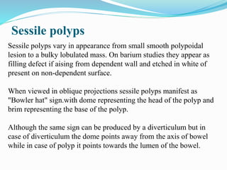
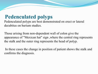
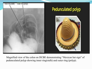
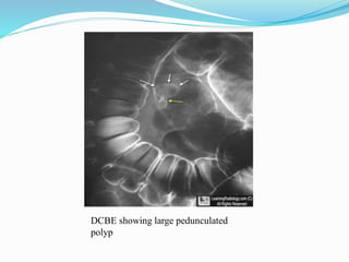
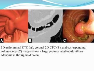
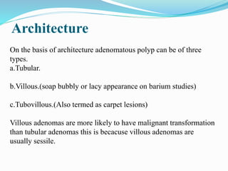
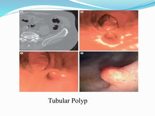
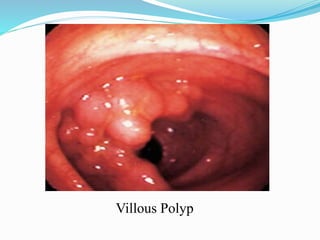
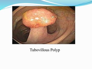
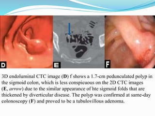
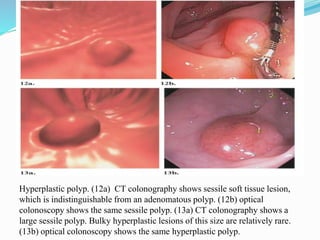
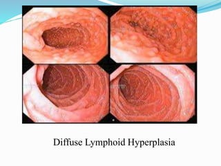
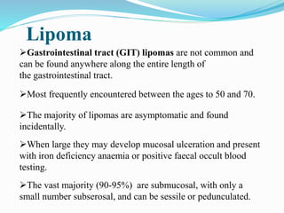
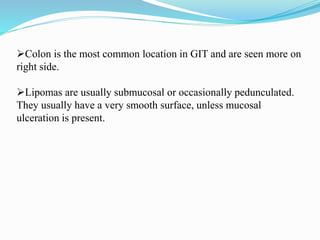
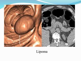
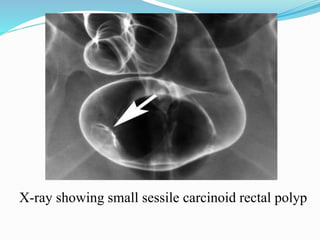
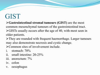
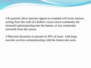
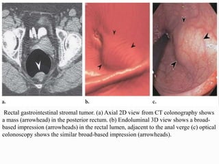
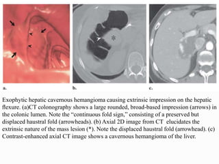
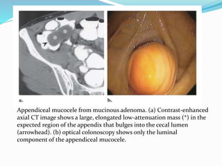
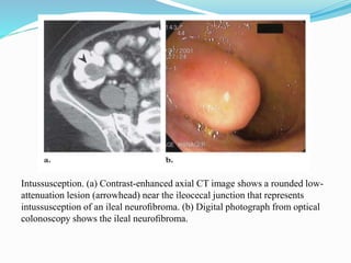
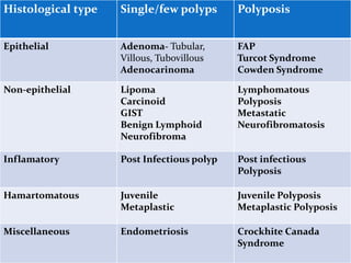
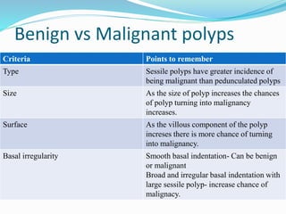
OSR
Dr. Yash Kumar Achantani
What is a polyp?
A GI polyp is defined as a mass of the mucosal surface
protruding into the lumen of the bowel.
Polyps can be neoplastic, nonneoplastic, or submucosal.
The majority of colorectal polyps are inflammatory or
metaplastic and usually 5mm or less in diameter and
have no malignant potential; the majority of larger lesions are
adenomatous polyps
Incidence
Incidence rises with age
• About 3% in 3rd decade
• 10% in 7th decade
• 26% in 9th decade
• About 11% overall in all ages
Most (60%) occur in rectum or sigmoid
Symptoms & Signs
Only a small proportion of polyps cause symptoms or signs. When
they do, the symptoms and signs usually are the result of bleeding
from the polyp and may include.
•Diarrhoea
•Abdominal pain (secondry to intususseption)
•Red blood mixed with stool
•Red blood on the surface of stools
•Black stools
•Weakness
•Light-headedness,
•Fainting, and
•Pale skin.
Radiological Diagnostic methods
1.Single contrast barium enema.
2.Double contrast barium enema.
3. CT colonograhy.
Single contrast barium enema
Procedure
During a single contrast study, the entire colon is filled with the
barium solution and then images are captured, so large growths can
be seen along the large intestine.
Sometimes, a balloon-like device is attached to the end of the enema
tube to keep the solution from leaking out while the X-ray is being
taken.
Patient is asked to move around to help the barium solution flow to
different sections of the colon while images are taken at multiple
angles.
Indications
1.Patient under 40 years of age with abdominal signs or symptoms
(pain, bloating, constipation, gynecologic mass, etc.) not
particularly suggestive for mucosal polyps, colitis, or bleeding.
2.Suspected diverticulitis.
3.Bowel not prepared but limited exam requested to verify or
exclude colon obstruction, volvulus, appendicitis, fistula, etc.
4.Uncooperative, disabled, very old, or very ill patient unable to
tolerate or perform the maneuvers for a double contrast study
Contraindications
1.Suspected acute perforation.
2.Acute, fulminating colitis.
3.Immediately after biopsy.
Normal SCBE showing normal
haustrations.
Large filling defect in sigmoid colon
with a typical lacy, reticulated surface
pattern, representing a villous
adenoma.
Adenomatous polyp seen in
ascending colon on SCBE
Double contrast barium enema
Procedure
In a double contrast study, also known as an air contrast study or
a double contrast barium enema, the barium solution is pumped
through the colon and then the entire colon is drained, leaving a
thin, residual layer of barium solution lining the colon wall.
Then, air is pumped through the rectum, inflating the colon for
better viewing.
The double contrast barium enema is actually more detailed and
can detect abnormalities that would otherwise be more subtle, such
as inflammatory bowel disease (IBD), slight bowel narrowing or
diverticulitis.
The test detects approximately 30-50 percent of polyps usually
found during a regular colonoscopy procedure.
Indications
1.Rectal bleeding - gross or occult.
2.Polyps or carcinoma - suspected or known .
3.Inflammatory bowel disease - suspected or known.
4.Patient over 40 years of age who can cooperate and turn over
without assistance.
Contraindications
1.Suspected acute perforation.
2.Acute, fulminating colitis.
3.Immediately after biopsy.
DCBE showing large pedunculated
polyp
DCBE Normal study showing large
bowel.
( A ) Double-contrast barium enema reveals a ring-like density etched-in-white
in the transverse colon representing a polyp viewed en face ( arrow ). ( B ) More
oblique view reveals a small pedunculated polyp ( arrow ).
Single Contrast vs. Double
Contrast Barium Enema
1.SCBE uses only barium as contrast while in DCBE both air and
barium act as contrast.
2. In DCBE, the process of inflating the colon with air is often
reported to be quite uncomfortable for patients which is not the case
in SCBE.
3. The double contrast barium enema also takes more time to
perform and is therefore not the preferred method for some patients.
4. In many cases, especially with older patients, a single contrast
barium enema is preferred.
5. DCBE has 20% better sensitivity than SCBE for detecting polyps
less than 1cm in size.
Problems with Barium enema
1.Thorough bowel preparation is a must lacking which the
radiograph can be misinterpreted.eg. presence of faecal matter can
mimic or mask polyp and is the most common reason for false
positive finding on barium enema.
2.Requires meticulous technique which may be uncomfortable for the
patient.
3.False negative errors can be caused by perceptive lapses, technical
problems, interpretive mistakes, or combination of these factors.
CT SCAN
Conventional CT
Conventional CT may show colonic polyps as intraluminal filling
defects that cause deformation of the contrast material–filled lumen.
Conventional abdominal CT is also a valuable tool in planning
surgery for colon cancer, which can be a complication of colonic
polyposis.
On CT scans, colon cancer appears as a soft tissue–attenuating mass
that narrows the colonic lumen. Colon cancer can also appear as
focal bowel thickening and luminal narrowing.
CT can also readily depict complications of colon cancer, such as
obstruction, perforation and fistula formation, and liver, lymph node,
and other distant metastases. Extraluminal spread is depicted as the
loss of fat planes between the colon and adjacent organs .
CT colonography (virtual CT
colonoscopy)
CT colonography typically refers to the evaluation of a cleansed
and gas-distended colon to detect polyps and masses.
A low-dose CT is performed in both the supine and prone
positions. The images are interpreted using specialized software by
viewing the axial, multiplanar reconstruction (MPR), and 3D
endoluminal images.
Thorough colonic cleansing is required to eliminate false-positive
scans caused by fecal residue.
Dry preparation by using sodium chloride cathartics is preferred to
a colonic lavage, to minimize the retention of fluid that might hide
polyps.
After colonic cleansing, the patient is placed in the left lateral
decubitus position on the CT table.
An endorectal tube is passed, and the large bowel is insufflated
with air or carbon dioxide using either manual insufflation or a
mechanical pump.
To minimize respiratory motion, a fast scanner is needed.
Multidetector-row CT is the preferred imaging modality.
3D virtual colonoscopic techniques have the appeal of truly
simulating conventional colonoscopy , Virtual colonoscopy does
not yet compete with colonoscopy in the demonstration of small
colonic polyps, Polyps smaller than 1 cm may not be detected.
CT has good sensitivity for the detection of clinically important
polyps.
CT may be helpful in less mobile and/or older persons, as well as
those in whom malignant transformation is suspected; therefore, it
is a useful adjunct to a contrast-enhanced examination.
Problems with CT Colonography
Adequate bowel preparation is absolutely vital for confident
detection of significant lesions because residual fecal material may
be indistinguishable from polyps or neoplasms, or it may obscure
polyps, making their detection impossible.
Other problems in detection of polyps with CT colonoscopy arise
with small, sessile polyps (< 1 cm).
Distinguishing polyps from haustral folds can also be difficult;
thus, a clean, well-distended colon visualized prone and supine is
important.
Endoluminal 3D view of colon and Axial 2D view
Classification of polyps
Polyps can be of following types
1.Mucosal
A. Neoplastic.
B. Non-neoplastic.
2. Submucosal.
Neoplastic mucosal lesions
Adenoma
1. Tubular
2. Tubulovillous
3. Villous
Adenocarcinoma (malignant polyp)
Tubular Polyps
Tubulovillous polyp
Tubovillous Adenoma
Fig 3a (Endoluminal view) and 3b
(colonoscopy image) showing
sessile lobulated polyp from a
colonic fold.
Fig 4a (Endoluminal view) and 4b
(Axial CT image) showing
pedunculated polyp with a well
defined stalk.
Colon cancer. A, Carcinoma in the cecum; B, pedunculated polyp; C, apple
core lesion; A’, B’, C’, corresponding barium enema x-rays
Non-neoplastic mucosal lesions
 Hyperplastic polyp.
 Juvenile polyp.
 Inflammatory polyp.
 Hamartomatous polyp.
Submucosal lesions
Lymphoid polyp
 Lipoma
Carcinoid tumor
 Gastrointestinal stromal tumor (and other mesen- chymal tumors)
 Hematogenous metastases.
Pathogeniesis of polyp
Polyps may not have any change or may undergo malignant
transformation.
Most of the colorectal malignancies arise from adenomatous
polyps.
The risk of malignant transformation is higher in adenomas more
than 1cm in size.
The transition from benign adenoma to carcinoma takes 7-10
years.
The malignant potential of adenomatous polyp has four major
determinants.
Size, presence of stalk, villous architecture and degree of cellular
atypia and dysplasia.
SIZE
On the basis of size the fate of a polyp can interpreted on the basis of
several studies as, the polyps of size
•Less than 5mm - no risk of malignancy.
•1-2cm- 5% risk of malignancy.
•More than 2cm- 50% risk of harboring malignancy.
Presence of stalk
On the basis of presence or absence of stalk there are two types of
polyps.
a. Stalk absent- Sessile.
b. Stalk present-Pedenculated.
Sessile polyp are at a higher risk of malignancy >50% than
pedunculated polyps.
In case a pedunculated polyp turns malignant the invasion if the
colonic wall by cancer is rare if size of stalk is more than 2cm.
Sessile polyps
Sessile polyps vary in appearance from small smooth polypoidal
lesion to a bulky lobulated mass. On barium studies they appear as
filling defect if aising from dependent wall and etched in white of
present on non-dependent surface.
When viewed in oblique projections sessile polyps manifest as
"Bowler hat" sign.with dome representing the head of the polyp and
brim representing the base of the polyp.
Although the same sign can be produced by a diverticulum but in
case of diverticulum the dome points away from the axis of bowel
while in case of polyp it points towards the lumen of the bowel.
Magnified view of the sigmoid colon on DCBE demonstrating “Bowler
hat sign” of sessile polyp seen obliquely (arrowhead) and diverticula en
face(arrow).
Pedenculated polyps
Pedenculated polyps are best demonstrated on erect or lateral
decubitus on barium studies.
Those arising from non-dependent wall of colon give the
appearance of "Mexican hat" sign ,where the central ring represents
the stalk and the outer ring represents the head of polyp.
In these cases the change in position of patient shows the stalk and
confirms the diagnosis.
Magnified view of the colon on DCBE demonstrating “Mexican hat sign” of
pedunculated polyp showing inner ring(stalk) and outer ring (polyp).
DCBE showing large pedunculated
polyp
3D endoluminal CTC (A), coronal 2D CTC (B), and corresponding
colonoscopy (C) images show a large pedunculated tubulovillous
adenoma in the sigmoid colon.
Architecture
On the basis of architecture adenomatous polyp can be of three
types.
a.Tubular.
b.Villous.(soap bubbly or lacy appearance on barium studies)
c.Tubovillous.(Also termed as carpet lesions)
Villous adenomas are more likely to have malignant transformation
than tubular adenomas this is becacuse villous adenomas are
usually sessile.
Tubular Polyp
Villous Polyp
Tubovillous Polyp
3D endoluminal CTC image (D) f shows a 1.7-cm pedunculated polyp in
the sigmoid colon, which is less conspicuous on the 2D CTC images
(E, arrow) due to the similar appearance of hte sigmoid folds that are
thickened by diverticular disease. The polyp was confirmed at same-day
colonoscopy (F) and proved to be a tubulovillous adenoma.
Adenomatous polyps
The most common type of polyp is the adenoma or adenomatous
polyp.
It is an important type of polyp not only because it is the most
common, but because it is the most common cause of colon cancer.
The likelihood that an adenoma will develop into (or has already
developed into) cancer is partially dependent on its size; the larger
the polyp, the more likely it is that the polyp is or will become
malignant (concern about the malignant potential increases with a
polyp greater than one centimeter in size).
It also matters if there is a single polyp or multiple polyps. Patients
with multiple polyps - even if they are not malignant when
examined under microscope are more likely to develop additional
polyps in the future that may become malignant.
Concern about this increasing malignant potential begins when
there are three or more polyps.
Finally, the malignant potential of an adenomatous polyp is related
to the manner in which the cells of the polyp organize themselves
as seen under the microscope. Cells that organize themselves into
tubular structures (tubular adenomas) are less likely to become
cancerous than cells that organize themselves into finger-like
structures (villous adenomas).
Most adenomatous polyps are considered sporadic, that is, they do
not stem from a recognized genetic mutation that is present at birth
(are not familial).
Nevertheless, the risk of having colon polyps greater than one
centimeter in size or developing colon cancer is two-fold greater if
a first degree relative has colon polyps greater than one centimeter
in size.
Therefore, there it is likely to be a genetic factor working even in
sporadic adenomatous polyps.
3D endoluminal CTC (G) and double-contrast BE (H and I) images from
three different patients show large pedunculated adenomas.
Genetic adenomatous polyp
syndromes
There are several familial, genetic conditions in which the
mutations or the development of mutations are programmed into
an individual's genes from before birth, passed down from
parent to child.
In the most common of these conditions, hundreds to thousands
of adenomatous polyps form (FAP) as a result of a mutation in
the APC gene.
It is important to recognize these polyposis syndromes and the
exact genetic abnormality that causes them, if possible since the
malignant potential of these polyps is much greater than that of
individuals without the genetic abnormality. (Eighty percent or
more of these patients develop colon cancer.)
Even though these syndromes are responsible for only a few
percent of all colon cancers, recognition of a polyposis syndrome
identifies patients in whom screening for additional polyps needs
to be done more frequently so that new polyps and cancers can be
discovered and treated early.
Because of the autosomal dominant mode of transmission of the
gene and its effects, only one parent needs to have the FAP gene
to pass on to his or her children, and therefore, there is a 50/50
chance that each of his or her children will have FAP.
There is an uncommon form of FAP in which the number of
polyps is less than classic FAP - less than 100--called attenuated
FAP.
Unlike FAP which is an autosomal dominant syndrome, attenuated
FAP is a recessive mutation so that an individual needs to inherit
one mutated gene from each parent to develop polyps and colon
cancer, and because of the rarity of the mutation, this occurs rarely.
Another syndrome of polyps and colon cancer is the MYH
polyposis syndrome.
Individuals with MYH polyposis develop less than 100 polyps at a
young age and are at high risk for developing colon cancer.
It is caused by mutations in a different gene than FAP, the MYH
gene; however, the mutation occurs sporadically due to
spontaneous mutations and, therefore, a hereditary pattern is not
apparent in parents, although it may be seen in siblings.
Because it is an autosomal recessive gene that requires a mutated
gene from each parent, the MYH polyposis syndrome is rare.
(a) Contrast-enhanced 2D axial CT image shows a large irregular soft-tissue mass in the
ascending colon (arrowheads). (b) Digital photograph from subsequent optical
colonoscopy shows the large mass seen in a, which proved to be malignant at histologic
evaluation. (c) Axial CT image shows multiple small polypoid lesions (arrowheads) (d)
optical colonoscopy shows multiple polyps in the transverse colon.
Familial
adenomatous
polyposis
syndrome with
adenocarcinoma.
A case of FAP showing multiple small polyps as seen on DCBE
Hyperplastic polyps
The second most common type of colon polyp is the hyperplastic
polyp.
It is important to recognize these polyps and to differentiate them
from adenomatous polyps since they have little or no potential to
become cancerous unless they are located in the proximal
(ascending colon), or show a particular histologic pattern under the
microscope (a serrated appearance).
Nevertheless, there are uncommon genetic syndromes in which
patients form many hyperplastic polyps.
These patients may be at a similar risk for developing colon
cancer as patients with multiple adenomatous polyps, particularly
if the polyps are large, serrated, located in the ascending colon,
and there is a family history of colon cancer.
Hyperplastic polyps may coexist with adenomatous polyps.
Mucous glands lined by a single layer of columnar epithelium.
Usually located in rectum.
Usually less than 5mm in diameter.
Hyperplastic polyp. (12a) CT colonography shows sessile soft tissue lesion,
which is indistinguishable from an adenomatous polyp. (12b) optical
colonoscopy shows the same sessile polyp. (13a) CT colonography shows a
large sessile polyp. Bulky hyperplastic lesions of this size are relatively rare.
(13b) optical colonoscopy shows the same hyperplastic polyp.
Juvenile Polyps
Classified as cystic hamartomas by some and inflammatory
retention cysts by others.
No malignant potential.
 Most occur as isolated colonic lesions in children less than 10
years.
 Most are solitary.
 Rectal bleeding is the most common symptom.
 Most occur in the rectum or sigmoid.
 Since they have a tendency to autoamputation, they are usually
not removed.
Lymphoid Polyp
Benign lymphoid polyps are a rare histologic entity and should
not be confused with malignant disease of the colon and rectum.
Although retention polyps are the single most common type of
colonic polyp in children, the presence of multiple clustered
polyps in the rectum should alert the radiologist to the possibility
of benign lymphoid polyps.
Diffuse Lymphoid Hyperplasia
Lipoma
Gastrointestinal tract (GIT) lipomas are not common and
can be found anywhere along the entire length of
the gastrointestinal tract.
Most frequently encountered between the ages to 50 and 70.
The majority of lipomas are asymptomatic and found
incidentally.
When large they may develop mucosal ulceration and present
with iron deficiency anaemia or positive faecal occult blood
testing.
The vast majority (90-95%) are submucosal, with only a
small number subserosal, and can be sessile or pedunculated.
Colon is the most common location in GIT and are seen more on
right side.
Lipomas are usually submucosal or occasionally pedunculated.
They usually have a very smooth surface, unless mucosal
ulceration is present.
Lipoma
CT Axial section showing Lipoma in Ascending
colon
CT Coronal section showing Lipoma in Ascending
colon
Carcinoid Tumors
Carcinoid tumours are a type of neuroendocrine tumour that can
occur in a number of locations.
Carcinoid tumours arise from endocrine amine precursor uptake
and decarboxylation (APUD) cells that can be found throughout the
gastrointestinal tract as well as other organs (e.g. lung).
In general they are slow growing tumours but are nevertheless
capable of metastasis.
Most colorectal carcinoids arise in the rectum, with fewer
occurring in the cecum.
Colorectal carcinoids do not exhibit specific gender
predilection.
The mean age of patients at diagnosis for colonic carcinoids is
50-60 years.
Carcinoids of the proximal colon are polypoid intraluminal
masses that are indistinguishable from polypoid adenomas or
adenocarcinomas.
Rectal carcinoids are most commonly small mural or polypoid
masses.
X-ray showing small sessile carcinoid rectal polyp
GIST
Gastrointestinal stromal tumours (GIST) are the most
common mesenchymal tumours of the gastrointestinal tract.
GISTs usually occurs after the age of 40, with most seen in
older patients.
They are rounded with frequent haemorrhage. Larger tumours
may also demonstrate necrosis and cystic change.
Common sites of involvement include.
i. stomach: 70%
ii. small intestine: 20-25%
iii. anorectum: 7%
iv. colon
v. oesophagus
In general, these tumours appear as rounded soft tissue masses,
arising from the wall of a hollow viscus (most commonly the
stomach) and projecting into the lumen, or less commonly
outwards from the serosa.
Mucosal ulceration is present in 50% of cases with large
necrotic cavities communicating with the lumen also seen.
Rectal gastrointestinal stromal tumor. (a) Axial 2D view from CT colonography shows
a mass (arrowhead) in the posterior rectum. (b) Endoluminal 3D view shows a broad-
based impression (arrowheads) in the rectal lumen, adjacent to the anal verge (c) optical
colonoscopy shows the similar broad-based impression (arrowheads).
Extrinsic lesions that can mimic
polyps
Impression from any extracolonic structure.
 Appendiceal lesion.
 Intussusception
Exophytic hepatic cavernous hemangioma causing extrinsic impression on the hepatic
flexure. (a)CT colonography shows a large rounded, broad-based impression (arrows) in
the colonic lumen. Note the “continuous fold sign,” consisting of a preserved but
displaced haustral fold (arrowheads). (b) Axial 2D image from CT elucidates the
extrinsic nature of the mass lesion (*). Note the displaced haustral fold (arrowhead). (c)
Contrast-enhanced axial CT image shows a cavernous hemangioma of the liver.
Appendiceal mucocele from mucinous adenoma. (a) Contrast-enhanced
axial CT image shows a large, elongated low-attenuation mass (*) in the
expected region of the appendix that bulges into the cecal lumen
(arrowhead). (b) optical colonoscopy shows only the luminal
component of the appendiceal mucocele.
Intussusception. (a) Contrast-enhanced axial CT image shows a rounded low-
attenuation lesion (arrowhead) near the ileocecal junction that represents
intussusception of an ileal neurofibroma. (b) Digital photograph from optical
colonoscopy shows the ileal neurofibroma.
Histological type Single/few polyps Polyposis
Epithelial Adenoma- Tubular,
Villous, Tubovillous
Adenocarinoma
FAP
Turcot Syndrome
Cowden Syndrome
Non-epithelial Lipoma
Carcinoid
GIST
Benign Lymphoid
Neurofibroma
Lymphomatous
Polyposis
Metastatic
Neurofibromatosis
Inflamatory Post Infectious polyp Post infectious
Polyposis
Hamartomatous Juvenile
Metaplastic
Juvenile Polyposis
Metaplastic Polyposis
Miscellaneous Endometriosis Crockhite Canada
Syndrome
Benign vs Malignant polyps
Criteria Points to remember
Type Sessile polyps have greater incidence of
being malignant than pedunculated polyps
Size As the size of polyp increases the chances
of polyp turning into malignancy
increases.
Surface As the villous component of the polyp
increses there is more chance of turning
into malignancy.
Basal irregularity Smooth basal indentation- Can be benign
or malignant
Broad and irregular basal indentation with
large sessile polyp- increase chance of
malignacy.
Imaging of Large Bowel Polyp

Imaging of Large Bowel Polyp

  • 1.
  • 2.
    What is apolyp? A GI polyp is defined as a mass of the mucosal surface protruding into the lumen of the bowel. Polyps can be neoplastic, nonneoplastic, or submucosal. The majority of colorectal polyps are inflammatory or metaplastic and usually 5mm or less in diameter and have no malignant potential; the majority of larger lesions are adenomatous polyps
  • 4.
    Incidence Incidence rises withage • About 3% in 3rd decade • 10% in 7th decade • 26% in 9th decade • About 11% overall in all ages Most (60%) occur in rectum or sigmoid
  • 5.
    Symptoms & Signs Onlya small proportion of polyps cause symptoms or signs. When they do, the symptoms and signs usually are the result of bleeding from the polyp and may include. •Diarrhoea •Abdominal pain (secondry to intususseption) •Red blood mixed with stool •Red blood on the surface of stools •Black stools •Weakness •Light-headedness, •Fainting, and •Pale skin.
  • 6.
    Radiological Diagnostic methods 1.Singlecontrast barium enema. 2.Double contrast barium enema. 3. CT colonograhy.
  • 7.
    Single contrast bariumenema Procedure During a single contrast study, the entire colon is filled with the barium solution and then images are captured, so large growths can be seen along the large intestine. Sometimes, a balloon-like device is attached to the end of the enema tube to keep the solution from leaking out while the X-ray is being taken. Patient is asked to move around to help the barium solution flow to different sections of the colon while images are taken at multiple angles.
  • 8.
    Indications 1.Patient under 40years of age with abdominal signs or symptoms (pain, bloating, constipation, gynecologic mass, etc.) not particularly suggestive for mucosal polyps, colitis, or bleeding. 2.Suspected diverticulitis. 3.Bowel not prepared but limited exam requested to verify or exclude colon obstruction, volvulus, appendicitis, fistula, etc. 4.Uncooperative, disabled, very old, or very ill patient unable to tolerate or perform the maneuvers for a double contrast study
  • 9.
    Contraindications 1.Suspected acute perforation. 2.Acute,fulminating colitis. 3.Immediately after biopsy. Normal SCBE showing normal haustrations.
  • 10.
    Large filling defectin sigmoid colon with a typical lacy, reticulated surface pattern, representing a villous adenoma. Adenomatous polyp seen in ascending colon on SCBE
  • 11.
    Double contrast bariumenema Procedure In a double contrast study, also known as an air contrast study or a double contrast barium enema, the barium solution is pumped through the colon and then the entire colon is drained, leaving a thin, residual layer of barium solution lining the colon wall. Then, air is pumped through the rectum, inflating the colon for better viewing. The double contrast barium enema is actually more detailed and can detect abnormalities that would otherwise be more subtle, such as inflammatory bowel disease (IBD), slight bowel narrowing or diverticulitis. The test detects approximately 30-50 percent of polyps usually found during a regular colonoscopy procedure.
  • 12.
    Indications 1.Rectal bleeding -gross or occult. 2.Polyps or carcinoma - suspected or known . 3.Inflammatory bowel disease - suspected or known. 4.Patient over 40 years of age who can cooperate and turn over without assistance.
  • 13.
    Contraindications 1.Suspected acute perforation. 2.Acute,fulminating colitis. 3.Immediately after biopsy.
  • 14.
    DCBE showing largepedunculated polyp DCBE Normal study showing large bowel.
  • 15.
    ( A )Double-contrast barium enema reveals a ring-like density etched-in-white in the transverse colon representing a polyp viewed en face ( arrow ). ( B ) More oblique view reveals a small pedunculated polyp ( arrow ).
  • 16.
    Single Contrast vs.Double Contrast Barium Enema 1.SCBE uses only barium as contrast while in DCBE both air and barium act as contrast. 2. In DCBE, the process of inflating the colon with air is often reported to be quite uncomfortable for patients which is not the case in SCBE. 3. The double contrast barium enema also takes more time to perform and is therefore not the preferred method for some patients. 4. In many cases, especially with older patients, a single contrast barium enema is preferred. 5. DCBE has 20% better sensitivity than SCBE for detecting polyps less than 1cm in size.
  • 17.
    Problems with Bariumenema 1.Thorough bowel preparation is a must lacking which the radiograph can be misinterpreted.eg. presence of faecal matter can mimic or mask polyp and is the most common reason for false positive finding on barium enema. 2.Requires meticulous technique which may be uncomfortable for the patient. 3.False negative errors can be caused by perceptive lapses, technical problems, interpretive mistakes, or combination of these factors.
  • 18.
    CT SCAN Conventional CT ConventionalCT may show colonic polyps as intraluminal filling defects that cause deformation of the contrast material–filled lumen. Conventional abdominal CT is also a valuable tool in planning surgery for colon cancer, which can be a complication of colonic polyposis. On CT scans, colon cancer appears as a soft tissue–attenuating mass that narrows the colonic lumen. Colon cancer can also appear as focal bowel thickening and luminal narrowing. CT can also readily depict complications of colon cancer, such as obstruction, perforation and fistula formation, and liver, lymph node, and other distant metastases. Extraluminal spread is depicted as the loss of fat planes between the colon and adjacent organs .
  • 19.
    CT colonography (virtualCT colonoscopy) CT colonography typically refers to the evaluation of a cleansed and gas-distended colon to detect polyps and masses. A low-dose CT is performed in both the supine and prone positions. The images are interpreted using specialized software by viewing the axial, multiplanar reconstruction (MPR), and 3D endoluminal images. Thorough colonic cleansing is required to eliminate false-positive scans caused by fecal residue.
  • 20.
    Dry preparation byusing sodium chloride cathartics is preferred to a colonic lavage, to minimize the retention of fluid that might hide polyps. After colonic cleansing, the patient is placed in the left lateral decubitus position on the CT table. An endorectal tube is passed, and the large bowel is insufflated with air or carbon dioxide using either manual insufflation or a mechanical pump. To minimize respiratory motion, a fast scanner is needed. Multidetector-row CT is the preferred imaging modality.
  • 21.
    3D virtual colonoscopictechniques have the appeal of truly simulating conventional colonoscopy , Virtual colonoscopy does not yet compete with colonoscopy in the demonstration of small colonic polyps, Polyps smaller than 1 cm may not be detected. CT has good sensitivity for the detection of clinically important polyps. CT may be helpful in less mobile and/or older persons, as well as those in whom malignant transformation is suspected; therefore, it is a useful adjunct to a contrast-enhanced examination.
  • 22.
    Problems with CTColonography Adequate bowel preparation is absolutely vital for confident detection of significant lesions because residual fecal material may be indistinguishable from polyps or neoplasms, or it may obscure polyps, making their detection impossible. Other problems in detection of polyps with CT colonoscopy arise with small, sessile polyps (< 1 cm). Distinguishing polyps from haustral folds can also be difficult; thus, a clean, well-distended colon visualized prone and supine is important.
  • 23.
    Endoluminal 3D viewof colon and Axial 2D view
  • 24.
    Classification of polyps Polypscan be of following types 1.Mucosal A. Neoplastic. B. Non-neoplastic. 2. Submucosal.
  • 26.
    Neoplastic mucosal lesions Adenoma 1.Tubular 2. Tubulovillous 3. Villous Adenocarcinoma (malignant polyp)
  • 27.
  • 28.
  • 29.
    Tubovillous Adenoma Fig 3a(Endoluminal view) and 3b (colonoscopy image) showing sessile lobulated polyp from a colonic fold. Fig 4a (Endoluminal view) and 4b (Axial CT image) showing pedunculated polyp with a well defined stalk.
  • 31.
    Colon cancer. A,Carcinoma in the cecum; B, pedunculated polyp; C, apple core lesion; A’, B’, C’, corresponding barium enema x-rays
  • 32.
    Non-neoplastic mucosal lesions Hyperplastic polyp.  Juvenile polyp.  Inflammatory polyp.  Hamartomatous polyp.
  • 33.
    Submucosal lesions Lymphoid polyp Lipoma Carcinoid tumor  Gastrointestinal stromal tumor (and other mesen- chymal tumors)  Hematogenous metastases.
  • 34.
    Pathogeniesis of polyp Polypsmay not have any change or may undergo malignant transformation. Most of the colorectal malignancies arise from adenomatous polyps. The risk of malignant transformation is higher in adenomas more than 1cm in size. The transition from benign adenoma to carcinoma takes 7-10 years. The malignant potential of adenomatous polyp has four major determinants. Size, presence of stalk, villous architecture and degree of cellular atypia and dysplasia.
  • 36.
    SIZE On the basisof size the fate of a polyp can interpreted on the basis of several studies as, the polyps of size •Less than 5mm - no risk of malignancy. •1-2cm- 5% risk of malignancy. •More than 2cm- 50% risk of harboring malignancy.
  • 37.
    Presence of stalk Onthe basis of presence or absence of stalk there are two types of polyps. a. Stalk absent- Sessile. b. Stalk present-Pedenculated. Sessile polyp are at a higher risk of malignancy >50% than pedunculated polyps. In case a pedunculated polyp turns malignant the invasion if the colonic wall by cancer is rare if size of stalk is more than 2cm.
  • 38.
    Sessile polyps Sessile polypsvary in appearance from small smooth polypoidal lesion to a bulky lobulated mass. On barium studies they appear as filling defect if aising from dependent wall and etched in white of present on non-dependent surface. When viewed in oblique projections sessile polyps manifest as "Bowler hat" sign.with dome representing the head of the polyp and brim representing the base of the polyp. Although the same sign can be produced by a diverticulum but in case of diverticulum the dome points away from the axis of bowel while in case of polyp it points towards the lumen of the bowel.
  • 39.
    Magnified view ofthe sigmoid colon on DCBE demonstrating “Bowler hat sign” of sessile polyp seen obliquely (arrowhead) and diverticula en face(arrow).
  • 40.
    Pedenculated polyps Pedenculated polypsare best demonstrated on erect or lateral decubitus on barium studies. Those arising from non-dependent wall of colon give the appearance of "Mexican hat" sign ,where the central ring represents the stalk and the outer ring represents the head of polyp. In these cases the change in position of patient shows the stalk and confirms the diagnosis.
  • 41.
    Magnified view ofthe colon on DCBE demonstrating “Mexican hat sign” of pedunculated polyp showing inner ring(stalk) and outer ring (polyp).
  • 42.
    DCBE showing largepedunculated polyp
  • 43.
    3D endoluminal CTC(A), coronal 2D CTC (B), and corresponding colonoscopy (C) images show a large pedunculated tubulovillous adenoma in the sigmoid colon.
  • 44.
    Architecture On the basisof architecture adenomatous polyp can be of three types. a.Tubular. b.Villous.(soap bubbly or lacy appearance on barium studies) c.Tubovillous.(Also termed as carpet lesions) Villous adenomas are more likely to have malignant transformation than tubular adenomas this is becacuse villous adenomas are usually sessile.
  • 45.
  • 46.
  • 47.
  • 48.
    3D endoluminal CTCimage (D) f shows a 1.7-cm pedunculated polyp in the sigmoid colon, which is less conspicuous on the 2D CTC images (E, arrow) due to the similar appearance of hte sigmoid folds that are thickened by diverticular disease. The polyp was confirmed at same-day colonoscopy (F) and proved to be a tubulovillous adenoma.
  • 49.
    Adenomatous polyps The mostcommon type of polyp is the adenoma or adenomatous polyp. It is an important type of polyp not only because it is the most common, but because it is the most common cause of colon cancer. The likelihood that an adenoma will develop into (or has already developed into) cancer is partially dependent on its size; the larger the polyp, the more likely it is that the polyp is or will become malignant (concern about the malignant potential increases with a polyp greater than one centimeter in size).
  • 50.
    It also mattersif there is a single polyp or multiple polyps. Patients with multiple polyps - even if they are not malignant when examined under microscope are more likely to develop additional polyps in the future that may become malignant. Concern about this increasing malignant potential begins when there are three or more polyps. Finally, the malignant potential of an adenomatous polyp is related to the manner in which the cells of the polyp organize themselves as seen under the microscope. Cells that organize themselves into tubular structures (tubular adenomas) are less likely to become cancerous than cells that organize themselves into finger-like structures (villous adenomas).
  • 51.
    Most adenomatous polypsare considered sporadic, that is, they do not stem from a recognized genetic mutation that is present at birth (are not familial). Nevertheless, the risk of having colon polyps greater than one centimeter in size or developing colon cancer is two-fold greater if a first degree relative has colon polyps greater than one centimeter in size. Therefore, there it is likely to be a genetic factor working even in sporadic adenomatous polyps.
  • 52.
    3D endoluminal CTC(G) and double-contrast BE (H and I) images from three different patients show large pedunculated adenomas.
  • 53.
    Genetic adenomatous polyp syndromes Thereare several familial, genetic conditions in which the mutations or the development of mutations are programmed into an individual's genes from before birth, passed down from parent to child. In the most common of these conditions, hundreds to thousands of adenomatous polyps form (FAP) as a result of a mutation in the APC gene.
  • 54.
    It is importantto recognize these polyposis syndromes and the exact genetic abnormality that causes them, if possible since the malignant potential of these polyps is much greater than that of individuals without the genetic abnormality. (Eighty percent or more of these patients develop colon cancer.) Even though these syndromes are responsible for only a few percent of all colon cancers, recognition of a polyposis syndrome identifies patients in whom screening for additional polyps needs to be done more frequently so that new polyps and cancers can be discovered and treated early. Because of the autosomal dominant mode of transmission of the gene and its effects, only one parent needs to have the FAP gene to pass on to his or her children, and therefore, there is a 50/50 chance that each of his or her children will have FAP.
  • 55.
    There is anuncommon form of FAP in which the number of polyps is less than classic FAP - less than 100--called attenuated FAP. Unlike FAP which is an autosomal dominant syndrome, attenuated FAP is a recessive mutation so that an individual needs to inherit one mutated gene from each parent to develop polyps and colon cancer, and because of the rarity of the mutation, this occurs rarely.
  • 56.
    Another syndrome ofpolyps and colon cancer is the MYH polyposis syndrome. Individuals with MYH polyposis develop less than 100 polyps at a young age and are at high risk for developing colon cancer. It is caused by mutations in a different gene than FAP, the MYH gene; however, the mutation occurs sporadically due to spontaneous mutations and, therefore, a hereditary pattern is not apparent in parents, although it may be seen in siblings. Because it is an autosomal recessive gene that requires a mutated gene from each parent, the MYH polyposis syndrome is rare.
  • 57.
    (a) Contrast-enhanced 2Daxial CT image shows a large irregular soft-tissue mass in the ascending colon (arrowheads). (b) Digital photograph from subsequent optical colonoscopy shows the large mass seen in a, which proved to be malignant at histologic evaluation. (c) Axial CT image shows multiple small polypoid lesions (arrowheads) (d) optical colonoscopy shows multiple polyps in the transverse colon. Familial adenomatous polyposis syndrome with adenocarcinoma.
  • 58.
    A case ofFAP showing multiple small polyps as seen on DCBE
  • 59.
    Hyperplastic polyps The secondmost common type of colon polyp is the hyperplastic polyp. It is important to recognize these polyps and to differentiate them from adenomatous polyps since they have little or no potential to become cancerous unless they are located in the proximal (ascending colon), or show a particular histologic pattern under the microscope (a serrated appearance). Nevertheless, there are uncommon genetic syndromes in which patients form many hyperplastic polyps.
  • 60.
    These patients maybe at a similar risk for developing colon cancer as patients with multiple adenomatous polyps, particularly if the polyps are large, serrated, located in the ascending colon, and there is a family history of colon cancer. Hyperplastic polyps may coexist with adenomatous polyps. Mucous glands lined by a single layer of columnar epithelium. Usually located in rectum. Usually less than 5mm in diameter.
  • 61.
    Hyperplastic polyp. (12a)CT colonography shows sessile soft tissue lesion, which is indistinguishable from an adenomatous polyp. (12b) optical colonoscopy shows the same sessile polyp. (13a) CT colonography shows a large sessile polyp. Bulky hyperplastic lesions of this size are relatively rare. (13b) optical colonoscopy shows the same hyperplastic polyp.
  • 62.
    Juvenile Polyps Classified ascystic hamartomas by some and inflammatory retention cysts by others. No malignant potential.  Most occur as isolated colonic lesions in children less than 10 years.  Most are solitary.  Rectal bleeding is the most common symptom.  Most occur in the rectum or sigmoid.  Since they have a tendency to autoamputation, they are usually not removed.
  • 63.
    Lymphoid Polyp Benign lymphoidpolyps are a rare histologic entity and should not be confused with malignant disease of the colon and rectum. Although retention polyps are the single most common type of colonic polyp in children, the presence of multiple clustered polyps in the rectum should alert the radiologist to the possibility of benign lymphoid polyps.
  • 64.
  • 65.
    Lipoma Gastrointestinal tract (GIT)lipomas are not common and can be found anywhere along the entire length of the gastrointestinal tract. Most frequently encountered between the ages to 50 and 70. The majority of lipomas are asymptomatic and found incidentally. When large they may develop mucosal ulceration and present with iron deficiency anaemia or positive faecal occult blood testing. The vast majority (90-95%) are submucosal, with only a small number subserosal, and can be sessile or pedunculated.
  • 66.
    Colon is themost common location in GIT and are seen more on right side. Lipomas are usually submucosal or occasionally pedunculated. They usually have a very smooth surface, unless mucosal ulceration is present.
  • 67.
  • 68.
    CT Axial sectionshowing Lipoma in Ascending colon
  • 69.
    CT Coronal sectionshowing Lipoma in Ascending colon
  • 70.
    Carcinoid Tumors Carcinoid tumoursare a type of neuroendocrine tumour that can occur in a number of locations. Carcinoid tumours arise from endocrine amine precursor uptake and decarboxylation (APUD) cells that can be found throughout the gastrointestinal tract as well as other organs (e.g. lung). In general they are slow growing tumours but are nevertheless capable of metastasis. Most colorectal carcinoids arise in the rectum, with fewer occurring in the cecum.
  • 71.
    Colorectal carcinoids donot exhibit specific gender predilection. The mean age of patients at diagnosis for colonic carcinoids is 50-60 years. Carcinoids of the proximal colon are polypoid intraluminal masses that are indistinguishable from polypoid adenomas or adenocarcinomas. Rectal carcinoids are most commonly small mural or polypoid masses.
  • 72.
    X-ray showing smallsessile carcinoid rectal polyp
  • 73.
    GIST Gastrointestinal stromal tumours(GIST) are the most common mesenchymal tumours of the gastrointestinal tract. GISTs usually occurs after the age of 40, with most seen in older patients. They are rounded with frequent haemorrhage. Larger tumours may also demonstrate necrosis and cystic change. Common sites of involvement include. i. stomach: 70% ii. small intestine: 20-25% iii. anorectum: 7% iv. colon v. oesophagus
  • 74.
    In general, thesetumours appear as rounded soft tissue masses, arising from the wall of a hollow viscus (most commonly the stomach) and projecting into the lumen, or less commonly outwards from the serosa. Mucosal ulceration is present in 50% of cases with large necrotic cavities communicating with the lumen also seen.
  • 75.
    Rectal gastrointestinal stromaltumor. (a) Axial 2D view from CT colonography shows a mass (arrowhead) in the posterior rectum. (b) Endoluminal 3D view shows a broad- based impression (arrowheads) in the rectal lumen, adjacent to the anal verge (c) optical colonoscopy shows the similar broad-based impression (arrowheads).
  • 76.
    Extrinsic lesions thatcan mimic polyps Impression from any extracolonic structure.  Appendiceal lesion.  Intussusception
  • 77.
    Exophytic hepatic cavernoushemangioma causing extrinsic impression on the hepatic flexure. (a)CT colonography shows a large rounded, broad-based impression (arrows) in the colonic lumen. Note the “continuous fold sign,” consisting of a preserved but displaced haustral fold (arrowheads). (b) Axial 2D image from CT elucidates the extrinsic nature of the mass lesion (*). Note the displaced haustral fold (arrowhead). (c) Contrast-enhanced axial CT image shows a cavernous hemangioma of the liver.
  • 78.
    Appendiceal mucocele frommucinous adenoma. (a) Contrast-enhanced axial CT image shows a large, elongated low-attenuation mass (*) in the expected region of the appendix that bulges into the cecal lumen (arrowhead). (b) optical colonoscopy shows only the luminal component of the appendiceal mucocele.
  • 79.
    Intussusception. (a) Contrast-enhancedaxial CT image shows a rounded low- attenuation lesion (arrowhead) near the ileocecal junction that represents intussusception of an ileal neurofibroma. (b) Digital photograph from optical colonoscopy shows the ileal neurofibroma.
  • 80.
    Histological type Single/fewpolyps Polyposis Epithelial Adenoma- Tubular, Villous, Tubovillous Adenocarinoma FAP Turcot Syndrome Cowden Syndrome Non-epithelial Lipoma Carcinoid GIST Benign Lymphoid Neurofibroma Lymphomatous Polyposis Metastatic Neurofibromatosis Inflamatory Post Infectious polyp Post infectious Polyposis Hamartomatous Juvenile Metaplastic Juvenile Polyposis Metaplastic Polyposis Miscellaneous Endometriosis Crockhite Canada Syndrome
  • 81.
    Benign vs Malignantpolyps Criteria Points to remember Type Sessile polyps have greater incidence of being malignant than pedunculated polyps Size As the size of polyp increases the chances of polyp turning into malignancy increases. Surface As the villous component of the polyp increses there is more chance of turning into malignancy. Basal irregularity Smooth basal indentation- Can be benign or malignant Broad and irregular basal indentation with large sessile polyp- increase chance of malignacy.