
Recommended
More Related Content
What's hot
What's hot (20)
Similar to Comparative study of Blood vessels of vertebrates
Similar to Comparative study of Blood vessels of vertebrates (20)
Recently uploaded
Recently uploaded (20)
Comparative study of Blood vessels of vertebrates
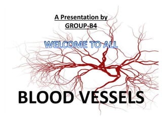
- 1. A Presentation by GROUP-B4 BLOOD VESSELS
- 2. BLOOD VESSEL: The blood vessels are the part of the circulatory system, and microcirculation, that transports blood throughout the whole body.
- 3. TYPES OF BLOOD VESSELS: There are three major types of blood vessels. They are as follows: (1)Arteries: The arteries carry the blood away from the heart. They are thick walled. (2)Capillaries: Capillaries are narrow- diameter tubes that can fit red blood cells in single-file lines and are the sites for the exchange of nutrients, waste, and oxygen with tissues at the cellular level. (3)Veins: The veins carry blood from the capillaries back toward the heart. They are thin walled and have valve to prevent the backflow of blood.
- 4. STRUCTURE: • The arteries and veins have three layers. The middle layer is thicker in the arteries than in the veins. • The inner layer, Tunica intima is the thinnest layer. It is a layer of simple squamous epithelium glued by a polysaccharide intercellular matrix and surrounded by a thin layer of subendothelial connective tissue. • The middle layer Tunica media is the thickest layer in arteries. It consists of elastic fiber, connective tissue, polysaccharide and elastic lamina. Veins only have internal elastic lamina. • The outer layer is Tunica adventitia and is the thickest layer in veins. • Capillaries consists of little more than a layer of endothelium and occasional connective tissue.
- 6. FUNCTION: A blood vessel’s main function is to transport blood around the body. Blood vessels also play a role in controlling blood pressure. Blood vessels are found throughout the body.
- 7. COMPERATIVE STUDY OF BLOOD VESSELS Pieces Arterial System:- Carry oxygenated Blood. Have thick and more elastic made up of Tunica intima, Tunica Media, Tunica externa Efferent Branchial Arteries:- Collects The blood from Capillaries of gill lamellae. Afferent Branchial Arteries Supply arterial branches to the anterior and posterior gill lamellae. Amphibians Arteries:- Carry oxygenated Blood. Have thick and more elastic walls Made up of three concentric layers ie Tunica intima, Tunica Media, Tunica externa. Arterial System can be divided in to:- Carotid arch:- Lower jaw and tongue , Orbit and brain Systemic arch:- Oesophageal, occipitovertebral, subclavian(Forelimbs) Pulmo-cutaneous arch:- to lung and skin and buccal cavity
- 9. Pieces and Amphibia Cont.. Capillaries Their walls are very thin. Only tunica intima is present. Exchange of foods, gases, waste takes place between blood and tissue Veins Carry deoxygenated blood Have thin, fibrous and less elastic walls, than arteries provided with valves. Venous system can be divided into Anterior Cardinal system Posterior Cardinal System Hepatic Portal System Cutaneous System. Capillaries Found abundantly in excessive metabolism sites. Their walls are very thin. Only tunica intima is present. Exchange of foods, gases, waste takes place between blood and tissue Veins Carry deoxygenated blood Have thin, fibrous and less elastic walls, than arteries provided with valves. Venous System can be divided into Pulmonary Veins Caval veins Renal portal vein Hepatic portal
- 10. Comparative Study of Blood Vessels of Reptiles and Aves Arterial system Three aortic arches (1 pulmonary 2 systemic) 1. One Pulmonary arch Lies ventrally arises from the right ventral side of the ventricle . Right PA goes to right lung and left PA goes to left lung. 1. Two Systemic arch Both the systemic arches arise directly from the cavum dorsale of the ventricle carrying oxygenated blood. Right and left communicates each other by Foramen of Panizzae. Arterial system Pulmonary aorta and systemic arch 1. Pulmonary aorta Arise from a single pulmonary aorta which passes ventral to aortic arch bifurcates. Each of these enters into lung 2. Aortic arch (systemic):- Arises from left ventricle. Left aortic arch is absent in birds Coronary arteries arise from right aortic arch
- 11. Reptile Some Extensions Common carotid arteries Common subclavian artery Anterior esophageal artery Dorsal Aorta is formed by union of right and left systemic arches , extends backwards mid dorsal line beneath the vertebral column it give rise to: Post esophageal, Gastric, Coeliac, hepatic, rectal iliac, caudal arteries etc Aves Simply the arterial system in Aves is similar that of reptiles.
- 12. Reptiles Birds
- 13. Reptiles VS Birds Venous System in Reptiles 1. Pulmonary:- brings oxygenated blood from lungs to arteries 2. Precaval :- Drains blood from Head, Neck, Shoulders, Forearm, and Thoracic wall Histologically each precaval is formed by four veins Jugular, Subclavian, Intercostal, laryngo-tracheal. 3. Postcaval :- collects blood from posterior body parts i.e. Kidneys Gonads and Liver 4.Hepatic Portal :- collects blood from alimentary canals, formed by hepatic portal vein and Porto- abdominal vein 5. Renal Portal:- Cloacal and rectal veins meet afferent renal veins Blood from hindlimb is collected bi internal and external iliac. Venous System in Birds 1. Pulmonary veins:- brings oxygenated blood from lungs to arteries 2. Precaval veins:- similar in lizard 3. Postcaval veins:-Coccygeo- Mesentric vein is the characteristic of Bird. Receive blood from cloaca and rectum. 4. Hepatic portal:- collects blood from Rectum, ileum, duodenum and gizzard. Coccygeo-Mesentric vein connects the two portal systems 5. Renal Portal: it is greatly reduced in the pigeon. Includes two Hypogastric or renal portal veins.
- 14. Mammals • Heart is four chambered. Blood circulation is double, closed and complete. • Histologically there are three distinct layers or tunics in the blood vessels of mammals:-, that form the walls of blood vessels.
- 15. Contd. • Tunica media is composed of smooth muscle, • Tunica externa is connective tissue (collagen and elastic fibers). • The elastic, connective tissue stretches and supports the blood vessels, while the smooth muscle layer helps regulate blood flow by altering vascular resistance through vasoconstriction and vasodilation. • Unlike veins and arteries, capillaries have only one tunic; this single layer of cells is the location of diffusion of oxygen and carbon dioxide between the endothelial cells and red blood cells, as well as the exchange site via endocytosis and exocytosis.
- 16. Veins ans Arteries in Mammals