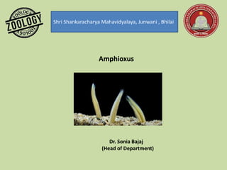
2. Lancelets/Amphioxus
Introduction
• Lancelets include 23 species of marine animals that make up the subphylum
Cephalochordata. Lancelets are also called amphioxus, which translates to
“both ends pointed,” because of the shape of their elongated bodies.
• The lancelets are capable of swimming, however, they spend most of their
time buried in sandy, shallow regions of the ocean. Adult lancelets retain the
pharyngeal slits, notochord, dorsal nerve cord, and post-anal tail, which are all
characteristic of chordates. Although lancelets have a brain-like swell at the
end of the notochord in the head region, it is not very highly developed.
• Habit and Habitat -Amphioxus is a fish like Marine chordate. Found in Shallow
water of sandy coast in tropical and temperate region.
• Burrowing and Nocturnal.
3.
4.
5.
6.
7. Wheel organ in amphioxus is part of oral hood.
• The wheel organ is located on the sides of the oral cavity of the amphioxus. They lines
the oral epithelium and contains large densed vesicles. They are made of the specialized
epithelium and contains the heterochromatin nuclei. They also contain microvilli in their
structure.
• They are the main structures involved in intake of food. The ‘wheel organ’ is present on
the sides of the oral cavity of amphioxus.
• The epithelial lining of the cavity forms ‘six to eight pairs’ of finger like folds which is
made of ciliated groove bound by a ciliated ridge.
• These structures are collectively called the wheel organ or the muller’s organs.
• As water enter the mouth of amphioxus, the wheel organ filters out food from the
water.
8. • These structures are collectively called the wheel organ or the muller’s organs.
• As water enter the mouth of amphioxus, the wheel organ filters out food from
the water
Muller Organ:
The epithelium (ectoderm) of the oral hood is projected to form six to eight pairs of
finger-like folds, each having a ciliated groove borded by ciliated ridge. These folds are
collectively known as wheel organ or Muller’s organ or rotatory organ. Outer of these,
the mid-dorsal groove is the largest and ends in a pit or depression in the roof of
buccal cavity. This is called Hatscheks groove and Hatscheks pit respectively. Both are
ciliated and glandular, and secretes mucus.
9. Physiology of Digestion:
Food:
Branchiostoma feeds on protozoans, diatoms, algae, desmids and other
organic particles suspended in sea water.
Feeding:
Branchiostoma is a ciliary feeder. Action of cilia of the pharynx causes a
current of water containing food. The current of water enters the mouth
and goes to the pharynx from where it passes through gill-clefts into an
atrial cavity, and then it goes out through an aperture of the atrial cavity
called atriopore. In feeding the oral hood is extended and oral cirri are
turned inwards, they prevent sand from entering the mouth.
Rotary movements of cilia of wheel organ direct water towards the
pharynx, but some food particles fall out of the water current, they are
mixed with mucus secreted by groove of Hatschek and passed back into
the pharynx. When the main water current passes through the
enterostome into the pharynx, mainly due to lateral cilia of gill-bars,
the suspended food particles fall on the gill-bars where they get
entangled in mucus secreted mainly by the endostyle and to some
extent by the pharyngeal epithehum.
Mucus secreted by endostyle is transferred to the lateral wall of the
pharynx by its lateral rows of cilia; the median row of long endostylar
cilia keeps supplying mucus to the lateral rows of cilia of the endostyle.
These cilia lash outwards throwing the muccus on the lateral wall of
pharynx.
10. The frontal cilia of gill- bars beat along the length of the bars in such a way that they
propel mucus from the lateral to the mid- dorsal side of the pharynx. In this way a
stream of mucus with food particles passes from the lower- side into epipharyngeal
groove. The cilia of epipharyngeal groove beat backwards moving the cord of mucus
and food into the oesophagus.
The peripharyngeal bands also collect and pass to the epipharyngeal groove any food
particles which fall out of the water current at the extreme anterior end of the
pharynx (where no gill-clefts are present). Food and mucus are not transferred from
the endostyle into the peri-pharyngeal bands, as is often stated.
The cord of food and mucus passes down the gut by action of cilia. It is moved from
the oesophagus into the midgut where a lateral tract of cilia directs it into the midgut
diverticulum, from here the cord is returned again into the midgut. The iliocolic or
iliocolonic ring rotates the cord of food causing the food and enzymes to mix, and
then the cord of food is moved into the hindgut.
Digestion:
Digestive enzymes are secreted by the epithelial cells of the gut and midgut diverticulum.
They are mixed with food as it passes along. Digestion starts in the midgut and is continued
in the hind-gut.
Besides this extracellular digestion, intracellular digestion also occurs in which food particles
are taken into the epithelial cells of the hindgut and digested there. Some papillae on the
floor of the atrium contain phagocytic cells which engulf food particles which may pass into
the atrial cavity. Absorption of digested food takes place mostly in the hindgut and to lesser
extent in the midgut.