
Recommended
More Related Content
What's hot
What's hot (20)
Similar to Blood circulatory system of earthworm
Similar to Blood circulatory system of earthworm (20)
More from SoniaBajaj10
More from SoniaBajaj10 (20)
Recently uploaded
Recently uploaded (20)
Blood circulatory system of earthworm
- 1. Shri Shankaracharya Mahavidyalaya, Junwani , Bhilai Dr. Sonia Bajaj (Head of Department) Circulatory System of Earthworm
- 2. Introduction • The blood vascular system of Pheretima is of closed type. • It consists of the blood vessels, hearts, loops, capillaries and the blood glands. Blood: • The blood of Pheretima is red colored due to the presence of a respiratory pigment hemoglobin in it. • The hemoglobin is not contained in the corpuscles like the vertebrates but it is found dissolved in the plasma. The plasma also contains many corpuscles which are colorless and nucleated. Blood Vessels: • The blood vessels are of two types collecting blood vessels and distributing blood vessels which are closed tubes with definite walls and they break up into capillaries to ramify in the different parts of the body. • The arrangement of blood vessels in the anterior thirteen segments is somewhat different from that behind the thirteen segment, i.e., in the region of intestine.
- 3. Therefore, for convenience the blood vessels can be described under the following two heads: A. Blood vessels and their arrangement in the segments behind 13th, i.e., intestinal region. B. Blood vessels and their arrangement in the anterior thirteen segments. A.Blood Vessels and their Arrangement in the Segments behind 13th, i.e., Intestinal Region: The blood vessels of this region include: 1. Median longitudinal blood vessels; 2. The intestinal blood plexus; 3. The commissural vessel; 4. The integumentary vessel; and 5. The nephridial vessels.
- 4. 1. Median Longitudinal Blood Vessels: (i) Dorsal Vessel: It runs mid-dorsally above the alimentary canal from the posterior to the anterior end of the body. It is the thickest vessel with contractile muscular walls visible from outside as a dark line through the thin and semi transparent body wall. The direction of flow of blood in this vessel remains from behind to forward (from posterior to anterior). It is contractile and pulsates rhythmically to force the blood from posterior to anterior side. In each segment it has a pair of valves internally which check the backward flow of blood. It is the main collecting vessel behind the 14th segment, but in front it distributes the blood. From the posterior segment up to the 14th segment it receives two pairs of dorso intestinal vessels from the intestine in each segment and a pair of commissural vessels from the sub-neural vessel. The commissural vessels form a loop behind each septum and they receive blood from the body wall, nephridia and prostrate glands. The commissural vessels also give out blood in each segment through a septo intestinal branch to the intestine.
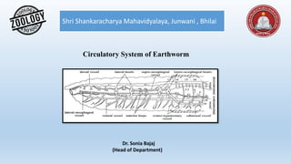
- 5. Fig- Pheritima –The Blood Vascular system in the anterior 13 segment in the body.
- 6. Fig- Pheritima –A part of body wall on the left side has been cut and reflected in order to blood vessels in position.
- 7. (ii) Ventral Vessel: • It is also a long vessel and runs ventrally below the alimentary canal and above the ventral nerve cord from second segment up to the last segment of the body. • It is thin- walled without muscles and valves. The direction of flow of blood in this vessel remains from anterior to the posterior side or from in front to backwards. It is a distributing vessel. It gives out a pair of ventrotegumentary vessels, one on each side in front of the septum in all segments. • The ventrotegumentary vessels run upwards along the body wall and supply blood to the body wall, integumentary nephridia, septal nephridia, gonads, seminal vesicles and spermathecae. • The ventral vessel also gives out a ventrointestinal vessel in each segment behind the 13th segment, these take blood to the lower part of the intestine. The branches in intestine form blood plexuses consisting of two networks in the intestinal wall. (iii) Sub-neural Vessel: • It is also a long but thin vessel extending from anterior 14th segment up to the last segment situated mid- ventrally below the ventral nerve cord. It is without muscular walls and internal valves. The direction of flow of blood in this vessel remains from anterior to posterior side and it is mainly a collecting vessel.
- 8. 2. Intestinal Blood Plexus: The intestine of Pheretima is richly supplied with blood capillaries which form a close network. The intestinal blood plexus consists of a close network of capillaries in the wall of intestine.In fact, there are two capillary networks in the intestine: (i) The external and (ii) The internal. The capillary network which is present at the outer surface of intestine is known as external plexus which receives blood from the ventral vessel through ventro intestinal and passes it on to the internal plexus. The capillary network which is present between the circular muscle layer of intestine and its internal epithelial lining is known as internal plexus which serves to absorb the nutrients from the gut and is connected with dorsal blood vessel through the dorso intestinal. 3. Commissural Vessels: These vessels connect the dorsal and sub-neural vessels. These vessels receive blood from nephridia, body wall and reproductive organs through capillaries and then they send it to dorsal blood vessel.
- 9. 4. Integumentary Vessels: These vessels coming from ventral vessels supply the blood to integument for aeration and the aerated blood is collected by numerous capillaries of commissural vessel in each segment. Thus, there is a close parallelism between venous and arterial capillaries throughout the body wall. 5. Nephridial Vessels: These vessels originate from the ventrotegumentary vessels of ventral vessel and supply the blood to the nephridia. B. Blood Vessels and their Arrangement in the Anterior 13 Segments: The blood vascular system in the first thirteen segments is modified considerably and differs markedly from that of the intestinal region. It consists of the following: 1. Median longitudinal vessels; 2. Hearts and anterior loops; 3. Blood vessels of the gut. The function of collecting blood from the anterior region of the gut is taken over by a new vessel supra- oesophageal, while the blood from the peripheral structures is collected by the right and left lateral oesophageal.
- 10. 1. Median Longitudinal Blood Vessels: (i) Dorsal vessel: This blood vessel becomes the distributing vessel in these segments instead of collecting vessel. Structurally, it retains its original identity as it was in the posterior segments. But is has neither dorsointestinals nor commissural vessels opening into it. It sends out all the collected blood from the posterior region of the body into hearts and the anterior region of the gut where it divides into three branches distributed over the pharyngeal bulb and the roof of the buccal chamber. However, it supplies to stomach, gizzard, oesophagus, pharynx and other related parts. (ii) Ventral vessel: This blood vessel remains distributing in these segments also but extends only up to the second segment. The ventrointestinals are absent, hence, it does not supply to the alimentary canal in this region. However, the ventrotegumentary vessels, a pair in each segment, supply blood to the integument, nephridia, septa and reproductive organs. (iii) Supra-oesophageal vessel: It is the shortest longitudinal vessel extending from 9th to 13th segment situated above the stomach. It receives blood from the lateral oesophageals by two pairs of anterior loops that encircle the stomach in the 10th and 11th segments. It sends its collected blood by the latero-oesophageal hearts in segments 12th and 13th to the ventral vessel.
- 11. (iv) Lateral esophageal: In fact, the sub-neural vessel bifurcates in the 14th segment to form two lateral oesophageals. These vessels are considerably thick and situated along the ventrolateral margins of alimentary canal in the anterior thirteen segments. These vessels are closely attached to the wall of the stomach from 10th to 13th segments and communicate with the ring vessels. But in the region of gizzard and further forwards, they remain free from the wall of the alimentary canal even though they receive branches from it in each segment. These vessels receive a pair of branches in each segment bringing blood from the body wall and the septum. They also collect blood from the reproductive organs and nephridia, thus, functioning like the sub-neural and commissural vessels of the posterior region, i.e., these are collecting vessels. 2. Hearts and Anterior Loops: In the posterior segments behind 13th the dorsal and ventral blood vessels have no direct connections but anteriorly both these vessels are connected together by 4 pairs of pulsatile hearts which are neurogenic, i.e., the heart beat originates in the nerve cells of the hearts. The hearts are contractile and encircle the alimentary canal, they are in the segments 7th, 9th, 12th and 13th. Fig- Pheritima –A- Lateral Heart ,B – Latero-oesophageal heart.
- 12. The hearts of segments 12th and 13th are joined above to both the dorsal and the oesophageal vessels, these are called latero-oesophageal hearts. These hearts have thick muscular walls and a pair of valves at each junction with the dorsal vessels and supra-oesophageal vessel, and another pair of valves at the ventral end. These valves allow blood to flow downwards only. The other hearts of the segment 7th and 9th are called lateral hearts. These connect the dorsal vessels to the ventral vessel. They have four pairs of valves which allow blood to flow only downwards. Besides four pairs of hearts there are two pairs of loop-like vessels in the 10th and 11th segments which connect the supra-oesophageal with the lateral oesophageals. These vessels are neither muscular nor pulsatile and are called anterior loops. These are devoid of valves. The blood from lateral oesophageals flows through these loops into supra-oesophageal which sends all its collected blood into ventral vessel through the hearts of 12th and 13th segments. 3. Blood Vessels of the Gut: On either side of stomach are situated ring-like vessels which connect the supra-oesophageal and lateral- oesophageal vessels. Through these vessels blood flows upwards from the lateral- oesophageals into the supra- oesophageal. Buccal cavity, pharynx and gizzard receive their blood supply from dorsal blood vessel directly.
- 13. Circulation of Blood: The blood collected by the dorsal vessel through the dorsointestinals, blood plexuses of intestine, and commissural is given out partly to the anterior alimentary canal, but mainly through the hearts to the ventral vessel. In the ventral vessel the blood flows forwards to the anterior region in front of the hearts, but the main portion of blood flows backwards, this is distributed through ventrotegumentaries to the body wall and the organs in the coelom, and through the ventrointestinal vessels to the alimentary canal. In other words all parts receive blood from the ventral vessel. From the ventral body wall blood is collected by the sub-neural which also receives some blood through the lateral-oesophageal from the anterior region. This blood passes from the sub-neural to the dorsal vessel through the commissurals. The lateral- oesophageals also send blood through the anterior loops to the supra- oesophageal vessel which then passes it through the latero-oesophageal hearts to the ventral vessel. Fig- Pheritima –A- T.S. through latero- oesophageal heart. B –T.S. through a segment on the left and through a segment on the right.
- 14. Fig- The course of circulation of blood in Pheretima
- 15. Functions: The blood distributes digested food to various body regions, and it collects waste substances like nitrogenous waste and Co2 which are given up to nephridia, skin and the coeiomic fluid. Respiration in almost all aquatic and terrestrial oligochaetes takes place by diffusion of gases through the integument which in larger forms contains a capillary network in the outer epidermal layer. In terrestrial species the film of moisture necessary for diffusion of gases is supplied by mucous glands, coeiomic fluid, and nephridial excretions. The hemoglobin of plasma extracts O2 from the capillaries of the skin, but there must be a moist skin where O2 can combine with hemoglobin to be transported by the blood. Hemoglobin is an efficient pigment and it can take up O2 either from the surrounding air or from an environment comparatively deficient in oxygen. Hence, earthworms can live in well aerated water and are not drowned. They can also live for several hours without O2, in this condition they probably carry on anaerobic respiration. Blood Glands: In the segments 4th, 5th and 6th above the pharyngeal mass are several groups of small rounded follicles of red colour, which are called blood glands. The follicles have a syncytial wall enclosing a capsule containing a mass of loose cells. The blood glands are connected with pharyngeal nephridia and with salivary glands. These glands manufacture blood corpuscles and hemoglobin. These glands are probably also excretory.
- 16. References- Modern text book – R.L.Kotpal Jantu Vigyan- S.M. Sexsena Jantu Vigyan- Dr.H.N. Baijal