
Recommended
More Related Content
What's hot
What's hot (20)
Similar to Placentation in mammal,classification of placenta and function
Similar to Placentation in mammal,classification of placenta and function (20)
More from SoniaBajaj10
More from SoniaBajaj10 (20)
Recently uploaded
Recently uploaded (20)
Placentation in mammal,classification of placenta and function
- 1. Shri Shankaracharya Mahavidyalaya,Junwani,Bhilai Placentation in mammal Dr. Sonia Bajaj (Head of Department)
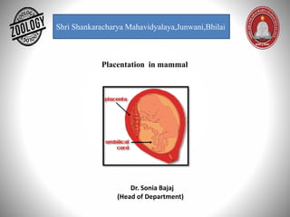
- 2. Placenta- The placenta is defined as an organ that develops during pregnancy in mammals. The placenta provides oxygen and nutrients to the growing fetus in the uterus of the mother. Circular disc with a diameter of 15-20 cm and thickness of about 2.5 cm at its center. • It thins off towards the edge. • It feels spongy and weight about 500 gm. • Proportion to the weight of the baby being roughly 1:6 at term and occupies about 30% of the uterine wall. • It presents two surfaces, fetal and maternal, and a peripheral margin. Fetal surface • Covered by the smooth and glistening amnion with the umbilical cord attached at or near its center. • Branches of the umbilical vessels are visible beneath the amnion. • The amnion can be peeled off from the underlying chorion except at the insertion of the cord. • At term, about four-fifths of the placenta is of fetal origin. Maternal surface- Rough and spongy Dull red colour. • A thin greyish, somewhat shaggy layer which is remnant of decidua basalis and has come away wit the placenta, may be visible. • 15-20 convex polygonal areas known as lobes cotyledons which are limited by fissures.
- 3. The decidua is the uterine lining (endometrium) during a pregnancy, which forms the maternal part of the placenta. It is formed under the influence of progesterone and forms highly characteristic cells. Structure- The part of the decidua that interacts with the trophoblast is the decidua basalis (also called decidua placentalis), while the decidua capsularis grows over the embryo on the luminal side, enclosing it into the endometrium. The remainder of the decidua is termed the decidua parietalis or deciduavera, and it will fuse with the decidua capsularis by the fourth month of gestation. Three morphologically distinct layers of the decidua basalis can then be described: Compact outer layer (stratum compactum) Intermediate layer (stratum spongiosum) Boundary layer adjacent to the myometrium (stratum basalis)
- 4. Within the decidua, occasional fibrinoid deposits form where the syncytiotrophoblast is damaged. The region of fibrinoid deposition where trophoblasts meet the compact portion of the decidua basalis is called Rohr's layer, while the fibrinoid deposits that occur between the compact and spongy layer of the decidua basalis is termed Nitabuch's layer. This layer is absent in placenta accreta. The decidua has a histologically- distinct appearance, displaying large polygonal decidual cells in the stroma. These are enlarged endometrial stromal cells, which resemble epithelium. Decidualization includes the process of differentiation of the spindle-shape stromal fibroblasts into the plump secretory decidual cells, which create a pericellular extracellular matrix rich in fibronectin and laminin (similar to epithelial cells). Vascularity, as well as vascular permeability, is enhanced in the decidualizing endometrium. Its leukocyte population is distinct, with the presence of large endometrial granular leukocytes being predominant, while polynuclear leukocytes and B cells are scant. The large granular lymphocytes (CD56 bright) are called "uterine NK cells" or "UNK cell in human.
- 5. Development- After ovulation in placental mammals, the endometrial lining becomes hypertrophic and vascular under the influence of Estrogens and progesterone. In animals exhibiting hemochorial placentation, the endometrium undergoes decidualization following implantation. If implantation does not occur, the secretory lining will be absorbed estrous cycle or shed (menstrual cycle). The decidua is shed with the placenta during parturition.
- 7. Types of Placenta A. BEHAVIOR DURING PARTURITION- 1.Deciduate type Placenta: The allantochorianic villi penetrate into uterine villi. They are intimately fused. Hence at the time of birth, the uterus is damaged. Bleeding occurs, the utrine wall enters into formation of placenta is called deciduas. 2. Non deciduate type placenta: The chorianic villi are simple projections, they lie in contact with uterus. They have a loose contact. There is no fusion. At the time of birth of embryo uterus is not damaged.
- 8. B. ON THE BASIS OF DISTRIBUTION OF VILLI:- According to the distribution of villi five kinds of placenta are seen. 1.Diffused type placenta: The villi are uniformly distributed on the surface of blastocyst, except at the extreme ends. Ex: Horse, pig. 2.Cotyledonary placenta: The villi are arranged in groups. Each group is called cotyledon. Each cotyledon fits into caruncles for uterus. Ex: Sheep, Cow, Deer. 3. Intermediate type Placenta: It is a rare type, it shows free villi on cotyledons. Hence it is called intermediate type placenta. Ex: Giraffe In these three types of placenta during perturition the foetus will not damage uterus. 4. Zonary placenta: The placenta takes the form of a complete or incomplete band of tissue surrounding the fetus.. Ex: Cat, Dog, Carnivores. 5. Discoid type placenta : A single placenta is formed and is discoid in shape. When the embryo Is growing It move saway from uterus hence the with look like a disc. Ex: Rabbit
- 9. C. BASING ON HISTOLOGY CONNECTION: According to number of layers of cells present between foetus and uterus blood supply the placenta Is classified into five types. a) Epithelio chorion placenta: The foetal chorion Is In contact with epithelium of the uterus hence it is called epithello chorion placenta. In between foetal, maternal parts six layers are present. Ex: Pig, Horse • Endothelium of mother blood vessel. • Maternal syndesmose connective tissue. • Epitheliurn of mother • Chorion of foetus. • Foetus connective tissue (syndesmose) • Endothellum of foetal blood vessel. If all the six layers are present the placenta is called epithellochorlal placenta.
- 10. b) Syndeumose chorial placenta: The allanto-chorianic with will pierce into the uterus of the mother, the chorion will come in contact with syndesmose of mother’s uterus. Hence it is called syndesmose chorial. Ex: Sheep, Cow. c) Endotheliochorial placenta: The chàrion of the foetus will come in contact with the endotheli of mother ‘s uterus, hence it is called endothelio-chorial placenta. Ex: Dog, Carnivores. d) Hemo-chorial placenta: The placental connections are more intimate. The chorion of foetus will float In the blood pools of mother’s uterus. Hence It Is called haemochorIal placenta. Ex: Bat, Man, Primates e) Hemo-endothelial placenta: Hence guinea-pig will float In mother’s blood. Hence it called hemo endothelial placenta. Ex: Rat, Rabbit,
- 11. D. LAYER THAT FROM YOLK SAC a) Yolk sac placenta- consisting of the vascular embryonic yolk sac wall intimately associated with the vascular maternal uterine or oviducal wall, and serving to nourish the embryo. Ex. Metatheria( Marsupials) b) Allantois Placenta -the allantois is intimately associated with the chorion, contributing blood vessels to that structure as it forms—in conjunction with the endometrium. Ex. Eutharian (Apis) c) Chorionic Placenta- The fetal part of the placenta is known as the chorion. The maternal component of the placenta is known as the decidua basalis. Oxygen and nutrients in the maternal blood in the intervillous spaces diffuse through the walls of the villi and enter the fetal capillaries. Ex. Primates
- 12. FUNCTIONS OF PLACENTA: 1.Placenta will form a physiological barrier between mother and foetus. It will possess foetal and maternal blood mixing. 2.Placenta allows the diffusion of monosaccharaides, amino adds, hormones, vitamins, oxygen, .carbon dioxide, water and other waste materials, because of this it supplies food, oxygen to foetus. 3.It works as an excretory organ of foetus. It releases the nitrogenous waste materials Into mother blood. 4.It works as an endocrine gland. It will secretes lactogen ,progesterone,etc. hormones. 5.The placenta will manufacture fructose from glucose.
- 13. References- Modern text book – R.L.Kotpal Jantu Vigyan- S.M. Sexsena Jantu Vigyan- Dr. H.N. Baijal