
Recommended
More Related Content
What's hot
What's hot (20)
Similar to Extra embryonic membrane in chick, Types, Developments, Functions
Similar to Extra embryonic membrane in chick, Types, Developments, Functions (20)
More from SoniaBajaj10
More from SoniaBajaj10 (20)
Recently uploaded
Recently uploaded (20)
Extra embryonic membrane in chick, Types, Developments, Functions
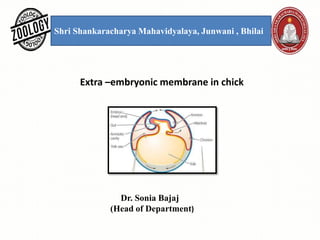
- 1. Shri Shankaracharya Mahavidyalaya, Junwani , Bhilai Extra –embryonic membrane in chick Dr. Sonia Bajaj (Head of Department)
- 2. Extra embryonic membrane: These are the membranes which do not form any part of the embryo proper but performs various functions which assist in the development of the embryo. These are discarded at the time of hatching. These membranes formed outside the embryo . Amniotes: These are the vertebrates group whose eggs contain extra embryonic membranes for protecting the embryo. They lay eggs on the land. Therefore ,in chick and other amniotes the following extra embryonic membrane develop in them for the protection, respiration and nutrition of the embryo. Example: Reptiles, Birds and Mammals. An amniotes: These are the vertebrates group whose eggs do not contain extra embryonic membranes during embryonic development. They lay eggs in the water. Example: Fish, Amphibian.
- 3. Extra embryonic membranes in chick In chick, the presence of an enormous amount of yolk and embryonic life to be spent within a shell is correlated with the development of extra-embryonic membranes. Original blastoderm is a small disc, which spreads by peripheral expansion and eventually covers the entire surface of the egg. But only the most central region is directly connected with the formation of the embryo proper. All the remainder of the blastoderm is extra-embryonic. There are four types of extra embryonic membranes in birds- 1. Yolk sac. 2. Amnion. 3. Chorion. 4. Allantois
- 4. Extra embryonic membrane: These are the membranes which do not form any part of the embryo proper but performs various functions which assist in the development of the embryo. These are discarded at the time of hatching. These membranes formed outside the embryo.
- 5. 1. Yolk sac: It is the first structure to develop among the embryonic membrane. In 16 hours stag, the embryonic entrones is situated over the yolk in the form of a flat and circular space.` • Formed from splanchnopleure (inner endoderm and outer mesoderm). • Well developed in the animals with megalecithal egg as reptiles, birds and Prototheria. • Formed completely on the 9 th day of incubation. In human it is vestigial. • The yolk –sac gradually grow over yolk to completely surround it. • The yolk is used up with the increase in size of the embryo and the yolk –sac gradually reduce in size.
- 6. Functions of Yolk sac: 1.It surrounds the yolk. Its main function is in digestion. It serve as extra embryonic gut. 2. It help in digestion of yolk and transfer the digested material to the developing embryo. 3. First respiratory organ in the embryo. 4. Form yolk sac placenta in the marsupials.
- 7. Development of yolk sac- In reptiles and birds, the somatopleure and splanchnopleure develop from the periphery of the blast disc. These usually spread peripherally over the yolk mass. Soon afterwards, the embryo undergoes series of folds, which appear all around the body of the embryo. These folds are termed as the body folds. The extra embryonic splanchnopleure (splanchnic mesoderm + endoderm) constantly spreads over the yolk mass and eventually yolk sac encloses the mass of yolk in a large measure. The yolk sac, however, not surrounds the yolk fully. A small passage is left on the ventral side for the embryo to absorb the remains of albumen at a later stage. Immediately with the formation of the yolk sac, the intra embryonic splanchnopleure is subjected to fold resembling with the more superficial body folds i.e., the intra embryonic folds. The intra embryonic folds give rise to walled digestive tract, or gut, in the body of the embryo. The middle of the embryonic gut remains open to the yolk beneath. At this level, yolk sac is connected to the digestive tract by a constricted yolk stalk.
- 8. 2. Amnion: Formed of somatopleure (inner ectoderm and outer mesoderm). It surrounds the embryo. In 13 somite stage of the embryonic development of chick, It appears after 30 hours of incubation. A amniotic cavity is present between the amnion membrane and the embryo, which filled with the amniotic fluid. In this fluid filled cavity embryo floats.
- 9. Function of Amnion: The fluid filled in the amniotic cavity forms a sort of pool for the embryo and therefore, performs the following functions- 1. It prevents the embryo from drying. 2. Protection of the embryo from the mechanical injury and desiccation. 3. It protects the embryo from external shock. 4. It prevents the embryo from sticking to the shell or embryonic membranes. 5. It also helps in the absorption of the albumen. 6. The amniotic fluid allows freedom of motion to the embryo . 7. It is working as a barrier of outer organisms. 8. In most mammals of a part of the chorion forms finger-like out-growths called chorionic villi. That penetrates into corresponding crypts or depressions in the wall of the uterus. It is at this site where exchange of substances occurs found in the inner cell mass begin to spread along the inner surface on the trophoblast and surround the internal cavity of the blastocyte in the same way as the endodermal cell in reptiles and birds surrounded the mass of uncleaved egg.
- 10. 3. Chorion: Formed of somatopleure (outer ectoderm and inner mesoderm). It forms the outermost boundary. Space between amnion and chorion is called chorionic cavity which further provides protection to the embryo. Function of chorion: 1. It is outer layer, therefore, it is the place where exchange of substance occurs between embryonic tissue in the maternal environment. 2. It acts as a respiratory organ and is in contact with the allantois. 3. In most mammals it develops villi which penetrate into the tissue of uterus wall and increase the absorptive area of chorion. 4. Blood vessels are developing after the formation of allantochorion and this is working as a lung. It takes O2, from atmosphere and removes CO2, to the atmosphere. 5. It provides calcium for skeleton of developing embryo by transportation of Ca by shell.
- 11. Development of Amnion & Chorion- The amnion and chorion are developed jointly as upward projecting folds, the amniotic folds of the extra embryonic somatopleure. The amniotic folds are named according to their location. They are amniotic head fold and amniotic tail fold. The amniotic fold first appears as a transverse fold in front of the head. It is called amniotic head fold. It grows upwards and then bends backwards, over the anterior end of the head and covers it as with a hood. Another fold develops behind the embryo. It is termed as amniotic tail fold. All these folds finally cover an embryo in two sheets of somatopleure. The inner somatopleuric sheet becomes the amnion and the outer, the chorion. The amnion consists of a layer of extra embryonic ectoderm on the inside and a layer of extra embryonic somatic mesoderm on the outside whereas, the chorion is made up of a layer of extra embryonic ectoderm on the outside and a layer of extra embryonic somatic mesoderm on the inside. The cavity between the amnion and the embryo is called the amniotic cavity. In between, the amnion and the chorion is the chorionic cavity or extra embryonic coelom.
- 12. 4. Allantois: Formation of Allantois membrane is starting in the 29 somatic stage of after 60 hours incubation. Formed of splanchnopleure (inner endoderm and outer splanchnic mesoderm). Connected with the hindgut of the embryo. Develops on the third day of incubation from the floor of the hind gut as a outgrowth.
- 13. Development of allantois -The allantois arises as a ventral outgrowth of the splanchnopleure from the hindgut on the third day of incubation. It slowly enlarges as holosac and expands inside exocoel. Its walls are formed of an outer splanchnic mesoderm and inner endoderm. The proximal part of the allantois forms a slender neck or the allantoic stalk with which it remains connected with the hindgut of the embryo. The distal part of the allantois expands and penetrates between the amnion and the yolk sac on one side and the chorion on the other side. By the middle of the incubation period, the allantois spreads all around the egg underneath the chorion. The mesoderm on the external surface of the allantois fuses with that of the chorion forming a conjoined chorio-allantoic membrane.
- 14. Function of allantois: The allontois is highly muscular from the beginning, As a result of the formation of the allanto-chorion the blood vessels become dispersed over the inner surface of the porous shell. Thus, it performs the following functions – 1. It acts as a respiratory organ. It is working as a lung and provides atmospheric O2, to the embryo by porous shell. It is working of the exchange of gases because it is situated near the porous shell. 2. It collects the embryonic villi and working as a excretory organ (urinary bladder) of embryo. 3. Also helps in digestion and nutrition from albumen and calcium of the shell. 4. It helps in removal of waste products, e.g, CO2, urea, etc. of metabolism from the embryo. 5.
- 15. Importance of foetal membranes: • The presence of amnion, allantois and chorion is an adaptation for terrestrial life of animals. Their presence enables the embryo and adult to live on land. • The presence of yolk-sac is for different purposes. It acts as a digestive and absorptive surface through which the embryo gets the yolk.
- 16. References- Modern text book – R.L.Kotpal Jantu Vigyan- S.M. Sexsena Jantu Vigyan- Dr.H.N. Baijal