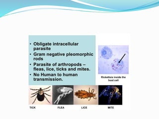
By- Sanju Sah
St. Xavier’s college, Maitighar, Kathmandu
Department of Microbiology
 Rickettsiae are small bacteria of genera Rickettsia,
orienta, Coxiella and Ehrlichia
 They are obligate intracellular parasite
 Primarily a parasite of arthropods such as fleas, lice,
ticks and mites in which organism propagate without
producing any disese
 Responsible for ranges of disease, typhus fever, spotted
fever and scrub typhus
 Typically manifest by fever, rashes and vasculitis except
Qfever, and ehrlichioses
Morphological chracteristics
 Rickettsia are pleomorphic coccobacilli, appearing
either as short rods (0.3umx1-2um) or as cocci) or as
cocci(0.3 um in diameter)
 They are non motile, non capsulated, and gram
negative though stain poorly.
 Readily visible under light microscope when stain with
giemsa, Casteneda, macchiavello, gimnez stain
Typhus fever group
 This consist of epidemic typhus, Brill-Zenser disease
and endemic typhus
 Epidemic typhus-
 The causative agent, R prowazekii, is carried from
man to man by infected louse, Pediculus humanus
corpans, a reserviour host.
 The lice get infected during blood meal on
rickettsiaemic patient.
 The organism multiply in gut and excreted in faeces in
3-5 days.
 When the louse feeds, its human victim scratches the
irritated site of of scalp, that leads to introduction of
infective faeces into the skin puncture
 A macular rash appears 4 to 7 days after the patient
become ill, first on trunk and axillary fold, and later on
spread to extremities
 Towards the second week of illness, the patient
develops cloudy state of conciousness( typhos
meaning smoke or cloud)
 Brill- Zinsser disease-
 People survive a primary infection of louse borne
typhus may develop a relatively milder reactivation of
latent infection many year later. This is referred to as
recrudescent typhus
Endemic Typhus
 Murine typhus (also called endemic typhus) is a form
of typhus transmitted by fleas (Xenopsylla cheopis),
usually on rats.
 Caused by a bacteria called Rickettsia typhi or R
mooseri
 People get sick with murine typhus when infected flea
feces are rubbed into cuts or scrapes in the skin
Rat flea
 Symptoms of murine typhus begin within 2 weeks after
contact with infected fleas. Signs and symptoms may
include:
 Fever and chills, Body aches and muscle pain, Loss of
appetite, Nausea, Vomiting, Stomach pain, Cough
 Rash (typically occurs around day 5 of illness)
 Most people will recover without treatment, but some cases
may be severe.
 When left untreated, severe illness can cause damage to
one or more organs including the liver, kidneys, heart,
lungs, and brain.
Spotted fever
 Rocky mountain Spotted fever/ siberian tick typhus
 These are many clinical similarities among tick borne
rickettsioses
 Patient become ill within 2 weeks of infection. Early
symptoms include fever, severe headache and myalgia,
often accompanied by anorexia, vomiting, abdominal
pain, diarrhoea, photophobia and cough.
 A maculopapular rash usually develop within 3-5 days,
first on extremities rather than trunk.
Rocky mountain spotted fever
Rickettsia rickettsii
Ticks
 Vascular damage in severe cases may result in
haemorrhagic rash, hypovolaemia, hypotensive shock,
non cardiogenic pulmonary edema and impairment of
CNS function.
 An eschar develop at the site of tick bite in all spotted
fever except rocky montain spotted fever

 Rickettsia sibirica is transmitted by ticks
(Dermacentor nuttalli, Dermacentor marginatus and
Haemaphysalis concinna).
 Symptom include fever, maculopapular rash,eschar,
has symptom regional adenopathy,
 Rickettsial pox- similar to other spotted fever
R sibrica
 Rickettsialpox is a mite-borne infectious illness
caused by bacteria of the genus R. akari.
 The bacteria are originally found in mice and cause
mites feeding on the mice (usually the house mouse)
to become infected.
 Humans will get rickettsialpox when receiving a bite
from an infected mite, not from the mice themselves.

Rickettsialpox
cultivation
 Unable to grow in cell free media
 Can be cultivated in hen embryo yolk sac, animals
(mouse, rat, guinea pig) or in cell culture
 When patient blood is inouculated intraperitonially to
guinea pig, it develops fever in animals.
 In case of R mooseri (R typhi) the rickettsia multiply
abudantly in the cytoplasm of serosal cells of testes
leading to scrotal swelling which is known as Neil
mooser reaction, the swollen cells are called “mooser
cell” and intracytoplasmic rickettsiae as “mooser
bodies”
 Rickettsiae grow in different parts of the cell in tissue
culture, those of the typhus group are usually found in
the cytoplasm, and those of the spotted fever group are
usually found in the nucleus.
Laboratory diagnosis
 Specimen- whole blood or emulsified blood
 Transportation-insulated container with enough CO2
at -70
 Use of dry nitrogen shipper
 If ticks or arthropods- tightly stoppered glass
containing moist cotton
Direct microscopic examination
 Presumptive diagnosis is based on finding rickettsia in
tissue or blood
 Poorly stained by gram stain
 Infected tissue may be stained by
 Macchiavello stain- bright red
 Casteneda stain- blue
 Geimsa stain- bluish purple
 In tissue smear, R rickettsii may be found within nuclei.
 Infected tissue section can be examined by
Immunofluorescence
Animal inouculation
 Isolation of rickettsiae is technically difficult and is of only
limited usefulness in diagnosis. It is also hazardous.
 Whole blood (or emulsified blood clot) is inoculated into
guinea pigs, mice, eggs, or appropriate cell culture lines.
 The inouculated are to be observed for 3 to 4 weeks
 In R rickettsii infection- fever , scrotal necrosis
 In R mooseri, R coronari, R akari- fever and inflammation
of tunica
 In R prowazekii- fever without testicular inflammation
Serological test
 Weil Felix reaction- Rickettsia and proteus share
antigen. Rickettsia of typhus fever group share antigen
with P vulgaris ox19, ox2 and srub typhus with P
mirabilis. So Proteus can bE used as antigen for
detection of antibody in serum.
 The most widely used serologic tests are indirect
immunofluorescence and enzyme immunoassays (see
earlier discussion)
 Antigen Detection in tissue smear, smear of
arthropods
Treatment
 Tetracyclines, preferably doxycycline, are effective
provided treatment is started early.

Rickettsia

  • 1.
    By- Sanju Sah St.Xavier’s college, Maitighar, Kathmandu Department of Microbiology
  • 2.
     Rickettsiae aresmall bacteria of genera Rickettsia, orienta, Coxiella and Ehrlichia  They are obligate intracellular parasite  Primarily a parasite of arthropods such as fleas, lice, ticks and mites in which organism propagate without producing any disese  Responsible for ranges of disease, typhus fever, spotted fever and scrub typhus  Typically manifest by fever, rashes and vasculitis except Qfever, and ehrlichioses
  • 3.
    Morphological chracteristics  Rickettsiaare pleomorphic coccobacilli, appearing either as short rods (0.3umx1-2um) or as cocci) or as cocci(0.3 um in diameter)  They are non motile, non capsulated, and gram negative though stain poorly.  Readily visible under light microscope when stain with giemsa, Casteneda, macchiavello, gimnez stain
  • 4.
    Typhus fever group This consist of epidemic typhus, Brill-Zenser disease and endemic typhus  Epidemic typhus-  The causative agent, R prowazekii, is carried from man to man by infected louse, Pediculus humanus corpans, a reserviour host.  The lice get infected during blood meal on rickettsiaemic patient.  The organism multiply in gut and excreted in faeces in 3-5 days.
  • 5.
     When thelouse feeds, its human victim scratches the irritated site of of scalp, that leads to introduction of infective faeces into the skin puncture  A macular rash appears 4 to 7 days after the patient become ill, first on trunk and axillary fold, and later on spread to extremities  Towards the second week of illness, the patient develops cloudy state of conciousness( typhos meaning smoke or cloud)
  • 6.
     Brill- Zinsserdisease-  People survive a primary infection of louse borne typhus may develop a relatively milder reactivation of latent infection many year later. This is referred to as recrudescent typhus
  • 7.
    Endemic Typhus  Murinetyphus (also called endemic typhus) is a form of typhus transmitted by fleas (Xenopsylla cheopis), usually on rats.  Caused by a bacteria called Rickettsia typhi or R mooseri  People get sick with murine typhus when infected flea feces are rubbed into cuts or scrapes in the skin
  • 8.
  • 9.
     Symptoms ofmurine typhus begin within 2 weeks after contact with infected fleas. Signs and symptoms may include:  Fever and chills, Body aches and muscle pain, Loss of appetite, Nausea, Vomiting, Stomach pain, Cough  Rash (typically occurs around day 5 of illness)  Most people will recover without treatment, but some cases may be severe.  When left untreated, severe illness can cause damage to one or more organs including the liver, kidneys, heart, lungs, and brain.
  • 10.
    Spotted fever  Rockymountain Spotted fever/ siberian tick typhus  These are many clinical similarities among tick borne rickettsioses  Patient become ill within 2 weeks of infection. Early symptoms include fever, severe headache and myalgia, often accompanied by anorexia, vomiting, abdominal pain, diarrhoea, photophobia and cough.  A maculopapular rash usually develop within 3-5 days, first on extremities rather than trunk.
  • 11.
  • 12.
  • 13.
  • 14.
     Vascular damagein severe cases may result in haemorrhagic rash, hypovolaemia, hypotensive shock, non cardiogenic pulmonary edema and impairment of CNS function.  An eschar develop at the site of tick bite in all spotted fever except rocky montain spotted fever 
  • 15.
     Rickettsia sibiricais transmitted by ticks (Dermacentor nuttalli, Dermacentor marginatus and Haemaphysalis concinna).  Symptom include fever, maculopapular rash,eschar, has symptom regional adenopathy,  Rickettsial pox- similar to other spotted fever
  • 16.
  • 18.
     Rickettsialpox isa mite-borne infectious illness caused by bacteria of the genus R. akari.  The bacteria are originally found in mice and cause mites feeding on the mice (usually the house mouse) to become infected.  Humans will get rickettsialpox when receiving a bite from an infected mite, not from the mice themselves. 
  • 19.
  • 20.
    cultivation  Unable togrow in cell free media  Can be cultivated in hen embryo yolk sac, animals (mouse, rat, guinea pig) or in cell culture  When patient blood is inouculated intraperitonially to guinea pig, it develops fever in animals.  In case of R mooseri (R typhi) the rickettsia multiply abudantly in the cytoplasm of serosal cells of testes leading to scrotal swelling which is known as Neil mooser reaction, the swollen cells are called “mooser cell” and intracytoplasmic rickettsiae as “mooser bodies”
  • 21.
     Rickettsiae growin different parts of the cell in tissue culture, those of the typhus group are usually found in the cytoplasm, and those of the spotted fever group are usually found in the nucleus.
  • 22.
    Laboratory diagnosis  Specimen-whole blood or emulsified blood  Transportation-insulated container with enough CO2 at -70  Use of dry nitrogen shipper  If ticks or arthropods- tightly stoppered glass containing moist cotton
  • 23.
    Direct microscopic examination Presumptive diagnosis is based on finding rickettsia in tissue or blood  Poorly stained by gram stain  Infected tissue may be stained by  Macchiavello stain- bright red  Casteneda stain- blue  Geimsa stain- bluish purple  In tissue smear, R rickettsii may be found within nuclei.  Infected tissue section can be examined by Immunofluorescence
  • 24.
    Animal inouculation  Isolationof rickettsiae is technically difficult and is of only limited usefulness in diagnosis. It is also hazardous.  Whole blood (or emulsified blood clot) is inoculated into guinea pigs, mice, eggs, or appropriate cell culture lines.  The inouculated are to be observed for 3 to 4 weeks  In R rickettsii infection- fever , scrotal necrosis  In R mooseri, R coronari, R akari- fever and inflammation of tunica  In R prowazekii- fever without testicular inflammation
  • 25.
    Serological test  WeilFelix reaction- Rickettsia and proteus share antigen. Rickettsia of typhus fever group share antigen with P vulgaris ox19, ox2 and srub typhus with P mirabilis. So Proteus can bE used as antigen for detection of antibody in serum.  The most widely used serologic tests are indirect immunofluorescence and enzyme immunoassays (see earlier discussion)  Antigen Detection in tissue smear, smear of arthropods
  • 27.
    Treatment  Tetracyclines, preferablydoxycycline, are effective provided treatment is started early.