Nervous Tissue Structure and Function Explained
•
30 likes•11,542 views

This document discusses the structure and function of nervous tissue. It begins by defining the basic components of nervous tissue: neurons, nerve processes (axons and dendrites), and neuroglia. It then describes in detail the anatomy and roles of neurons, glial cells like astrocytes and oligodendrocytes, myelination of axons, and synaptic transmission of nerve impulses between neurons. In summary, it provides an overview of the key cell types in the nervous system and how they enable neural signaling and communication.

Recommended
More Related Content
What's hot
What's hot (20)
Similar to Nervous Tissue Structure and Function Explained
Similar to Nervous Tissue Structure and Function Explained (20)
More from Dr. Devi Shankar
More from Dr. Devi Shankar (17)
Recently uploaded
Recently uploaded (20)
Nervous Tissue Structure and Function Explained
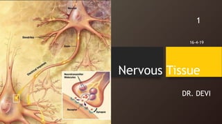
- 2. Contents • Neuron • Nerve cell processes • Synapses And impulse transmission • The neuroglia • Myelin sheath 2
- 3. The Neuron • Special property of irritability & conductivity • Nerve cells are capable of receiving the information from external and internal environment (Pain, touch, temperature, pressure) • This information is relayed to CNS by nerve cell processes • CNS integrates and interprets this information and commands are distributed to effector tissue via nerve cells • information is also stored in the brain for future reference in the form of memory 3
- 4. Nervous tissue composed of : • Nerve cell ( Neuron) • Nerve cell processes • Neuroglia 4
- 5. THE NEURON • Functional cell unit of nervous tissue • Highly specialized to carry information in the form of electric signals from one cell to another • Cell body & nerve cell process 5
- 6. Nerve cell body • Typical pale staining euchromatic nucleus with prominent dark nucleolus • Cytoplasm contain prominent basophilic material celled – Nissl bodies • It is an aggregation of RER and free ribosomes • Cell body contains all other organelles • cytoskeleton formed by microtubules and microfilaments 6
- 7. 7• Neurons in old age may show presence of lipofuscin pigment (neuronal lysosomes) • Size 10um- 120um • Neurons don’t divide after birth hence, their number doesn’t increase during life time
- 8. Nerve cell processes • Elongated cytoplasmic processes take origin from the cell body • These process may travel long distances from neuron • Two types of nerve cell processes - single axon & multiple dendrites 8
- 9. Dendrites • Short, multiple • Each of these may branch extensively to form “ dendritic tree” • They are involved in receiving information from other cells – input portions of neuron • Cytoplasm of dendrites contains Nissl bodies, microtubules, microfilaments and other organelles 9
- 10. Axon • Nerve cell processes that send information in the form of electrical signals away from nerve cell body to another • Usually one axon for each cell body • Originate from a conical region – Axon hillock • End in terminal branching pattern • Axolemma • Axoplasm 10
- 11. • Axoplasm contains neurofilament and microtubules , but lack Nissl bodies and Golgi apparatus • Nissl substance is also absent from the region of axon hillock • Axons are surrounded by myelin sheath • Axonal transport system 11
- 12. Differences between axon and dendrites Axon • Single ,long, thin process • Terminates away from cell body • Rarely branches • Uniform diameter • Smooth surface • Free of nissl bodies • Nerve impulse travel away from the cell body Dendrites • Multiple, short, thick ,tapering process • Terminate near cell body • Highly branching • Dendritic tree • Thickness reduces as it divides • Surface not smooth • Contain Nissl bodies • Nerve impulse travel towards CB 12
- 13. The Neuroglia • CNS contains no connective tissue component • smeller • Supporting cells • Function : fill-up the space, provide support, ensheathment , facilitates during nerve transmission • 5-50 times more than neurons • Cannot generate impulses 13
- 14. 14• Neuroglial cells of PNS: 1. Schwann cells 2. Satellite cells • Neuroglial cells of CNS: 1. Ependymal cells 2. Astrocytes 3. Oligodendrocytes 4. Microglia
- 15. Ependymal cells • Arranged in a single layer • Ciliated columnar or cuboidal cells • Line the ventricles of brain and central canal of spinal cord • Function : responsible for the formation of CSF 15
- 16. Astrocytes • Star shaped, • Protoplasmic astrocyte- abundant cytoplasm present, found in grey matter, end-feet/ vascular feet • Fibrous astrocytes- long , fewer in number, found in white matter • Functions : help in providing nutrients to neurons, formation of blood-brain barrier, maintaining chemical environment for generation of impulse, metabolism of neurotransmitter , migration of neurons at the time of brain development 16
- 17. Oligodendrocytes 17 • Small round or oval cells • Few cytoplasmic process • Found in CNS • Functions; produce myelin sheath around axons in CNS, form supportive network around CNS neurons
- 18. Microglia • Involved in phagocytic activity within CNS, can engulf invading microorganisms • Rarely found • Small cells with tortuous process 18
- 19. Schwann cells • Flattened cells with flattened nucleus surrounded by abundant cytoplasm • Present in PNS only • Functions : produce myelin sheath, participate in regulation of PNS axon 19
- 20. 20
- 21. Satellite cells • Surround the nerve cells of ganglia • flattened cells • Prominent nuclei • Present in PNS • Functions: insulates and support neurons of ganglia, also provide pathways for metabolic exchange • 21
- 22. Classification of Neurons • Functionally, three types: sensory, motor, interneuron • Classified on the basis of the shape of the cell body, which is depending on the number and orientation of cell process arising from it 22
- 23. 23 • Unipolar – only one process present, found in mesencephalic nucleus of trigeminal nerve • Pseudounipolar neurons –one process that extends from cell body and bifurcates forming a T- shape, one branch extends to CNS and other to PNS , found in dorsal root ganglion & cranial nerve ganglia
- 24. 24 • Bipolar neurons : they have two process , found in retina & ganglion of VII CN • Multipolar neurons- have only one axon, many dendrites (satellite/star like) or pyramidal/triangular , most common type, found in spinal cord , cerebrum, cerebellum; motor neurons and interneurons
- 25. Synapse • Specialized region of contact between two neurons • At the synapse nerve impulse is transmitted from one neuron to other • Transmission of impulse is mediated through the release of chemical substances at the synapse 25
- 26. • Presynaptic neuron • Postsynaptic neuron • Types of synapses : 1. Axodendritic 2. Axosomatic 3. Axoaxomic 4. Dendrodendritic 26
- 27. Structure of a Synapse • Axodendritic –most common • Parts- presynaptic part, synaptic cleft, postsynaptic part 27
- 28. 28 Presynaptic part -Contains mitochondria and thickened cytolemma. Axon terminal contains many presynaptic vesicles containing neurotransmitters Synaptic cleft- 20-5-um space, filled with interstitial fluid. Post synaptic part of the synapse consist of thickened region on the dendrite of the postsynaptic neuron
- 29. Impulse transmission • Neurotransmitter diffuses across synaptic cleft • Acts on plasma membrane of postsynaptic neuron to produce postsynaptic potential • Presynaptic electrical signal -> chemical signal-> electrical signal • Chemical synapse, Electrical synapse 29
- 30. Myelin sheath • Insulating sheath surrounding axons of central peripheral nervous system • In PNS Schwann cells form myelin sheath around axons • In CNS the Oligodendrocytes form myelin sheath around axons • Neurolemmal sheath/ Schwann cell sheath 30
- 31. Nodes of Ranvier • Along the given axon, myelin is formed in discrete units each unit being formed by one glial cell. • Between adjacent segments of myelin along each axon a gap occurs, where axon is not covered by myelin, these gap are called nodes of Ranvier • The segment of myelin between two nodes is called inter- node, which is produced by single Schwann cell 31 Nodes of Ranvier
- 32. Clinical applications • Tumors of glial cells: glial cells capable of cell division , may produce tumors • Gliomas: tumors of the glial cells of CNS • Schwannoma: tumors of the Schwann cells • Medulloblastoma 32
- 33. • Demyelination : degenerative process that erodes away the myelin sheath that normally protects nerve fibers , it exposes the nerve fibers and cause problems with nerve impulse conduction that may affect physical systems 33
- 34. “ ” THANK YOU 34