SlideShare a Scribd company logo
1 of 68
1 | C O R N E R S T O N E L E A D E R S H I P A C A D E M Y - B O Y S A - L E V E L B I O N O T E S
1 | E D I T E D b y K I N G S O Y E K W O k i n g s o y e 8 8 @ y a h o o . c o m 0 7 0 1 2 1 1 9 6 6
THE CHEMICALS OF LIFE
These are compounds needed to maintain life of living organisms
Cells, tissues and organs are composed of chemicals, many of which are identical with those found in
nonliving matter. Chemical compounds are divided into
(i) Organic compounds, include all complex compounds of carbon namely; carbohydrates, lipids,
proteins and nucleic acids.
(ii) Inorganic compounds, include; Acids,bases,salts and water.
ACIDS, BASES AND SALTS.
(i) Acid: Is a compound which dissociate in water to liberate a hydrogen ion. For example:
Hydrochloric acid, Sulphuric acid. The strength of an acid is determined by the extentto which it dissociates
in aqueous solutions. The acidity of a solution is expressed as its pH. A pH of 7 represents neutrality,pH
value less than 7 is acidic and pH greater than 7 is Alkaline.
(ii) A base:is a compound which can combine with hydrogen ions liberated by dissociation of an acid.
For example; Sodium hydrogen carbonate. In so doing it a base acts as a buffer (compound which resist
change in pH on dilution or addition of small amounts acid or Alkali). PH of body fluids should be kept as
constant as possible because, cells and tissues can only function properly at around neutrality, they cannot
tolerate fluctuations of pH.
In case of increased acidity, the NaHCO3 combines with free hydrogen ions as shown above if alkalinity is
increased, it reacts with free hydroxyl ions to form carbonate ions and water.
)
(
2
)
(
2
)
(
)
(
3 l
aq
aq
aq O
H
CO
H
HCO 


 

In the human body, bi-carbonate ions play a minor role as buffers. A more important role is by the
appropriate salts e.g. K3 PO4,Na3 PO4 etc.
These combine with hydrogen ions to form Di-hydrogen phosphate.





 )
(
4
2
2
)
(
4
)
( aq
aq
aq PO
H
HPO
H
Question. Explain howthe following are involved in maintaining pH of body fluids.
(a) Chemical acid-base buffer systems (08 marks)
(b) The respiratory centre 07 marks
(c) The kidneys (05 marks
APPROACH:
(i) Protein Buffer System
-Haemoglobin buffers inside red blood cells
-Plasma protein buffers
-Amino acid buffers by all proteins
Haemoglobinbuffer system
H+
+ Hb HHb
During acidosis(low pH), the haemoglobin (Hb) in red blood cells binds with H+ to increase intracellularpH;
During alkalosis (high pH) and in presence of oxygen, H ions are released from haemoglobin, which decreases
intracellularpH;
2 | C O R N E R S T O N E L E A D E R S H I P A C A D E M Y - B O Y S A - L E V E L B I O N O T E S
2 | E D I T E D b y K I N G S O Y E K W O k i n g s o y e 8 8 @ y a h o o . c o m 0 7 0 1 2 1 1 9 6 6
Plasma protein and Amino acid buffer systems
During acidosis, exposed amino group of amino acids like histidine for plasma proteins like albumin accept H
+To decrease acidity;
During alkalosis,the exposed carboxyl group of amino acids can release H to increase acidity;
(ii) Carbonic Acid - Bicarbonate Buffer System
During acidosis(Low pH), NaHCO3 dissociates to form bicarbonate ions which combine with excess hydrogen
ions to form carbonic acid; which dissociates into water and carbon dioxide,which can be eliminated by the
respiratory system as pH is increased to the norm;
During alkalosis(High pH), carbon dioxide reacts with water to form carbonic acid (H2CO3) which dissociate
to form bicarbonate ionsand hydrogen ions;which lower the pH to the norm;
OR
(iii) Phosphate Buffer System
When pH increases(alkalosis), dihydrogenphosphate dissociates into hydrogen ions and monohydrogen
Phosphate ions; to lowerpH to the norm;
When pH decreases (acidosis), hydrogenions react with monohydrogen phosphate ions to form dihydrogen
phosphate; to increase pH to the norm;
-
(refer to proteins for more information)
(iii) Salts: In the body, some of the common solutes found dissolved in water are mineral salts,
compounds of a metal with nonmetal or non-metallic radical, e.g.sodium chloride. Some nutrients are need
by the body in large amounts and are called Macronutrients while other nutrients are needed in small
amounts thus called micronutrients.
The functions of theses mineral salts and their relative derivatives are varied, but their roles can be summed
up thus;
(i) As constituents of various chemicals.
(ii) As constituents of structures.
(iii) As constituents of enzymes.
(iv) As metabolic activators.
(v) As constituents of certain pigments.
(vi) As determinants of the anion – cation balance in cells.
(vii) As determinants of osmotic pressure.
Question: account for the different functions ofmineral salts in organisms
General functions ofmineral salts (summary)
- Formation of tissues such as a bone and teeth.
- Essential for clothing of blood
- Function as enzyme activators and components of enzymes.
- Formation of hydrochloric acid in stomach.
3 | C O R N E R S T O N E L E A D E R S H I P A C A D E M Y - B O Y S A - L E V E L B I O N O T E S
3 | E D I T E D b y K I N G S O Y E K W O k i n g s o y e 8 8 @ y a h o o . c o m 0 7 0 1 2 1 1 9 6 6
- Maintenance of water balance
- Maintenance of onion/cation balance
- Components of haemoglobin, myoglobin and cytochromes.
- Components of nuclei acids / DNA and RNA.
- Involved in transfer of energy in ATP
- Important in muscle and nuclear activity.
- Needed for proper heart functioning
- Important for amino acid formation and muscle growth.
- Important for wound healing
- Formation of hormones.
- Preventing infection and body immunity.
Inorganic ions and their importance in plants and animals.
Macro element Function Notes and deficiency
Nitrate NO3
-
Ammonium
NH4+
(i) Nitrogen is a component of amino acids,
proteins, vitamins, coenzymes, nucleotides and
chlorophyll: (ii) some hormones contain nitrogen
e.g. Auxins in plants and insulin in animals
In plants causes chlorosis
(yellowing of leaves)and stunted
growth.
Phosphate
PO43-
Orthophosphate
H2PO42-
(i) A component of nucleotide, ATP and
some proteins used in phosphorylation
of sugars in respiration.
(ii) Constituent of bones and teeth.
(iii) Component of cell membranes in form
of phospholipids.
In plants leads to stunted growth,
especially of roots, and
formation of dull, dark green
leaves: in animals, canresult into
bone malformation called
crickets.
Sulphate
SO42-
Sulphur is a component of some proteins and
certain coenzymes e.g. acetyl coenzyme A
Sulphur forms important bridges
between the polypeptide chains
of some proteins, giving them
their tertiary structure.
Deficiency causes chlorosis and
poor root development.
Potassium K+
(i) Maintains electrical, osmotic and anion
/ cation balance across cell memb
ranes.
(ii) Involved in
Potassium plays role in
transmission of impulses.
1
4 | C O R N E R S T O N E L E A D E R S H I P A C A D E M Y - B O Y S A - L E V E L B I O N O T E S
4 | E D I T E D b y K I N G S O Y E K W O k i n g s o y e 8 8 @ y a h o o . c o m 0 7 0 1 2 1 1 9 6 6
active transport of certain materials across cell
membrane. (iii) Co factor in photosynthesis and
respiration. (iv) Constituent of sap vacuoles in plants,
maintains turgidity.
Deficiency leads to yellow edged
leaves and premature death.
Calcium Ca2+ In plants: (i) component of middle lamella. (ii) Aids
translocation of carbohydrates and amino acids. In
animals, (i) Constituent of bones, teeth and shells. (ii)
Blood clotting. (iii) Muscle contraction.
In plants, death of growing points.
In animals causes rickets.
Sodium Na+ (i) Maintains electrical, osmotic and anion / cation
balance across membranes. (ii) Active transport across
membranes. (iii) Constituent of sap vacuole.
(i) Muscular clumps. (ii)
Deficiency rare in plants.
Chlorine Cl-
(i) Maintains electrical, osmotic and anion / cation
balance across membranes. (ii) Formation of
hydrochloric acid in gastric juice. (iii) transport of
carbon dioxide by blood (chloride shift)
Causes muscular clumps in
animals.
Magnesium
Mg2+
(i) Constituent of chlorophyll. (ii) Activator of some
enzymes e.g. ATPase. (iii) Component of bone and
teeth.
Chlorosis in plants.
Iron
Fe2+
(i) Constituent of electron carriers e.g.
cytochromes, needed in respiration and
photosynthesis.
(ii) A constituent of certain enzymes e.g.
peroxidases, dehydrogenases,
decarboxylases.
(iii) Required in synthesis of chlorophyll.
(iv) Forms part of haem group in respiratory
pigments such as
haemoglobin,,heamoerythrin, myoglobin.
Chlorosis in plants and Aneamia in
animals.
Micronutrients / trace elements
Magnesium
Mn2+
(i) Activator of certain ennzymes eg phosphatases.
(ii) Growth factor ion bone development
Leaves with mottled with grey
Bone deformation
Copper
Cu2+
(i) Constituent of some enzymes e.g.
Cytochrome oxidase and tyrosinase.
(ii) Component of haem ocyanin (a
respiratory pigment)
Young shoots die back at an early
stage.
Iodine
I-
Constituent of hormone thyroxin. Not required in higher plants.
Causes cretinism in children and
goiter in adults.
Essential for
metamorphic changes in
some vertebrates.
Cobalt
Co2+
(i) Constituent of Vitamin B12, important in synthesis
of RNA,nucleoprotein and red blood cells.
Anemia
5 | C O R N E R S T O N E L E A D E R S H I P A C A D E M Y - B O Y S A - L E V E L B I O N O T E S
5 | E D I T E D b y K I N G S O Y E K W O k i n g s o y e 8 8 @ y a h o o . c o m 0 7 0 1 2 1 1 9 6 6
Zinc
Zn2+
(i) Activator of certain enzymes, e.g.
carbonic anhydrase.
(ii) Required in plants for leaf formation,
synthesis of auxins and alcoholic
fermentation.
Carbonic anhydrase is important in
transportation of carbon dioxide in
vertebrates’ blood. In plants,
produces malformes and
sometimes mottled leaves.
Molybdenum
Mo4+
Mo5+
(i) Reduction of nitrate to nitrite in formation of
amino acids.
Reduction in crop yields. Not vital
in most animals.
2
Boron
BO33+
B4O2+
(i) Required for uptake of Ca2+
by roots.
(ii) Aids the germination of pollen grains
and mitotic division in meristems.
Not required by animals.
Death of young shoots and
abnormal growth.
Fluorine
F-
Component of teeth and bones. Not required by most plants.
Teeth decay
QUESTION: OUTLINE THE ROLE OF MINERALS AND IONS IN BIOLOGICAL SYSTEMS.
1) They are components of smaller molecules e.g. phosphorus is contained in ATP and iodine is
contained in thyroxin, etc.
2) They are constituents of large molecules e.g. proteins contain nitrogen and sulphur, phospholipids
contain phosphorus, nucleic acids contain nitrogen and phosphorus, etc.
3) They are components of pigments e.g. haemoglobin and cytochromes which contain ion,
chlorophyll contain magnesium, etc.
4) They are metabolic activators e.g. activates glucose before it is broken down in cell respiration,
calcium ions activate ATPase enzyme during muscle contraction.
5) They determine the anion, cation balance e.g. Na+
,K+
and Ca+
are important in transmission of
impulses and muscle contraction.
6) They determine the osmotic pressure and water potential so that it does not fluctuate beyond
narrow limits e.g. Na+
, K+
and Cl-
are involved in water balance in the kidneys.
7) They are constituents of structures in cell membranes, cell walls, bones, enamel and shells.
CHECK UP:
By now you should be able to:
(i) Describe properties of acids, bases and salts.
(ii) Explain the role of acids, bases and salts in maintaining a stable internal environment for
physiological processes.
WATER
Water is by far the most abundant component of organisms. Individual human cell contains about 80%
water,and the whole body is made up of over 60%. Life originated in water and today numerous organisms
make their home in it. Water provides the medium in which all biochemical reactions take place and has
played a major role in the evolution of biological systems.
6 | C O R N E R S T O N E L E A D E R S H I P A C A D E M Y - B O Y S A - L E V E L B I O N O T E S
6 | E D I T E D b y K I N G S O Y E K W O k i n g s o y e 8 8 @ y a h o o . c o m 0 7 0 1 2 1 1 9 6 6
Some Biologically important functions of water.
All organisms Plants Animals
Structure- high water content of
cells.
Osmosis and turgidity, ( cell
enlargement, guard cell
mechanism, support)
Transport in vascular system,
lymphatic and excretory system.
Solvent and medium
for
diffusion
Reagent in photosynthesis. Osmoregulation.
Reagent for hydrolysis Transpiratio n Cooling by evaporation, such as
sweating and panting.
Support for aquatic organisms. Translocation of inorganic ions
and organic compounds.
Lubrication, as in joints.
Fertilization by
swimming gametes
Germination of seeds, swelling
and breaking open of the testa and
further development.
Support, Hydrostaatic skeleton e.g.
annelid worms.
Dispersal of seeds, gametes and
larval stages of aquatic organisms
and seeds of some terrestrial
species e.g. coconuts.
Protection; e.g. mucus, tears
Migration in ocean currents
The importance of water as a medium for life comes from four of its properties:
(a) Its solvent properties.
Water’s properties as a solvent depend on the fact that it is a polar molecule (the distribution of electric
charge is such that the centers of negative and positive charges are separated by a short distance). Because
structure of a water molecule; instead of being arranged in a straight line, the hydrogen and oxygen atoms
are situated asymmetrically (V – shape). The molecule as a whole shows polarity, Because Oxygen part
has a net negative charge and hydrogen parts a net positive charge.
Consider: if sodium chloride is placed in water, having both positive and negative charges,water attracts
both ions of the molecule breaking it and clustering around it. Because of this:
7 | C O R N E R S T O N E L E A D E R S H I P A C A D E M Y - B O Y S A - L E V E L B I O N O T E S
7 | E D I T E D b y K I N G S O Y E K W O k i n g s o y e 8 8 @ y a h o o . c o m 0 7 0 1 2 1 1 9 6 6
(i) Water is a good solvent, ionic solids and polar molecules readily dissolve in it , this is of a very
biological importance because all chemical reactions taking place in a cell occur in aqueous
solution.
(ii) Water molecules associate with each other (positive hydrogen atom of one molecule maybe
attracted to negative oxygen atom of another) forming hydrogen bonds important in holding
organic molecules together.
(b) Thermal properties:
Heat capacity, is the amount of heat required to raise the temperature of 1g by 1o
C. Water has very high
heat capacity compared with other liquids. Therefore a large increase in heat results into a comparatively
small rise in temperature of water. This implies water is good at maintaining its temperature constant
irrespective of changes in temperature of the surrounding environment. This is biologically important
because;
(i) Biochemical processes proceed in very narrow range of temperatures,and most organisms cannot
tolerate wide variations of temperature
(ii) High thermal capacity of water keeps the temperature constant, thus making it an ideal for plant
and animal life.
(iii) Water has remarkably high boiling point, thus hardly evaporate.
(iv) It provides a sustainable habitat for aquatic organism the habitat does not easily get frozen in cold
weather.
(v) It maintains an effective transport medium of materials in the body since it maintains its liquid form
even on the cold weather.
(vi) Aquatic organisms can survive even in winter because the lower part of the water bodies hardly
freezes
8 | C O R N E R S T O N E L E A D E R S H I P A C A D E M Y - B O Y S A - L E V E L B I O N O T E S
8 | E D I T E D b y K I N G S O Y E K W O k i n g s o y e 8 8 @ y a h o o . c o m 0 7 0 1 2 1 1 9 6 6
(c) Surface tension.
This is the force that causes the surface of liquid to contact so that it occupies the least possible area. It is
due to inward – acting cohesive forces between molecules at the surface being caused by polarity of water.
Waterhashigh surface tension and molecules dissolved in watertend to lower its surface tension and collect
at the interface between liquid and other phases. This is biologically important in:
(i) Development of plasma membrane and movement of molecules across it.
(ii) Movement of water up the capillary like vessels and tracheid in the stems of plants
(iii) Enables the surface film to support and provide habitat for certain aquatic organisms.
(d) Freezing properties:
When water is cooled below a certain temperature, its volume increases and its density decreases. This
means ice tends to float than to sink. When the temperature drops the coldest water is at the surface and
being less dense than the slightly warmer water lower down, tends to remain at the surface. So ice forms at
the surface fist, and bottom later. Organisms which live towards the bottom of fresh water lakes are
protected from freezing increasing their survival.
QUESTION: HOW DO THE PROPERTIES OF WATER RELATE TO ITS BIOLOGICAL ROLE?
1 Water is transparent and this allows light penetration in aquatic habitats to enable photosynthesis
of aquatic autotrophs and visibility of aquatic animals.
2 Water has a low viscosity and this al lows for smooth flow of water and other dissolved substances
in an aquatic medium for easy transport.
3 It has a high surface tension providing support to aquatic organisms and allowing movement of
living organisms on water surface.
4 Has a high latent heat of vaporization hence a cooling effect on the body surface since evaporation
of water from the body of an organism draws out excess heat.
5 It has a high boiling point thus provides a stable habitat and medium since a lot of heat which is not
normally provided in the natural environment is needed to boil the water.
6 It has a high latent heat of fusion and hence a low freezing point thus providing a wide range of
temperature for survival of aquatic organisms since it prevents freezing of cells and cellular
components.
7 It has a high specific heat capacity which minimizes drastic temperature changes in biological
systems and provides a constant external environment for many plant cells and aquatic organisms.
8 It has a maximum density at 4o
C hence ice floats on top of water insulating the water below hence
increasing the chances of survival of aquatic organisms below the ice.
9 Water is liquid at room temperature providing a liquid medium for living organisms and metabolic
reactions and a medium of transport.
9 | C O R N E R S T O N E L E A D E R S H I P A C A D E M Y - B O Y S A - L E V E L B I O N O T E S
9 | E D I T E D b y K I N G S O Y E K W O k i n g s o y e 8 8 @ y a h o o . c o m 0 7 0 1 2 1 1 9 6 6
10 It has high adhesive and cohesive forces creating enough capillarity forces for transport in narrow
tubes of biological systems.
11 It is a universal solvent hence providing a medium for biochemical reactions.
12 Water is a polar molecule allowing solubility of polar substances, ionization or dissociation of
biochemical substances.
13 Water is incompressible thus providing support in hydrostatic skeleton and herbaceous stems.
14 Water is neutral hence does not alter the pH of cellular components on their environment.
15 A water molecule is relatively small for easy and fast transport across a membrane.
CHECK UP:
By now you should be able to:
(i) Describe the molecular structure of water.
(ii) State f unctions of water.
(iii) Explain the importance of water as a solvent.
(iv) Relate the water properties to its role in the life of organisms.
A. CARBOHYDRATES
These are food substances which contain elements; carbon, hydrogen and Oxygen. They have a general
formula C x (H2O) y, where Xand Y are variable numbers. Their name Hydrate of carbon is derived from
the fact that Hydrogen and oxygen are present in the same proportions as in water, namely two hydrogen
atoms per oxygen atom. In addition:
(i) All carbohydrates are either aldehydes or ketones.
(ii) All contain severalhydroxyl groups.
Question Howis carbon uniquely suited to its role as the main element in living organisms.
APPROACH
 Great strength of the carbon-to-carbon bond ensures that carbon-based molecules are very stable.
 Great strength of the carbon-to-hydrogen bond ensures that the hydrocarbon skeleton, typical of
carbon-based organic moleculesis very stable.
 Ability of carbon to form repetitive bonds with itself/catenation leads to; enormously varying
lengths of chains,enormously varying extent of branching, enormously varying length of branches,
enormously varying forms of ringed compounds,ensuring that carbon-based molecules can form
a variety of shapes.
 Great stability of the +4 oxidation state of carbon owing to its very small atomic size, ensures high
stability of carbon-based molecules formed in the +4 oxidation state.
10 | C O R N E R S T O N E L E A D E R S H I P A C A D E M Y - B O Y S A - L E V E L B I O N O T E S
10 | E D I T E D b y K I N G S O Y E K W O k i n g s o y e 8 8 @ y a h o o . c o m 0 7 0 1 2 1 1 9 6 6
 Ability of carbon to formmultiple bondswith itself and other elements, leads to a wide variety of
carbon-based molecules.
 Ability of carbon to form stable covalent bonds with other elements, ensures that carbon-based
molecules contain a variety of elements
Carbohydrates are divided into three main classes namely; (i) Monosaccharaides (ii) Disaccharides (iii)
polysaccharides.
1. Monosaccharaides;
These are single sugar units. Their generalformula is (CH2O) n ., Where n is variable number.
Properties:(i) sweet. (ii) Soluble in water. (iii) Crystalline. (iv) Low molecular weight
Classification:
They are class ified according to number (n) of carbon atoms. Example: (i) Trioses, 3 carbon atoms, e.g.
Glyceraldehyde. (ii) Pentoses,5 carbon atoms, e.g.Ribose, Deoxyribose. (iii) Hexoses,6 carbonatoms e.g.
Glucose, Fructose, Galactose.
Aldoses and ketoses:In monosaccharaides,all carbon atoms except one having a hydroxyl group attached,
the remaining carbon is either part of an aldehyde group, and the monosaccharide is called aldose or aldo
sugar, or is part of a keto group, monosaccharide called ketose or keto sugar. Aldoses include:
Glyceraldehyde, Ribose, and Glucose. And Ketoses include: Dihdroxyacetone, Ribulose, and fructose.
Trioses (
C3H6O3 )
Pentoses (
C5H10O5)
Hexoses (
C6H12O6 )
Aldoses ( - CHO)
( Aldo – sugar)
Glyceraldehyde Ribose Glucose,
Galactose
Ketoses ( C=O )
(keto sugars)
Dihdroxyacetone Ribulose Fructose
Glyceraldehyde Dihydroxyacetone.
11 | C O R N E R S T O N E L E A D E R S H I P A C A D E M Y - B O Y S A - L E V E L B I O N O T E S
11 | E D I T E D b y K I N G S O Y E K W O k i n g s o y e 8 8 @ y a h o o . c o m 0 7 0 1 2 1 1 9 6 6
Open chain and ring forms.
The open chain form can be straight, Because of bond angles between carbon atoms, hexoses and pentoses,
b end to form a stable ring structures. In hexoses like glucose, First carbon atom combines with the oxygen
on carbon atom five to give a six – membered ring. Note that oxygen is part of the ring and one carbon
atom, carbon atom number 6, sticks up out of the ring.
In Pentoses, the first carbon atom combines with oxygen atom on the fourth carbon atom forming a five –
membered ring. These ring structures are the forms used to make disaccharides and polysaccharides.
Ribose open chain Ribose with five – membered ring
Glucose:Is a common monosaccharide, It a hexose sugar ( C6H12O6 ). Has open chain structure and ring
structure. In Ring structure, has two isomers namely; Alpha and Beta isomers. The hydroxyl group on
carbon atom 1 canproject below the ring forming Alpha glucose or above the ring forming the Beta glucose.
Alpha glucose is used in making polysaccharide starch, Beta glucose is used in making polysaccharide
cellulose.
Open chain form  glucose  Glucose
12 | C O R N E R S T O N E L E A D E R S H I P A C A D E M Y - B O Y S A - L E V E L B I O N O T E S
12 | E D I T E D b y K I N G S O Y E K W O k i n g s o y e 8 8 @ y a h o o . c o m 0 7 0 1 2 1 1 9 6 6
Formation of a ring molecule of beta- D-glucose
Functions of monosaccharaides:
Monosaccharides Examples Functions
Trioses Glyceraldehyde
Dihdroxyacetone
Intermediates in respiration (Glycolysis) , Photosynthesis (dark
reactions) and other branches of carbohydrate metabolism.
Pentoses Ribose
Deoxyribose
Ribulose
(i) Synthesis of nucleic acids.
(ii) Synthesis of some coenzymes (Ribose is used in
synthesis of NAD and NADP).
(iii) Synthesis of ATP requires ribose.
(iv) Ribulose biphosphate is the CO2 acce[ptor in
photosynthesis, made from ribulose.
Hexoses Glucose
Fructose
Galactose
(i) Source of energy when oxidized in respiration. (ii) Synthesis
of disaccharides. (iii) Synthesis of polysaccharides
13 | C O R N E R S T O N E L E A D E R S H I P A C A D E M Y - B O Y S A - L E V E L B I O N O T E S
13 | E D I T E D b y K I N G S O Y E K W O k i n g s o y e 8 8 @ y a h o o . c o m 0 7 0 1 2 1 1 9 6 6
2. Disaccharides:
These are double sugar units formed when two monosaccharaides usually hexoses combine by means of a
chemical reaction called condensation. This means removal of water. The bond formed between two
monosaccharaides as a result of condensation is called Glycosidic bond and it normally forms between
carbon atom 1 and 4 of neighboring units ( 1,4 bond or 1,4 linkage ). The monosaccharide molecules are
called residues once they have been linked. Maltose molecule contains two glucose residues. Common
disaccharides are;
(i) Maltose (Glucose + Glucose).
(ii) Lactose (Galactose + Glucose).
(iii) Sucrose (Fructose + Glucose).
Formation of Maltose molecule (disaccharide) from Glucose molecules (monosaccharaides)
Addition of water to a disaccharide under suitable conditions, splits into monosaccharaides and this called
Hydrolysis (splitting by water). Likes monosaccharaides,disaccharides have the following characteristics;
 They are small molecules with a low molecular mass
 They are readily soluble in water
 Have a sweet taste
 They exist in crystalline form
 They are made by combining two monosaccharides in a condensation reaction
Formation ofa sucrose molecule
14 | C O R N E R S T O N E L E A D E R S H I P A C A D E M Y - B O Y S A - L E V E L B I O N O T E S
14 | E D I T E D b y K I N G S O Y E K W O k i n g s o y e 8 8 @ y a h o o . c o m 0 7 0 1 2 1 1 9 6 6
Formation ofa maltose molecule
+ H2O
Reducing sugars: All monosaccharaides and some disaccharides (Maltose and Lactose) are reducing
sugars, meaning they can carry out a type of chemical reaction called Reduction. Sucrose is the only
common non reducing sugar.Two common testsfor reducing sugars; (i)Benedict’stest.(ii)Fehling’stest,
make use of the ability of these sugars to reduce copper from a valence of 2 to a valence of 1. Both tests
involve use of alkaline blue solution of copper (II) sulphate which is reduced to insoluble brick-red
precipitate copper (I) oxide.
Equation showing reduction reaction involving a reducing sugar and test reagent
FUNCTIONS OF DISSACCHARIDES
i. They are food reserves in organisms and when they are hydrolysed to monosaccharide and used in
cell metabolism.
ii. They are the main forms of transport of organic substances in the phloem.
iii. Storage materials in some plants like sugar canes.
iv. They are energy reserves.
NB: Starch is converted in plants to maltose so as;-
15 | C O R N E R S T O N E L E A D E R S H I P A C A D E M Y - B O Y S A - L E V E L B I O N O T E S
15 | E D I T E D b y K I N G S O Y E K W O k i n g s o y e 8 8 @ y a h o o . c o m 0 7 0 1 2 1 1 9 6 6
i. To be transported easily because it’s soluble in water.
ii. Maltose is less reactive hence won’t be used.
3. Polysaccharides:
Polysaccharides are formed when monosaccharaides polymerize in a condensation reaction between two
hydroxyl groups, resulting in a covalent interaction called a glycosidic linkage. They are normally used for
food storage because;-
i. They can be hydrolyzed to sugars when required for production.
ii. They fold into compact shapes which cannot diffuse out of the cells.
iii. They exert no osmotic or chemical influence on the cell.
iv. They have large sizes that make them insoluble in water.
v. They are non-diffusible i.e. they don’t leave sites of storage
vi. Making structures compact e.g. cellulose.
The monosaccharide polymer may be branched, unbranched or coiled. In cells they exists as granules or
grains.
Examples ofpolysaccharides include:
(a) Starch: [A storage polysaccharides in plants]
It is a main food storage form of plants and it is a polymer of  glucose it exists as grains in the cells .it is
mainly found in seeds, leaves and tubers.
It comprises of amylose 20%,Amylopectin 79% together with other substances(1%)e.g.phosphate..Starch
has two components:
(i) Amylose, this has straight chain structure consisting of several thousand α-glucose residues joined by
α (1-4) glycosidic bonds. Soluble starches are mainly made up of amylose. They do not dissolve, but form
colloidal suspensions. Amylose is a polymer of α-glucose molecules that form unbranched molecule wound
in a helix. The bore of the helix is large enough to trap iodine molecules and produce the blue complex
during iodine tests for starch using iodine solution in potassium iodide,
16 | C O R N E R S T O N E L E A D E R S H I P A C A D E M Y - B O Y S A - L E V E L B I O N O T E S
16 | E D I T E D b y K I N G S O Y E K W O k i n g s o y e 8 8 @ y a h o o . c o m 0 7 0 1 2 1 1 9 6 6
Amylose helix
(iii) Amylopectin, is compact with many branches, formed by 1, 6 glycosidic bonds. It has up to twice
as many glucose molecules as amylose. Just like in amylose, the unbranched chain of amylopectin
has molecules of α-glucose held together by 1-4 glycosidic bonds, but the side branches are held
together by α(1-6) glycosidic bonds. Amylopectin also forms colloidal suspension in water.
The two components of starch fit together into a compact 3-D structure in which the branches of
amylopectin are entangled by the helices of amylose
In iodine (potassium iodide solution);
(i) Amylose suspension gives a blue black colour.
(ii) Amylopectin Suspension gives a red – violet colour. Starchmolecules accumulate to form
starch grains found in; many plant cells (chloroplast of leaves), Storage organs (tubers),
seeds of cereals and legumes.
Formation of one branch in amylopectin
17 | C O R N E R S T O N E L E A D E R S H I P A C A D E M Y - B O Y S A - L E V E L B I O N O T E S
17 | E D I T E D b y K I N G S O Y E K W O k i n g s o y e 8 8 @ y a h o o . c o m 0 7 0 1 2 1 1 9 6 6
Differences between amylose and amylopectin
Amylose Amylopectin
Stains blue with iodine Stains red-purple with iodine
Has a low RMM, maximum 500000 Has a high RMM up to 500000
Has between 200 to 500 glucose units Has between 5000 to 100000 glucose units
Has unbranched helical chain Has branched chain
Has 1,4 gylcosidic bonds Has both 1,4 and 1,6 gylcosidic bonds
Has 20% composition in starch molecule Has 79% composition in starch molecule
Adaptations of starch as a storage compound:
- It is in soluble in water, therefore it exerts no osmotic influence in the cell.
- It exerts no chemical influence in the cells due to its insoluble chemical nature.
- It can fold into compact shape, thus minimizing the space occupied.
- It can easily be converted back to glucose by hydrolysis. The glucose can be used in
respiration.
(b) Glycogen:[Highly branched storage polysaccharides in animals and also fungi]
This is an animal equivalent of starch, being a storage polysaccharide made from α-glucose, many fungi
also store it. In invertebrates, it’s stored mainly in liver and muscles, both centers of high metabolic
activity, where it provides a useful energy reserve. Similar to amylopectin, but shows more branching.
18 | C O R N E R S T O N E L E A D E R S H I P A C A D E M Y - B O Y S A - L E V E L B I O N O T E S
18 | E D I T E D b y K I N G S O Y E K W O k i n g s o y e 8 8 @ y a h o o . c o m 0 7 0 1 2 1 1 9 6 6
A molecule ofglycogen
(c) Cellulose:[A structural polysaccharide in plants]
Cellulose is a polymer of β-glucose monomers, joined by β-1,4glycosidic linkages.In plants, cellulose is
the major component of the cell wall. The geometry of the linkage is such that each glucose monomer in
the chain is flipped in relation to the adjacent monomer. The flipped orientation is important because
(i) It generates a linear molecule, rather than the helix seen in starch; and
(ii) It permits multiple hydrogen bonds to form between adjacent, parallel strands of cellulose. As a
result, cellulose forms long, parallel strandsthat are joined by hydrogen bonds. The linked cellulose
fibers are strong and give the cell structural support.
Structure
It is a polymer with straight chains of β-glucose units held by glycosidic bonds with the OHgroup projecting
out wards from each chain forming cross linkages of hydrogen bonds with adjacent chains.
The cross linking binds the chains together which associate to form micro fibrils that are arranged in larger
bundles to form macro fibrils.
Note: Starch lacks the structural properties possessed by cellulose because it lacks cross linkages.
The stability of cellulose makes it difficult to digest and therefore not a good source of food to animals
except those which have cellulase producing microorganisms which live in them in a symbiotic way e.g.
in the rumen of cattle, goats, sheep, etc.
Formation of Cellulose from Beta glucose.
Question:
What properties of glycogen makes it a
betterstoragecarbohydrate in animals
than starch
19 | C O R N E R S T O N E L E A D E R S H I P A C A D E M Y - B O Y S A - L E V E L B I O N O T E S
19 | E D I T E D b y K I N G S O Y E K W O k i n g s o y e 8 8 @ y a h o o . c o m 0 7 0 1 2 1 1 9 6 6
The plant cell wall is permeable to water because the matrix has minute water channels through which
diffusion occurs. Salts, sugars, and other polar molar molecules can easily disuse through these minute
channels. Cellulose is hydrophilic and this makes the cell walls always saturated with water.
Structure of cellulose showing Hydrogen bonds.
Question: Explain why cellulose formsa structural carbohydrate while starch does not (07 marks)
Approach
Cellulose is made up long chains of glucose linked by β-1–4 glycosidic bonds; This results in the
glucose molecules being alternately one way up, then the other; This means that chains lying side
20 | C O R N E R S T O N E L E A D E R S H I P A C A D E M Y - B O Y S A - L E V E L B I O N O T E S
20 | E D I T E D b y K I N G S O Y E K W O k i n g s o y e 8 8 @ y a h o o . c o m 0 7 0 1 2 1 1 9 6 6
by side can form hydrogen bonds with each other, making microfibrils; Starch, on the other hand,
is made up of long chains of glucose linked by α 1–4 glycosidic bonds; It twists into a spiral, and
one starch molecule does not form bonds with others; so no fibrils are formed; Moreover, it is
difficult to break down β 1–4 glycosidic bonds;so not many organisms can digest cell walls;
Uses of cellulose
 Rayon produced form cellulose extracted from wood is used in the manufacture of tyre
cords.
 Cotton is used in the manufacture of fibres and closes.
 Cellophane used in packaging is produced from cellulose.
 Paper is a product of cellulose.
 Celluloid used in photographic films is also a derivative of cellulose.
DIFFERENCES BETWEENCELLULOSE ANDGLYCOGEN
CELLULOSE GLYCOGEN
Polymer of ß-glucose Polymer of ∞-glucose
Monomers rote alternately at1800
to eachother No rotation of adjacent monomers
Straight chains, with cross-linking due to
hydrogen bonds
Branched chains
ß-1,4 linkages ∞-1,4 and 1,6 linkages
forms micro fibrils Does not form micro fibrils
hydroxyl groups project outwards Hydroxyl groups project inwards
performs structural role not for storage Performs storage role, no structural role
found in plants Found in animals & fungi
feworganisms can digest cellulose/ Difficult to
digest
Many organisms can digest cellulose/easily
digested
HOW DOES THE STRUCTURE OF CELLULOSE RELATE TO ITS ROLES
® The cross linking binds the chains together offering much tensile strength.
® The micro fibrils in cell walls are arranged in several layers offering protection to the plant
cell preventing it from bursting when water enters by osmosis.
® The arrangement of micro fibrils in cell walls contributes to turgidity hence offering
support.
® The parallel layers of cellulose are fully permeable to water and solutes.
® The arrangement of micro fibrils determines the shape of the cells and hence plant organs
since it determines the direction in which cells expand as they grow.
21 | C O R N E R S T O N E L E A D E R S H I P A C A D E M Y - B O Y S A - L E V E L B I O N O T E S
21 | E D I T E D b y K I N G S O Y E K W O k i n g s o y e 8 8 @ y a h o o . c o m 0 7 0 1 2 1 1 9 6 6
® The glycosidic bonds holding the β-glucose units in cellulose can be broken down in
presence of enzyme cellulase so that a free glucose molecule can be respired.
(d) Chitin: [A structural polysaccharide in fungi and animals].
Chitin is similar to cellulose, but instead of consisting of glucose monomers, the monosaccharide involved
is one called N-acetylglucosamine (NAG). NAGhas an aminoacyl group (NHOCCH3) at Carbon 2 of the
glucose molecule, therefore cellulose is very similar to chitin.
These NAG monomers are joined by β-1, 4-glycosidic linkages. As in cellulose, the geometry of these
bonds results in every other residue being flipped in orientation. Like the glucose monomers in cellulose,
the NAG subunits in chitin form hydrogen bonds between adjacent strands. The result is a tough sheet
animals. It is, for example, the most important that provides stiffness and protection. Chitin is a
polysaccharide that stiffens the cell walls of fungi. It is also found in a few types of protists and in many
component of the external skeletons of insects and crustaceans.
(e) Peptidoglycan:[A Structural Polysaccharide in Bacteria]
Peptidoglycan is the most complex of the polysaccharides discussed thus far. It has a long backbone formed
by two types of monosaccharaides that alternate with each other and are linked by β-1,4-glycosidic
linkages. Most bacteria,like all plants, have cell walls. But unlike plants, in bacteria the ability to produce
cellulose is extremely rare. Instead,peptidoglycan gives bacterial cell walls strength and firmness.
(f) INULUN(α-fructose )
This is a major storage form of carbohydrates in plants like Dahlia (Jerusalem artichoke). It is a polymer of
fructose molecules .It comprises 1, 2 glycosidic bonds and has unbranched chain of fructose molecules.
Question: Describe the Structural features of carbohydrates accounting for wide variety of
polysaccharides
Approach
® Both pentoses and hexoses can be used to make polysaccharides
® Two types of linkage 1,4 and 1,6 glycosidic bonds occur between sugar units leading
to branching.
® Lengths of chains vary enormously
® Lengths of branches vary enormously
® Extent of branching varies enormously
® Monosaccharides have alpha and beta isomeric ring forms.
22 | C O R N E R S T O N E L E A D E R S H I P A C A D E M Y - B O Y S A - L E V E L B I O N O T E S
22 | E D I T E D b y K I N G S O Y E K W O k i n g s o y e 8 8 @ y a h o o . c o m 0 7 0 1 2 1 1 9 6 6
® Sugars may be ketoses or aldoses
® Aldehyde, ketone,and hydroxyl groups of sugars, makethem highly reactive molecules.
(g) Mucopolysaccharides:
These are carbohydrate derivatives derived from a combination of sugar molecules and amino
acids in a condensation reaction. This group includes hyaruronic acid which forms part of the
matrix of vertebrae connecting tissue. Heparin, an anti-coagulant also contains muco-
polysaccharides. They contain amino sugars and are found in;
(i) The basement membrane of epithelium
(ii) The matrix of connective tissues
(iii) Synovial fluid in joints of vertebrates
(iv) Matrix of bone and cartilage.
(v) Vitreous humor of the eye
LIPIDS
Lipids include Fats (solids at room temperature) and Oils (Liquids at room temperature). Like
carbohydrates, lipids contain Carbon, Hydrogen and oxygen BUT a lipid molecule contain smaller
proportion of oxygen than carbohydrate molecule.
Constituents oflipids. Lipids are made up of;
(a) Fatty acids
These contain the acidic group –COOH (Carboxyl group). Named so because larger molecules in the series
occur in fats. General formula of R.COOH where R is a hydrogen or group (-CH3 , -C2H5 etc increasing
by –CH2 for each subsequent member of the series ). Fatty acids have a long chain of long chain of carbon
and hydrogen atoms forming a hydrocarbon tail. The tail determines many properties of lipids including
solubility in water. The tails are Hydrophobic (Hydro,water; Phabos,fear), water hating. Fatty acids can
be: (i) Saturated, lack double bonds. (ii) Unsaturated,Contain one or more double bonds (C=C ).
(b) Glycerol:
The formula of glycerol is C3H8O3,This shows no
variation and is the same in all lipids. Glycerol has
three hydroxyl groups of which can condense with
a fatty acid. Usually all the three undergo
condensation and the lipid formed is called a
triglyceride.
Formation oftriglycerides
Lipids are made of fatty acids and glycerol. Glycerol has 3 OH groups and each combines with a separate
fatty acid to form a lipid chemically known as a triglyceride. This is a condensation reaction that leads to
liberation of 3 water molecules.
23 | C O R N E R S T O N E L E A D E R S H I P A C A D E M Y - B O Y S A - L E V E L B I O N O T E S
23 | E D I T E D b y K I N G S O Y E K W O k i n g s o y e 8 8 @ y a h o o . c o m 0 7 0 1 2 1 1 9 6 6
Metabolic water is formed and an ester bond is formed between the glycerol and fatty acid. Since the
glycerol possesses 3 hydroxyl groups to which 3 fatty acids attach themselves, 3 water molecules are
formed. In this reaction, the fatty acids may all be the same or different, saturated or unsaturated.
Formation of Triglyceride from glycerol and fatty acids.
Characteristicsof lipids.
1. They are made up of carbon, hydrogen and oxygen. They are composed of 3 fatty acids (glycerol
alcohol), 3 CH groups. The general formula of glycerol is C3H8O2. The fatty acids contain the
carboxylic acid/carboxyl group (-COOH) and a hydrocarbon group.
24 | C O R N E R S T O N E L E A D E R S H I P A C A D E M Y - B O Y S A - L E V E L B I O N O T E S
24 | E D I T E D b y K I N G S O Y E K W O k i n g s o y e 8 8 @ y a h o o . c o m 0 7 0 1 2 1 1 9 6 6
2. They have a high proportion of hydrocarbon groups in their molecules.
3. They are insoluble in H2O but soluble in organic solvents e.g. benzene and chloroform alcohol.
This is because lipids have a low O2 and the numbers of polar hydroxyl groups are few and this
decreases their solubility in H2O.
4. They are either solids (fats) or liquids (oils) at room temperature.
5. The higher the proportion of unsaturated fatty acids, the lower will be their melting points.
6. They are non-polar and this makes them effective storage materials.
7. They are less dense than H2O and this helps the aquatic organism to float on H2O.
8. They have a function group COOH composed of fatty acids and glycerol.
FATTY ACIDS
Fatty acids are long molecules with a polar, hydrophilic end and a non-polar, hydrophobic "tail". The
hydrocarbon chain can be from 14 to 22 CH2 units long. The hydrocarbon chain is sometimes called an R
group, so the formula of a fatty acid can be written as R-COOH.
All occurring fatty acids can be saturated or unsaturated
Unsaturatedfatty acids are ones where the hydrocarbon part contains one or more double bonds e.g. linoleic
acid and oleic acid .The saturated don’t contain double bonds. The saturated fatty acids have a high boiling
or melting points compared to the unsaturated of the same molecular mass.
Fatty acids Formula Unsaturated Sources
Butyric acid C3H17COOH Unsaturated Butter
Linolenic acid C17H31COOH Saturated Lin seeds
Oleic acid C17H33COOH Unsaturated All fat
Palmitic acid C15H31COOH Unsaturated Palm oils
25 | C O R N E R S T O N E L E A D E R S H I P A C A D E M Y - B O Y S A - L E V E L B I O N O T E S
25 | E D I T E D b y K I N G S O Y E K W O k i n g s o y e 8 8 @ y a h o o . c o m 0 7 0 1 2 1 1 9 6 6
Stearic acid C17H35COOH Saturated Adipose fats
Arachidonic acid C19H39COOH Saturated Peanut oils
Cerotic C25H51COOH Saturated Wool oil.
Illustration of different fatty acids
NB:
There are some fatty acids which cannot be synthesized in the body. Therefore, they have to be obtained
from the diet and these are called essential fatty acids. Their deficiency results into retarded growth,
reproductive deficiency and kidney failure.
The body synthesizes some fatty acids and they are synthesized from compounds of proteins and
carbohydrate metabolism.
FUNCTIONS OF LIPIDS.
i) They serve as energy sources or stores and yield much more energy than carbohydrates i.e. 1g of
lipids yields 38kJ or 9k calon oxidation while that the carbohydrates of 1g of carbohydrates
yields 17kJ OR 4k Cal.
ii) In animals, it is found below the skin and acts as an insulator since it’s a poor conductor of heat.
It is most found in animals living in cold climates and aquatic mammals have very thick
subcutaneous fat called BLUBBER.
iii) Fats provide H2O to desert animals when oxidized e.g. on camels where they are heavily
deposited in the hump.
26 | C O R N E R S T O N E L E A D E R S H I P A C A D E M Y - B O Y S A - L E V E L B I O N O T E S
26 | E D I T E D b y K I N G S O Y E K W O k i n g s o y e 8 8 @ y a h o o . c o m 0 7 0 1 2 1 1 9 6 6
iv) They are constituents of the plasma membrane when they combine with phospholic acids to form
phospholipids. They are constituents of the waxy cuticle of plants and insects giving water
repelling properties making them cuticle waterproof Since they are insoluble in water
v) They are used to protect delicate organs of the body by offering cushion
vi) Buoyancy function in that they help the aquatic organism to float and the oils on bird feathers
help the aquatic varieties to float.
vii) Normal functioning of the brain.
SUITABILLITYOF LIPIDS OVER GLYCOGEN FOR STORAGE
- Lipids yield more metabolic water on oxidation important for survival of desert animals
- Fat insulates the body against heat loss whereas glycogen does not.
- Fats stored around delicate organs protect them from mechanical/physical damage.
- Fats store fat-soluble vitamins A, D, E and K
- Fats are more compact than glycogen and so occupy less space
- Lipids are lighter than glycogen hence contribute less weight to organism and structures
- Lipids are store and yield more energy than glycogen
- Lipids are less dense than water and so aid buoyancy of aquatic animals
QUESTION: Discuss the structural and physiological functions oflipids (10 marks)
Structural:
i) They are components of the plasma/cell membrane.
ii) They form subcutaneous fat in the dermis of the skin hence insulating the body since they
are poor conductors of heat.
iii) They are components of the waxy cuticle in plants and insects there by preventing water
loss (desiccation).
iv) They form a component of the myelin sheath of nerves hence playing a role in the
transmission of impulses.
iv) They protect delicate organs e.g. the heart and kidney from injury.
v) They coat on fur of animals enabling it to repel water which would otherwise wet the
organism. vii) They are component of adipose tissue.
Physiological:
i) They provide energy through oxidation.
ii) They are solvents for fat soluble vitamins (ADEK).
iii) They are a good source of metabolic water to desert animals, young birds and reptiles
while still in their shells.
iv) They are a constituent of the brown adipose tissue which provides heat for temperature
regulation (thermogenesis).
Other functions:
i) Some lipids provide a scent in plants which attracts insects for
pollination.
ii) Wax is used by bees to construct honey combs.
iii) Wax from bees is used in the manufacture of candles.
LIPID DERIVATIVES
27 | C O R N E R S T O N E L E A D E R S H I P A C A D E M Y - B O Y S A - L E V E L B I O N O T E S
27 | E D I T E D b y K I N G S O Y E K W O k i n g s o y e 8 8 @ y a h o o . c o m 0 7 0 1 2 1 1 9 6 6
Phospholipids
Phospholipids consist of a glycerol that is linked to a phosphate group and two hydrocarbon chains of fatty
acids. Has phosphate head which is Hydrophilic (water loving) and water hating tails. This is important in
formation of cell membranes.
The fatty acid chains have no electrical charge and so are not attracted to the dipoles of water molecules
They are said to be hydrophobic.
The phosphate group has an electrical charge and is attracted to water molecules. It is hydrophilic.
In water, a group of phospholipid molecules therefore arranges itself into a bilayer, with the hydrophilic
heads facing outwards into the water and the hydrophobic tails facing inwards, therefore avoiding contact
with water
Formation of a phospholipid.
How the phospholipids arrange themselvesin water environment
28 | C O R N E R S T O N E L E A D E R S H I P A C A D E M Y - B O Y S A - L E V E L B I O N O T E S
28 | E D I T E D b y K I N G S O Y E K W O k i n g s o y e 8 8 @ y a h o o . c o m 0 7 0 1 2 1 1 9 6 6
Glycolipid:
Are associations of lipids with carbohydrates. The carbohydrate forms a polar head to the molecule.
Glycolipids like phospholipids are found in membranes.
Waxes:
Waxes are formed by combination of fatty acids with alcohol other than glycerol. Alcohol is much larger
than glycerol therefore waxes have more complex chemical structure. Waxes are important for; (i)
Waterproofing in plants and animals. (ii) Form Storage compounds in few organisms like castor oil and in
fish.
Steroids:
Steroids are a family of lipids distinguished by the bulky, four-ring structure. The various steroids differ
from one another by the functional groups or side groups attachedto different carbons in those hydrophobic
rings. Cholesterol is common steroid in animals. It’s important for:
i) Limit uncontrolled leakage of small molecules (ions and water) in and out of the plasma
membranes.
ii) Constituent of myelin, facilitates movement of impulse along neuron.
iii) Formation of steroid hormones and bile salts.
How cholesterol causes arthesclerosis
Cholesterol is produced by the liver and is used as the starting point for the synthesis of other steroid
molecules. The major source of cholesterol is diet and many dairy products are rich in cholesterol or fatty
acids from which cholesterol can be synthesized. Thyroxin stimulates cholesterol production in the liver
and also increases the rate of excretion of bile. Excessive amounts of cholesterol in blood can be harmful.
It can be deposited in walls of arteries leading to arthesclerosis and increased risk of formation of a blood
clot which may block blood vessels, a condition known as thrombosis. This is often fatal if it occurs in the
coronary artery in the wall of the heart (coronary thrombosis or heart attack) or brain (cerebralthrombosis).
Although cholesterol is harmful in excess,it is essential to have some in the diet for reasons stated
B. PROTEINS
Proteins contains Carbon, hydrogen, oxygen and Nitrogen but sometimes sulphur and phosphorus.
Proteins are built up from amino acids.
AMINO ACIDS
These are groups of many chemicals of which around 20 occur in proteins. They contain an amino
group (NH2) and a carboxyl group (COOH). Most amino acids have one of each and are therefore
neutral but a few have more amino groups than carboxyl making them alkaline or may have more
carboxyl than amino groups making them acidic.
29 | C O R N E R S T O N E L E A D E R S H I P A C A D E M Y - B O Y S A - L E V E L B I O N O T E S
29 | E D I T E D b y K I N G S O Y E K W O k i n g s o y e 8 8 @ y a h o o . c o m 0 7 0 1 2 1 1 9 6 6
Structure of amino acids
Where R is a variable Amino acids are soluble in
water and ionize to form ions.
The carboxyl end of the amino acid is acidic in nature. It will ionize in water to give H+. This will
make the COOH group negatively charged.
The amino end (NH2) is basic in nature. It attracts the H+ in solution making it positively charged.
The ion is now dipolar i.e. having a negative and a positive pole. Such ions are called zwitter ions
i.e. the negative and positive charges exactly balance and the amino acid ion has no overall charge
i.e.
An Illustration of the Amphoteric Nature of Amino Acids
Zwitter ion (no overall charge) Therefore in acidic solutions, an amino acid acts like a base and in alkaline
solutions, it acts as an acid. In neutral conditions found in the cytoplasm of most living organisms, the
amino acid acts as both.
Amino acids therefore show both acidic and basic properties i.e. they are amphoteric.
The overall charge of the amino acid depends on the pH of the solution.
At some characteristic pH, the amino acid has no overall electric charge i.e. it exists as a zwitter ion. This
pH is called the isoelectric point of an amino acid.
If the pH falls below the iso-electric point i.e. the solution becomes more acidic, H+
are taken up by the
carboxyl ion. This reduces the concentration of the H+
in solution making the solution less acidic and the
amino acid gains an overall positive charge.
If the pH rises above the iso-electric point i.e. it becomes less acidic or more alkaline, hydrogen ions are
lost by the amino group. This increases the concentration of free H+
in the solution making it more acidic
and the amino acid gains an overall negative charge. Therefore being amphoteric, amino acids are buffers.
NOTE:a buffer solution is one which resists the tendency to alter its pH even when small amounts of acid
or base are added to it.
30 | C O R N E R S T O N E L E A D E R S H I P A C A D E M Y - B O Y S A - L E V E L B I O N O T E S
30 | E D I T E D b y K I N G S O Y E K W O k i n g s o y e 8 8 @ y a h o o . c o m 0 7 0 1 2 1 1 9 6 6
All amino acids have; (i) Central carbon
atom. (ii) Amino group (NH2). (iii)
Hydrogen atom. (iv) Carboxyl group
(COOH). (v) R group, this varies from one
amino acid to another. The simplest amino
acid is glycine in which R is a hydrogen
atom.
Neutral zwitterion form of an amino acid
-NH2, being a
base, possess a
high affinity for
H+
ions.
The acidic
COOH
dissociates,
releasing H+
which can attach
to the basic
amino group,
giving it a
positive charge.
Amino acids polymerize when a bond forms between the carboxyl group of one amino acid and the amino
group of another. The C-N covalent bond that results from this condensation reaction is called a peptide
bond. When a water molecule is removed in the condensation reaction, the carboxyl group is converted to
a carbonyl functional group (C=O) in the resulting polymer, and the amino group is reduced to an N-H.
Peptide bonds are stable because a pair of valence electrons on the nitrogen is partially shared in the C-N
bond. When fewer than 50 amino acids are linked polymer is called an oligopeptide (“few peptides”) ora
peptide.Polymersthat contain 50 or more amino acids are called polypeptides (“manypeptides”). Protein
is used to describe any chain of amino acid residues, complete, functional form of the molecule.
Formation of a dipeptide by a condensation reaction between amino acids.
31 | C O R N E R S T O N E L E A D E R S H I P A C A D E M Y - B O Y S A - L E V E L B I O N O T E S
31 | E D I T E D b y K I N G S O Y E K W O k i n g s o y e 8 8 @ y a h o o . c o m 0 7 0 1 2 1 1 9 6 6
There are three key points to note about the peptide bonded backbone:
i) R-group orientation; the side chains in each residue extend out from the backbone, making it
possible for them to interact with each other and with water.
ii) DirectionalityThere is an amino group (NH3+
)on one end of the backbone (N-terminus,oramino-
terminus) and a carboxyl group (COO−) on the other (C-terminus, or carboxylterminus. ).
iii) Flexibility although the peptide bond itself cannot rotate because of its double-bond nature, the
single bonds on either side of the peptide bond can rotate
.
Questions: How do amino acids act as buffer solutions?
Amino acids make the proteins serve as buffers; The amphoteric nature of amino acids is useful
biologically asitmeansthatthey serve asbuffersin solution resistingchangesin PH;The bufferaction
dependson concentrationof hydrogenions;Whenan acidisi.e more hydrogenions(H+),the hydrogen
ions(H+) are accepted by the amino acids making thempositively charged; hence reducing the H+
concentration of the solution; When a base is added, or a decrease in H+ concentration of the solution,
the amino acid releases H+ to the solution;
Properties:
(a) Functional Groups Affect Reactivity:
Severalof the side chains found in amino acids contain carboxyl, sulfhydryl, hydroxyl, or amino functional
groups. These participate in chemical reactions. For example, amino acids with a sulfhydryl group (SH) in
their side chains can form disulfide (S-S) bonds that help link different parts of large proteins. Such bonds
naturally form between the proteins in your hair; curly hair contains many cross-links and straight hair far
fewer. Some amino acids contain side chains that rarely participate in chemical reactions.
(b) The Polarity of Side Chains Affects Solubility:
The nature of its R-group affects the polarity, and thus the solubility, of an amino acid in water. Nonpolar
side chains lack charged or highly electronegative atoms capable of forming hydrogen bonds with water.
These R-groups are hydrophobic. Polar or charged side chains interact readily with water and are
hydrophilic. Amino acid side chains distinguish the different amino acids and can be grouped into four
general types:
(i) Acidic,side chain has a negative charge,has lost a proton.
(ii) Basic, has a positive charge, has taken on a proton.
(iii) Uncharged polar, side chain uncharged, has an oxygen atom, the highly electronegative oxygen
will result in a polar covalent bond and thus is uncharged polar.
(iv) Nonpolar. Side chain has no charge and has no oxygen atom.
Structure of proteins.
Each protein contain a characteristic three dimensional shape, its conformation. There are four separate
levels of structure and organization as shown.
32 | C O R N E R S T O N E L E A D E R S H I P A C A D E M Y - B O Y S A - L E V E L B I O N O T E S
32 | E D I T E D b y K I N G S O Y E K W O k i n g s o y e 8 8 @ y a h o o . c o m 0 7 0 1 2 1 1 9 6 6
(a) Primary structure.
This is the sequence ofamino acids in polypeptide chain. The sequence of amino acids of a protein dictates
its biological function. Primary structure is also fundamental to the higher levels of protein structure:
secondary, tertiary, and quaternary.
An illustration of a chain of amino acids in a polypeptide
(c) Secondary structure.
This involves folding or twisting of the polypeptide chains into a spiral shape or beta-pleated shape. It is
maintained by many ionic bonds which are formed between neighbouring COO- and NH3+ groups.
i) Spiral shape; proteins in this shape take the form of an alpha helix. They are hard but
stretchable. The etc. keratin is helical structure is maintained by hydrogen bonds. Examples of
such proteins include; keratin, collagen, found in hair, beaks,nails, feathers,claws, horns, etc.
ii) Beta pleated sheets: These result from the folding of two or more adjacently joined parallel
polypeptide chains which are stabilized by hydrogen bonds. The segments of a peptide chain
bend 180° and then fold in the same plane.
They then get arranged into sheets. Proteins with this structure are very stable and rigid
(unstretchable) e.gfibroin protein found in silk. Beta pleated sheetshave a flat zig-zag structure.
They commonly occur in insoluble structuralproteins but also in parts of some soluble proteins
Secondary structure is created in part by hydrogen bonding between components of the peptide-bonded
backbone. They are distinctively shaped sections of proteins that are stabilized largely by hydrogen bonding
that occurs between the oxygen on the C=O group of one amino acid residue (oxygen has a partial negative
charge due to its high electronegativity) and the hydrogen on the N-H groups of another (has a partial
positive charge because it is bonded to nitrogen, which has high electronegativity).
An α-helix A β-pleated sheet
33 | C O R N E R S T O N E L E A D E R S H I P A C A D E M Y - B O Y S A - L E V E L B I O N O T E S
33 | E D I T E D b y K I N G S O Y E K W O k i n g s o y e 8 8 @ y a h o o . c o m 0 7 0 1 2 1 1 9 6 6
(d) Tertiary structure.
TERTIARY STRUCTURE
This is the 3 dimensional structure formed by the folding up of a whole polypeptide chain. Every protein
has a unique tertiary structure, which is responsible for its properties and function. For example the shape
of the active site in an enzyme is due to its tertiary structure. The tertiary structure is held together by bonds
between the R groups of the amino acids in the protein, and so depends on what the sequence of amino
acids is.
Tertiary (Overall) structure, is as result from interactions between R-groups or between R-groups and the
backbone. These side chains can be involved in a wide variety of bonds and interactions. In addition, the
amino acid residues that interact with one another are often far apart in the linear sequence. Because each
contact between R-groups causes the peptide-bonded backbone to bend and fold, each contributes to the
distinctive three dimensional shape of a polypeptide. Five types of interactions involving side chains are
particularly important:
(i) Hydrogen bonding: Hydrogen bonds form between polar R-groups and opposite partial charges
either in the peptide backbone or other R-groups.
(ii) Hydrophobic interactions: In an aqueous solution, water molecules interact with the hydrophilic
polar side chains of a polypeptide and force the hydrophobic nonpolar side chains to coalesce into globular
masses. When these nonpolar R-groups come together, the surrounding water molecules form more
hydrogen bonds with each other, increasing the stability of their own interactions.
(iii) Van der Waals interactions: Once hydrophobic side chains are close to one another, their
association is further stabilized by electrical attractions known as van der Waals interactions because the
constant motion of electrons gives molecules a tiny asymmetry in charge that changeswith time. If nonpolar
molecules get extremely close to each other,the minute partial charge on one molecule induces an opposite
partial charge in the nearby molecule and causesanattraction.Although the interaction is very weakrelative
to covalent bonds or even hydrogen bonds, a large number of van der Waals attractions can significantly
increase the stability of the structure.
(iv) Covalent bonding:Covalent bonds can form between the side chains of two cysteines through a
reaction between the sulfhydryl groups. These disulfide (“two-sulfur”) bonds are frequently referred to as
bridges, because they create strong links between distinct regions of the same polypeptide or two separate
polypeptides.
(v) Ionic bonding: Ionic bonds form between groups that have full and opposing charges,such as the
ionized acidic and basic side chains.
The overall shape of many proteins depends in part on the presence of secondary structures like α-helices
and β-pleated sheets. Thus, tertiary structure depends on both primary and secondary structures.
34 | C O R N E R S T O N E L E A D E R S H I P A C A D E M Y - B O Y S A - L E V E L B I O N O T E S
34 | E D I T E D b y K I N G S O Y E K W O k i n g s o y e 8 8 @ y a h o o . c o m 0 7 0 1 2 1 1 9 6 6
An illustration showing the forces that stabilize the tertiary structure of a protein
(d) Quaternary structure:
The combination of multiple polypeptides, referred to as subunits, gives a protein quaternary structure.
The individual polypeptides are held together by the same types of bonds and interactions found in the
tertiary level of structure. The key thing to note is that protein structure is hierarchical. Quaternary structure
is basedon tertiary structure,which is based in part on secondarywhich is basedon primary. The quartenary
structure refers to the way in which the polypeptides in secondary structure clump together. E.g
haemoglobin has four polypeptide chains of two different types.
Tertiary structure Quaternary structure
35 | C O R N E R S T O N E L E A D E R S H I P A C A D E M Y - B O Y S A - L E V E L B I O N O T E S
35 | E D I T E D b y K I N G S O Y E K W O k i n g s o y e 8 8 @ y a h o o . c o m 0 7 0 1 2 1 1 9 6 6
Question: Give an account of the structure of proteins. (12 marks)
Approach:
There are 3 main protein structures i.e. primary structure, secondary structure and tertiary
structure.
Primary structure:
It is a sequence of amino acids in a polypeptide chain. It is made up of 2 polypeptide chains held
together by di-sulphide bridges. The sequences of amino acids of a protein dictate its biological
functions. Examples of primary structures are insulin and lysosomes.
Secondary structure:
This involves folding or twisting of the polypeptide chains into a spiral shape or beta-pleated shape.
It is maintained bymany ionic bonds which are formed between neighbouring COO- and NH2
groups.
(i) Spiral shape; proteins in this shape take the form of an alpha helix. They are hard but
stretchable. The helical structure is maintained by hydrogen bonds. Examples of such proteins
include; keratin, collagen, etc. keratin is found in hair,beaks, nails, feathers, claws, horns, etc.
(i) Beta pleated sheets:
These result from the folding of two or more adjacently joined parallel polypeptide chains which
are stabilized by hydrogen bonds. They then get arranged into sheets. Proteins with this structure
are very stable and rigid (unstretchable) e.g fibroin protein found in silk. Beta pleated sheets have
a flat zig zag structure. They commonly occur in insoluble structural proteins but also in parts of
some soluble proteins.
Tertiary structures:
The polypeptide chain coils extensively forming a compact globular shape. This structure is
maintained by interaction of the four types of bonds i.e. ionic, hydrogen, di sulphide bonds and
hydrophobic interactions.
The hydrophobic interactions are quantitatively the most important and occur when a protein folds
to shield the hydrophobic side groups from the aqueous surrounding and at the same time exposing
hydrophobic side chains.
Quaternary structure:
It is a combination of several polypeptide chains clumped together and associate with non-protein
parts to form complex proteins e.g. in haemoglobin.
Classification ofproteins
Because ofthe complexity of protein molecules, and their diversity of function, it is very difficult to classify
them into a single, well defined fashion. Three alternative methods include:
(a) Classification according to structure.
Type Nature Function
36 | C O R N E R S T O N E L E A D E R S H I P A C A D E M Y - B O Y S A - L E V E L B I O N O T E S
36 | E D I T E D b y K I N G S O Y E K W O k i n g s o y e 8 8 @ y a h o o . c o m 0 7 0 1 2 1 1 9 6 6
Fibrous (i) Secondary structure most important,
little/no tertiary structure. (ii) Insoluble in
water. (iii) Physically tough. (iv) Long
parallel polypeptide chains cross linked at
intervals forming long fibres or sheets.
Structural functions in cells and organisms:
(i) Collagen (tendons, bone, connective
tissue). (ii) Myosin in muscle. (iii) Silk in
spider’s web. (iv) Keratin (hairs, nails,
feathers,horn)
Globular (i) Tertiary structure most important. (ii)
Polypeptide chains tightly folded to form
spherical shape. (iii) Easily soluble
Form enzymes, antibodies and
some hormones.
Intermediate Fibrous but soluble Fibrinogen (forms insoluble fibrin when
blood clots)
(b) Classification according to composition.
They can be:
i) Simple, Only amino acids form their structure.
ii) Conjugated, complex compounds consisting of globular proteins and tightly bound non – protein
material (prosthetic group).
Conjugated
protein
Prosthetic group Location
Phosphoprotein Phospheric acid Casein in milk. Vitellin of egg yolk
Glycoprotein Carbohydrate Membrane structure, mucin in saliva
Nucleoprotein Nucleic acid Component of viruses, chromosomes
Chromoprotein Pigment Heamoglobin, phytochrome, chytochrome
Lipoprotein Lipid Membrane structure
Flavoprotein FAD (flavin adenine
dinucleotide)
Electron transport chain in respiration.
Metalprotein Metal E.g. Nitrate reductase.
(c) Protein classification according to function.
Type Examples,Occurrence and function.
Structural (i) Collagen,component of connective tissue, bone, tendon, cartilage. (ii) Keratin: Skin,
feathers, hairs, nails, horns. (iii) Elastin: Elastic connective tissue (ligaments). (iv) Viral
coat proteins,wraps up nucleic acid of virus.
Enzymes (i) Ribulose bisphosphate carboxylase, catalyses carboxylation. (ii) Glutamine Synthetase,
catalyses synthesis of glutamine from glutamic acid + ammonia. (iii) Trypsin , catalyses
hydrolysis of proteins.
Hormones (i) Insulin and Glucagon, Regulate sugar metabolism. (ii) ACTH, stimulates growth and
activity of adrenalcortex.
Pigment (i) Haemoglobin, transports oxygen in blood. (ii) Myoglobin, stores oxygen in muscles.
Transport Serum albumin, Transport of fatty acids and lipids in blood.
Protective (i) Antibodies, complexes with foreign proteins. (ii) Fibrinogen, forms fibrin in blood
clotting. (iii) Thrombin, involved in blood clotting.
37 | C O R N E R S T O N E L E A D E R S H I P A C A D E M Y - B O Y S A - L E V E L B I O N O T E S
37 | E D I T E D b y K I N G S O Y E K W O k i n g s o y e 8 8 @ y a h o o . c o m 0 7 0 1 2 1 1 9 6 6
Contractile (i) Myosin, moving filaments in myofibrils of muscles. (ii) Actin, Stationary filaments in
myofibrils of muscles.
Storage (i) Ovalbumin, Egg white protein. (ii) Casein, Milk protein.
Toxins (i) Snake venon, enzymes. (ii) Diphtheria toxin, made by diphtheria bacteria.
Denaturation and renaturation ofproteins.
Denaturation is the loss of specific three dimensional shape of a protein molecule. This is due to the
breaking of the weak hydrogen and ionic bonds that support it. The peptide and disulphide bonds remain
intact but the strong  3 dimensional structure is altered which is important in the function of the protein
i.e. It can no longer perform its biological function when a protein refolds into its original 3 dimensional
structure after denaturation, it is said to be renatured and this happens when suitable conditions are availed.
This proves that tertiary structures are determined purely by primary structures.
AGENTS THAT CAUSE DENATURINGOF PROTEINS.
Factor Explanation Example
Heat or radiation Causes the atoms of the proteins to vibrate
to more (Increased kinetic energy)
Thus breaking hydrogen and ionic bonds
Coagulation of albumen (boiling eggs
makes the white more fibrous and hence
insoluble)
Acid Addition H+
in acid which combine with
COO-groups on amino acids and form
COOH. Ionic bonds are broken.
The souring of milk by acid e.g.
lactobacillus bacterium produces lactic
acid.
Bases
Reduces no of H+ ions causes NH

3 groups
to lose H+
ions and from NH2. Ionic bonds
are hence broken.
Inorganic
chemicals
The ions of heavy metals such as Hg and
silver are highly electropositive. They
combine with COO-groups and disrupt
ionic bonds. Similarly, highly
electronegative ions e.g. cyanide (CN-
)
combine with NH

3
groups and disrupt
ionic bonds.
Many enzymes are inhibited being
denatured in presence of certain ions e.g.
cytochrome oxidase (respiratory
enzymes) is inhibited by cyanide (CN-
)
Salts cuddling milk.
Organic chemicals Organic solvents alter hydrogen bonding
with in a protein
Alcohol denatures certain bacteria
proteins. This is what makes it useful for
sterilization
Mechanical force Physical movement may break hydrogen
bonds.
Stretching the hair breaks the H2 bonds
in the Keratin helix. The helix is
extended and hair stretches. It releases,
the hair returns to the normal length. If
38 | C O R N E R S T O N E L E A D E R S H I P A C A D E M Y - B O Y S A - L E V E L B I O N O T E S
38 | E D I T E D b y K I N G S O Y E K W O k i n g s o y e 8 8 @ y a h o o . c o m 0 7 0 1 2 1 1 9 6 6
however wetted and dried under tension,
it keeps its new length.
Heavy metals Cations (Positively charged ions) of heavy
metals form strong bond with carboxyl
groups (negatively charged) of R-groups
and disrupt the ionic bonds. Reduce
protein’s polarity and increases its
insolubility. This causes the protein to
participate out of solution.
Renaturation: This is the spontaneous refolding of the protein into its original structure after denaturation
under suitable conditions.
Illustration of the denaturation of proteins
Differences between Globular and Fibrous proteins.
Fibrous Globular
Repetitive regular sequences of amino acids Irregular amino sequence.
Actual sequences may vary slightly between 2
examples of the some proteins.
Never varies between 2 examples of the same protein
Stable structure to heat. Relatively unstable to heat structure.
.
Insoluble. Soluble forms colloidal suspensions.
Support and structural functions keratin,
collagen , myosin ,actin
Metabolic function.
Example includes collagen and keratin. Examples all enzymes, some hormones e.g. insulin
and haemoglobin
Enzymes, hormones, antibodies.
Secondary structures is vital Tertiary structure is vital
Arranged in parallel chains. Arranged in coiled forms.
Chain length varies. Chain length is specific
39 | C O R N E R S T O N E L E A D E R S H I P A C A D E M Y - B O Y S A - L E V E L B I O N O T E S
39 | E D I T E D b y K I N G S O Y E K W O k i n g s o y e 8 8 @ y a h o o . c o m 0 7 0 1 2 1 1 9 6 6
QUESTION: HOW DOES THE MOLECULAR STRUCTURE OF PROTEINS RELATE TO THEIR
ROLES?
i) Some proteins have a structural function, these are fibrous proteins with a secondary structure
insoluble in water and physically tough e.g. collagen in connective tissues, bone, tendons and cartilage.
Other structural proteins include keratin in feathers,nails, hair, horns, beaks and skin.
ii) Some proteins function as enzymes. These have a globular structure and are soluble in water e.g.
digestive enzymes like pepsin, respiratory and photosynthetic enzymes.
iii) Some proteins function ashormones regulating metabolic processes.These are globular and soluble
in water e.g. insulin which regulates metabolic activity.
iv) Some proteins functions as respiratory pigment. These are globular proteins with a quaternary
structure that increases their surface area for transport or storage of respiratory gases e.g. haemoglobin
which transports oxygen in blood and myoglobin that stores oxygen in muscles.
v) Some proteins are involved in transport and are globular with primary or tertiary structures e.g.
serum albumen that transports fatty acids and lipids in blood.
vi) Some proteins are involved in immunological responses hence protecting the body. These are
globular e.g. antibodies, fibrinogen and thrombin.
vii) Some proteins are contractile e.g. they are fibrous with a secondary structure e.g. myosin and actin
filaments in muscles.
viii)Storage proteins are toxins and soluble and water with a globular structure e.g. snake venom,
bacteria toxins, etc.
ix) Some proteins are insoluble in water e.g. ovalbumin that occurs in egg white, casein in milk, etc.
x) Globular proteins form colloidal suspensions that hold molecules in position within cells e.g.
proteins in the cytoplasm of most cells where they are soluble in water and have a large surface area.
xi) Globular proteins in blood are buffers since they are soluble in water.
ENZYMES
These are biological catalysts, protein in nature which alter the speed of chemical reactions in living cells
but remain unchanged at the end of the reaction.
NOTE
Most enzymes speed up chemical reactions. Most enzymes are important because in their absence reactions
in the cell would be too slow to sustain life.
CHARACTERISTICS OF ENZYMES
® All are globular proteins
® They are very efficient i.e. a very small amount of catalystsbring about the change of large amount
of substrates .
® Enzymes lower the activation energy of the reactions they catalyze
® They are specific in nature i.e. an enzyme will catalyze only a single reaction e.g. Catalase will
only catalyze the decomposition of hydrogen peroxide
® The catalysed reactions are reversible
® Their activity is affected by pH i.e. they work in a narrow range of pH
® They are denatured by high temperatures
® They lower the activation energy of the reactions they catalyze
® They possess active sites where the reactions take place. These sites have specific shape
40 | C O R N E R S T O N E L E A D E R S H I P A C A D E M Y - B O Y S A - L E V E L B I O N O T E S
40 | E D I T E D b y K I N G S O Y E K W O k i n g s o y e 8 8 @ y a h o o . c o m 0 7 0 1 2 1 1 9 6 6
MOMENCLATURE OF ENZYMES
Normally an enzyme is named by attaching the suffix “ase” to the name of the substrate on which it acts
for example
i. Proteins - protease.
ii. Lipids - lipase
iii. Maltose - maltase
iv. Sucrose - sucrase.
CLASSIFICATION OF ENZYMES (UNEB 2016 P2)
Enzymes group Type of RxN (Role) Enzyme example
Oxi-reductases They catalyse biological oxidation and
reduction reaction.
Dehydrogenase, oxidase
Transferases Transfer of a chemical group from one
substance to another.
Transaminases
Phosphorylases
Hydrolases Catalyse reactions involving addition
of water (hydrolysis) and loss of water
molecules (condensation)
Proteases,Lipases
Lyases These catalyse breakdown of chemical
bonds without addition of water or a
new group to the free bonds
Decarboxylase
Carboxylase
Isomerases These catalyse the rearrangement of
groups within a molecule.
Isomerases
Phosphoglumutase
Ligases Formation of bonds between two
molecules using energy derived from
the breakdown of ATP
Phosphokinases.
ENZYME AND ACTIVATION ENERGY
Before a reaction can take place, it must overcome an energy barrier by exceeding its activation energy.
Enzymes work by lowering the activation energy and thus permit the reaction to occur more readily.
As heat is often the source of activation energy, enzymes often dispense with the need for this heat and so
allow reaction to take place at lower temperatures. Many reactions which would not ordinarily occur at the
temperature of an organism do so readily in the presence of enzymes
MECHANISM OF ENZYME ACTION
41 | C O R N E R S T O N E L E A D E R S H I P A C A D E M Y - B O Y S A - L E V E L B I O N O T E S
41 | E D I T E D b y K I N G S O Y E K W O k i n g s o y e 8 8 @ y a h o o . c o m 0 7 0 1 2 1 1 9 6 6
Enzymes generally work by lowering the activation energy needed to initiate the reaction. They make it
easy for a reaction to take place than it would without them. They lower the activation energy through a
number of mechanisms described below.
LOCK ANDKEY HYPOTHESIS
This hypothesis is based on the fact that all enzymes have a complex globular shape with a specific surface
configuration consisting of a specific site known as active sites which substrates of similar complementary
configuration can fit just as a key fits into a lock.
In this hypothesis, an analogy is made where the lock represents the enzyme and the key represents the
substrate. It’s assumedthat once in the active site, reactions occur which results in the formation of enzyme
substrate- complex and later the enzyme product complex after which the products are released and active
sites set free for other reactions.
An illustration of the lock and key hypothesis of enzyme action
ADVANTAGES OF LOCK AND KEY HYPOTHESIS
It explains the following;
a) The specificity of enzymes. Only substrates with complementary shapes to the active site can fit in
the active site of the enzyme.
b) Why enzymes can be used over and over again. This is because after the enzyme product complex
the active site of the enzymes is left free.
c) Why to some extent the role of enzyme controlled reactions is limited by increasing the concentration
of substrates. This is because all the active sites of the enzyme being used.
d) Why and how enzyme can be inhibited (see enzyme inhibition)
INDUCED FIT HYPOTHESIS
It’s an alternative mechanisms proposed in line with more recentevidence that the lock and key are actually
not static but may change their shapes during combination in that the two fit each other properly.
The enzyme initially has a binding configuration which attracts the substrate on binding to the enzyme, the
substrate disturbs the shape of the enzyme and causes it to assume a new configuration that is catalytically
active and affect the shape of the enzyme, thus lowering its activation energy.
Illustration of the induced fit hypothesis of enzyme action
42 | C O R N E R S T O N E L E A D E R S H I P A C A D E M Y - B O Y S A - L E V E L B I O N O T E S
42 | E D I T E D b y K I N G S O Y E K W O k i n g s o y e 8 8 @ y a h o o . c o m 0 7 0 1 2 1 1 9 6 6
FACTORS WHICH AFFECT ENYME ACTIVITY /REACTION
ENZYME CONCETRATION
Provided that the substrate concentration is maintained at a high level and other conditions e.g. pH and
temperature are kept constant, the rate of the reaction is proportional to the enzyme concentration. As the
enzyme concentration increases so will the rate of enzymatic reaction.
Comparison of rate of reaction between enzyme catalyzed and uncatalyzed reaction.
 Catalysed reaction  Uncatalysed reaction
 Maximum rate is high  Maximum rate is low
 Rate of product formation remains
constant after a given substrate
concentration.
 Rate of product formation continues to
increase
 Rate of product formation is higher at all
substrate concentrations
 Rate of product formation is lower at all
substrate concentrations
 Highest/maximum rate achieved at a
lower substrate concentration
 Highest/maximum rate achieved at a
higher substrate concentration
 Rate of product formation initially
increases rapidly.
 Rate of product formation initially
increases gradually.
SUBSTRATE CONCENTRATION
For a given enzyme concentration the rate of an enzymatic reaction increase with increasing substrate
concentration. The active sites of the enzyme molecule at any given moment are virtually saturated with
the substrate. Therefore in such circumstances any extra substrate have to wait until the enzyme substrate
complex has dissociated into products and feed enzymes therefore at high substrate levels, both enzyme
concentration and the time it takes for dissociation of the enzyme concentration and the time it takes for
dissociation of the enzyme substrate molecules limit the ratio of the reaction.
43 | C O R N E R S T O N E L E A D E R S H I P A C A D E M Y - B O Y S A - L E V E L B I O N O T E S
43 | E D I T E D b y K I N G S O Y E K W O k i n g s o y e 8 8 @ y a h o o . c o m 0 7 0 1 2 1 1 9 6 6
TEMPERATURE
An increase in temperature affects the rate of enzyme controlled reaction in two ways
As the temperature increases, the kinetic energy of the substrate and enzymes increases and so they move
faster. The faster these molecules move, the more often they collide with one another and the greater the
rate of reaction. The temp that promotes maximum activity is known as optimum temperature.
If the temperature increases above this level then a decrease in the rate of reaction occurs despite the
increasing frequency of collisions. This is because the secondary and tertiary structure of enzyme has been
disrupted and the enzyme is said to be denatured. In effect,the enzyme unfolds and the active site precise
structure is lost as a result of breaking the bonds which are most sensitive to temperature like hydrogen
bonds and hydrophobic interactions. If the temperature is reduced to near or below the optimum range,
enzymes are inactivated. They will regain their catalytic influence when higher temperature are restored
The temperature that promotes maximum activities is referred to as optimum temperature. If temperature
is moved as above this level thus decrease in the rate of reaction occurs despite the increasing frequency of
collision.
44 | C O R N E R S T O N E L E A D E R S H I P A C A D E M Y - B O Y S A - L E V E L B I O N O T E S
44 | E D I T E D b y K I N G S O Y E K W O k i n g s o y e 8 8 @ y a h o o . c o m 0 7 0 1 2 1 1 9 6 6
PH
Under conditions of constant temperature every enzyme function must be efficient over a narrow pH range.
The optimum pH is that at which the maximum rate of reaction occurs. As pH decreases, acidity and
concentration of H+
ions increases .This increases the positive charge in the medium .The change in pH
alter the ionic charge of the acidic or basic groups therefore disrupting ionic bond which maintains the
specific shape of the enzyme including its active site. If extremes of pH are encountered by an enzyme,
then it will be denatured.
NB:
Enzymes work best on specific pH ranges however most of them work best in neutral pH values.
Consider the enzymes below and their corresponding pH values.
Enzyme Optimum pH
Pepsin 2.0
Sucrase 4.5
Enterokinase 5.5
Salivary amylase 5.8
Catalase 7.6
Pancreatic lipase 9.0
Arginase 9.1
ENZYME INHIBITORS
Inhibitors slow down or stop the enzyme catalysed reactions. Inhibitors can be competitive or
noncompetitive. Noncompetitive inhibitors may be reversible or irreversible.
COMPETITIVE INBIBITORS
45 | C O R N E R S T O N E L E A D E R S H I P A C A D E M Y - B O Y S A - L E V E L B I O N O T E S
45 | E D I T E D b y K I N G S O Y E K W O k i n g s o y e 8 8 @ y a h o o . c o m 0 7 0 1 2 1 1 9 6 6
A competitive inhibitor is usually a substrate closely related to the normal substrate in both the chemical
composition and structure.
When both the substrate and inhibitor molecules are present,they compete for the active site of the enzyme
thereby slowing down the rate of reaction.
For this reason, the degree of inhibition depends on the relative concentration of both the substrate and the
inhibitor and if the concentration of the substrate is high there is no much problems and this offers a means
of reducing the inhibitors.
If the concentration of the inhibitors is high, the reaction is slowed down, competitive inhibitors is not
permanent and can be reversed. An example is in the oxidation of succinic acid. The enzyme catalyzing
this reaction is succinic dehydrogenase. However, malonic acid has a molecular configuration similar to
that of succinic acid if it’s added to the system; the rate of reaction is reduced. This is because malonic acid
is so similar to succinic acid that it fits in the active site of the enzyme. Thereby competing with the normal
substrate for the active site. When it is attached by the enzyme, it prevents the normal substrate from doing
so. This is known as competitive inhibition.
NB: The Degree of Inhibition by a competitive
inhibitor is less if the ratio of enzyme-to-inhibitor is high as shown below.
NON –COMPETITIVE INBIBITORS
This is the type of inhibition in which inhibitors attach themselves to the enzyme molecule outside the
active site thereby preventing the enzyme from carrying out its normal catalytic action. It brings about
inhibition by causing the enzyme to change the shape in some way i.e. there is a conformational change or
an allosteric effect thereby preventing the active site from fitting the substrate.
Examples of noncompetitive inhibitors.
46 | C O R N E R S T O N E L E A D E R S H I P A C A D E M Y - B O Y S A - L E V E L B I O N O T E S
46 | E D I T E D b y K I N G S O Y E K W O k i n g s o y e 8 8 @ y a h o o . c o m 0 7 0 1 2 1 1 9 6 6
 Cyanide, arsenic nerve gases,and heavy metals like Zinc, mercury and silver, etc.
Unlike competitive inhibition, the degree of inhibition by non-competitive inhibitors is never reduce by
increasing the number of substrate molecules. Non-competitive inhibitors can be divided into major
categories i.e. non-competitive reversible inhibitor.
a). Non-Competitive Reversible Inhibition
This type of inhibitor has no structural similarity to the substrate and combines with an enzyme at a point
other than the active site. It does not affect the ability of the substrate to bind to the enzyme but it makes it
impossible for catalysis to take place. The rate of reaction decreases with increasing inhibitor concentration
and when the inhibitor saturation is reached,the rate of reaction will be almost nil
b). Non-Competitive Irreversible Inbibition
This is where once the inhibitor molecule has combined with the enzyme it leaves it permanently damaged
and unable to carry out its catalytic function .they cannot be detached even if the enzyme were to break
onto fragments.
c) End product inhibition
This takes place in some metabolic pathways when the end product combines with the first enzyme
controlling the first step of the pathway for its production so that further formation of the end product is
slowed or stopped. This is an example of negative feedback which is important in regulation of body
processes.
Explain the changes in the graph shown below
47 | C O R N E R S T O N E L E A D E R S H I P A C A D E M Y - B O Y S A - L E V E L B I O N O T E S
47 | E D I T E D b y K I N G S O Y E K W O k i n g s o y e 8 8 @ y a h o o . c o m 0 7 0 1 2 1 1 9 6 6
Question
Using examples, distinguish between competitive and non-competitive inhibitors (6 marks)
Competitive Non competitive
Inhibitor is similar in structure to the substrate Different in structure
Attach at the active site. Attaches at another site, not the active site.
Rate increases with increase in substrate
concentration.
Substrate concentration has no effect on rate of
reaction
Compete with the substrate for the active site Does not compete with the substrate for the active
site.
Does not alter shape of the active site Alters the shape of the active site
For example, malonic acid and succinate For example cyanide in the respiratory chain for
cytochrome oxidase
@ 1 mark, total 6 marks
Application of enzyme inhibitors
 As antibiotics eg sulphonamides
 In organophosphate insecticides
 Chemotherapy
ALLOSTERIC ENZYMES
48 | C O R N E R S T O N E L E A D E R S H I P A C A D E M Y - B O Y S A - L E V E L B I O N O T E S
48 | E D I T E D b y K I N G S O Y E K W O k i n g s o y e 8 8 @ y a h o o . c o m 0 7 0 1 2 1 1 9 6 6
One of the commonest ways of regulating metabolic pathways in the body cells is by means of allosteric
enzymes .These are enzymes which are designed to change shape they are regulated by enzymes which act
as noncompetitive inhibitors . These compounds bind to the enzyme at specific sites well away from the
active site. They modify the enzyme activity by causing a reversible change in the structure of the enzymes
active site.
The word allosteric means different shapes and its characteristics of such enzymes that exist in 2 forms i.e.
and active form and an inactive form.
The inactive form of an enzyme is shaped in such a way that the substrate will not bind into the active site.
For the enzyme to work it must be transformed into the active form and this means/involves changing its
shape to that the substrate will fit into the active site.
NB:
Allosteric enzymes can be inhibited by certain molecules which combine with another part of the enzyme
other than the active sites such inhibits prevents the enzyme form changing into the active form. .
Question: Explain how allosteric inhibition occurs (3 marks)
Inhibitor binds at a site away from the active site; and cause a reversible change in the structure of
enzyme’s active site; which affects the ability of substrate to bind with the enzyme;
@ 1 mark, total 3 marks
c) State one way in which enzyme inhibition has been put to use in man. (1 mark)
 As antibiotics eg sulphonamides
 In organophosphate insecticides
 Chemotherapy
IMPORTANCE OF ENZYME INHIBITORS
i. They provide important information about shapes and properties of the active sites of enzymes.
ii. They can be used to block particular reaction thereby enabling bio chemists to construct metabolic
pathways.
iii. Can be used in medicine and agriculture for drugs and pesticides respectively.
iv. They control metabolic pathways by regulating the stages e.g. end point inhibition and allosteric
inhibition.
ENZYME CO-FACTORS.
These are non-protein substancesrequired by enzymes for their efficient activity. Co-factorsmay vary from
simple inorganic ions to complex organic molecules and may either remain unchanged at the end of the
reaction or be regenerated by a later process
The co-factors fall under the following groups.
i. Prosthetic group.
ii. Co-enzymes
iii. Activators.
PROSTHETIC GROUP
These are non-protein groups that are permanently attachedon to the enzyme. They function by transferring
atoms from the active site of enzymes to other sites e.g.FAD( Flavine Adenine dinucleotide) thiscontains
49 | C O R N E R S T O N E L E A D E R S H I P A C A D E M Y - B O Y S A - L E V E L B I O N O T E S
49 | E D I T E D b y K I N G S O Y E K W O k i n g s o y e 8 8 @ y a h o o . c o m 0 7 0 1 2 1 1 9 6 6
riboflavin(vit B2) the function of which is to accept hydrogen .FAD is concerned with cell oxidation
pathways and is part of the respiratory chain in respiration .
ACTIVATORS (INORGANIC IONS)
These are inorganic chemicals like metal ions e.g. Zn2+,
Cu2+,
Mg2+
Cl-
etc. which mould the enzyme or
substrate into a shape that an enzyme substrate complex canbe formed easily thereby increasing the chances
of a chemical reaction e.g.
 The activity of salivary amylase is increased by the presence of chloride ions. Enzyme
thrombokinase which converts
 Prothrombin to thrombin during blood clotting is activated by calcium ions
Co enzymes
These are non-protein organic molecules which act as co-factors but donot remain attached to the enzyme
during the reaction. All co enzymes are derived from vitamins e.g. NAD is derived from nicotinic acid, a
member of vitamin B complex, NADP, Co enzyme A.
NAD acts as a co enzyme to dehydrogenase by acting as a hydrogen acceptor. It can exist in both reduced
and oxidized form.
NUCLEIC ACIDS
Examples:1. Deoxyribonucleic acid (DNA) 2. Ribonucleic acid (RNA)
GENERAL STRUCTURE OF NUCLEIC ACIDS – (Describe the structure of nucleic acids /a nucleic
acid)
Nucleic acids are polymers made of subunits called nucleotides.
A nucleotide is made up of three molecules:
Phosphate group
Pentose sugar – either Deoxyribose (in DNA) or Ribose (in RNA)
Nitrogen base – any purine (Adenine, Guanine) or pyrimidine (Cytosine and either Thymine in DNA or
Uracil in RNA)
Nucleoside forms when a pentose sugar joins an organic base by condensation reaction (a water molecule
is lost).
Nucleotide forms when a nucleoside (pentose sugar + organic base) joins a phosphate by loss of second
water molecule.
The sugar-phosphate-sugar backbone is formed when the 3’ carbon on one sugar joins to the 5’ carbon on
the next sugar by phosphodiester bonds repeatedly to form a polynucleotide (long chain of nucleotides)
with organic bases protruding sideways from sugars.
COMPONENTS OF NUCLEOTIDES
MOLECULE DNA RNA
50 | C O R N E R S T O N E L E A D E R S H I P A C A D E M Y - B O Y S A - L E V E L B I O N O T E S
50 | E D I T E D b y K I N G S O Y E K W O k i n g s o y e 8 8 @ y a h o o . c o m 0 7 0 1 2 1 1 9 6 6
Pentose sugar
Phosphoric
acid
Purines
(Double ringed
organic bases)
Pyrimidines
(Single ringed
organic bases)
51 | C O R N E R S T O N E L E A D E R S H I P A C A D E M Y - B O Y S A - L E V E L B I O N O T E S
51 | E D I T E D b y K I N G S O Y E K W O k i n g s o y e 8 8 @ y a h o o . c o m 0 7 0 1 2 1 1 9 6 6
DNA STRUCTURE ACCORDING TO WATSON AND CRICK
 DNA is a very large polymer of nucleotides.
 A DNA nucleotide is made up of three molecules:
1. Phosphate group
2. Deoxyribose sugar
3. Nitrogen base - either adenine (A), guanine (G), thymine (T) or cytosine (C).
 The polynucleotide stands are antiparallel (face in opposite directions) i.e. one runs from 3’ to 5’
direction while another runs from 5’ to 3’ direction.
 Untwisted DNA is ladder-like, in which the sugar-phosphate backbones represent the handrails
while the nitrogen base pairs represent the rungs.
 Twisted DNA forms a double helix of major and minor grooves.
 The sugar-phosphate-sugar backbone is held by covalent phosphodiester bonds, while the
nitrogen bases from the two strands form weak hydrogen bonds by complimentary base pairing
i.e. A with T, C with G.
 A section of the DNA helix with few nitrogenous base(less than 10 bases) forms a minor groove,
while the section of the helix with ten or more nitrogenous bases is called the major groove.
 The major groove is about 34A wide, while the minor groove is 3.4A wide.
52 | C O R N E R S T O N E L E A D E R S H I P A C A D E M Y - B O Y S A - L E V E L B I O N O T E S
52 | E D I T E D b y K I N G S O Y E K W O k i n g s o y e 8 8 @ y a h o o . c o m 0 7 0 1 2 1 1 9 6 6
Characteristics ofDNA as a genetic material
 Consistency of DNAcontent in the nucleus. Diploid nuclei from cells in any speciesand atdifferent
stages of mitosis all contain the same quantity of DNA.
 The gamete nuclei contain half the quantity as expected.
 Unlike other cell components, DNA remains stable and intact as a large molecule.
 DNA is not metabolized at any stage.
 DNA has the capacity to mutate. Mutagens like U.V. light bring about changes in the DNA
molecule which acts as a basis for new material of inheritance. Mutation is however limited and
does not change the whole organism.
 Presence of DNA in chromosomes which are the materials of hereditary.
 Ability of DNA to replicate.
ADAPTATIONS OF DNAFOR ITS FUNCTIONS
 Sugar-phosphate backbone is held together by strong covalent phosphodiester bonds to provide
stability.
 The two sugar-phosphate backbones are antiparallel which enables purine and pyrimidine nitrogen
bases to project towards each other for complimentary pairing.
 Sugar-phosphate backbones are two / it is double stranded to provide stability.
 The two sugar-phosphate backbones form a double helix to protect bases/hydrogen bonds.
 Long/large molecule for storage of much information.
 Double helical structure makes the molecule compact to fit in the nucleus.
 Base sequence allows information to be stored.
 Double stranded for replication to occur semiconservatively/ strands can act as templates.
 There is complementary base pairing / A-T and G-C for accurate replication/identical copies can
be made;
 Weak hydrogen bonds enable unzipping/separation of strands to occur readily.
 There are many hydrogen bonds which increase stability of DNA molecule.
THEORIES OF DNA REPLICATION
DNA replication is the process by which the parent DNA molecule makes another copy of itself.
1. Fragmentation hypothesis (Dispersive hypothesis)
The parent DNA molecule breaks into segments and new nucleotides fill in the gaps precisely.
Chemicals of life @king soyekwo 2019

More Related Content

What's hot

Final presantation mike
Final presantation mikeFinal presantation mike
Final presantation mikemrjanucik
 
Free radicals and reactive oxygen species
Free radicals and reactive oxygen speciesFree radicals and reactive oxygen species
Free radicals and reactive oxygen speciesDeepa Devkota
 
Free redical vs antioxident
Free redical vs antioxidentFree redical vs antioxident
Free redical vs antioxidentrakesh6132
 
Free radicals and antioxidants
Free radicals and antioxidantsFree radicals and antioxidants
Free radicals and antioxidantsNavpreetSingh132
 
Free radicals
Free radicalsFree radicals
Free radicalsakif12345
 
Free Radical and Antioxidant
Free Radical and AntioxidantFree Radical and Antioxidant
Free Radical and AntioxidantMd Sadakatul Bari
 
Hardik free radical
Hardik  free radicalHardik  free radical
Hardik free radicalhardikdave61
 
Response and tolerance strategies of microorganisms to oxidative
Response and tolerance strategies  of microorganisms to oxidativeResponse and tolerance strategies  of microorganisms to oxidative
Response and tolerance strategies of microorganisms to oxidativeKumar Purushotam
 
Anti oxidants 1
Anti oxidants 1Anti oxidants 1
Anti oxidants 1barlevmo
 
FREE RADICALS & ANTIOXIDANTS
FREE RADICALS & ANTIOXIDANTS  FREE RADICALS & ANTIOXIDANTS
FREE RADICALS & ANTIOXIDANTS YESANNA
 
Antioxidants
AntioxidantsAntioxidants
AntioxidantsGaurav Kr
 
Free radicals and antioxidants
Free radicals and antioxidantsFree radicals and antioxidants
Free radicals and antioxidantssantoshi sahu
 

What's hot (20)

Antioxidants (1)
Antioxidants (1)Antioxidants (1)
Antioxidants (1)
 
Final presantation mike
Final presantation mikeFinal presantation mike
Final presantation mike
 
Free radicals and reactive oxygen species
Free radicals and reactive oxygen speciesFree radicals and reactive oxygen species
Free radicals and reactive oxygen species
 
Oxidative stress
Oxidative stressOxidative stress
Oxidative stress
 
Free redical vs antioxident
Free redical vs antioxidentFree redical vs antioxident
Free redical vs antioxident
 
Free radicals and antioxidants
Free radicals and antioxidantsFree radicals and antioxidants
Free radicals and antioxidants
 
Role of metal ions
Role of metal ionsRole of metal ions
Role of metal ions
 
Free Radicals
Free RadicalsFree Radicals
Free Radicals
 
Lipid peroxidation
Lipid peroxidation Lipid peroxidation
Lipid peroxidation
 
Free radicals
Free radicalsFree radicals
Free radicals
 
Antioxidents
AntioxidentsAntioxidents
Antioxidents
 
Free Radical and Antioxidant
Free Radical and AntioxidantFree Radical and Antioxidant
Free Radical and Antioxidant
 
Hardik free radical
Hardik  free radicalHardik  free radical
Hardik free radical
 
Karbohidrat 1 edit
Karbohidrat 1 editKarbohidrat 1 edit
Karbohidrat 1 edit
 
Response and tolerance strategies of microorganisms to oxidative
Response and tolerance strategies  of microorganisms to oxidativeResponse and tolerance strategies  of microorganisms to oxidative
Response and tolerance strategies of microorganisms to oxidative
 
Anti oxidants 1
Anti oxidants 1Anti oxidants 1
Anti oxidants 1
 
FREE RADICALS & ANTIOXIDANTS
FREE RADICALS & ANTIOXIDANTS  FREE RADICALS & ANTIOXIDANTS
FREE RADICALS & ANTIOXIDANTS
 
Antioxidant medical
Antioxidant medicalAntioxidant medical
Antioxidant medical
 
Antioxidants
AntioxidantsAntioxidants
Antioxidants
 
Free radicals and antioxidants
Free radicals and antioxidantsFree radicals and antioxidants
Free radicals and antioxidants
 

Similar to Chemicals of life @king soyekwo 2019

Bioinorganic Chemistry.pptx
Bioinorganic Chemistry.pptxBioinorganic Chemistry.pptx
Bioinorganic Chemistry.pptxAanshuSingh2
 
05 elements _ros__org._comp
05 elements _ros__org._comp05 elements _ros__org._comp
05 elements _ros__org._compMUBOSScz
 
Basic Inorganic chemistry part 3 Bioinorganic chemistry (1).pptx
Basic Inorganic chemistry part 3 Bioinorganic chemistry (1).pptxBasic Inorganic chemistry part 3 Bioinorganic chemistry (1).pptx
Basic Inorganic chemistry part 3 Bioinorganic chemistry (1).pptxMUHAMMEDBAWAYUSUF
 
Major extra and intra cellular electrolytes
Major extra and intra cellular electrolytesMajor extra and intra cellular electrolytes
Major extra and intra cellular electrolytesDipali Kulkarni
 
4 - Biological Molecules.docx
4 - Biological Molecules.docx4 - Biological Molecules.docx
4 - Biological Molecules.docxAmberAmeerAli
 
Pharmaceutical Inorganic Chemistry -B Pharmacy First Year -First semester -PI...
Pharmaceutical Inorganic Chemistry -B Pharmacy First Year -First semester -PI...Pharmaceutical Inorganic Chemistry -B Pharmacy First Year -First semester -PI...
Pharmaceutical Inorganic Chemistry -B Pharmacy First Year -First semester -PI...manjusha kareppa
 
BodyChemistry.ppt
BodyChemistry.pptBodyChemistry.ppt
BodyChemistry.pptkeepin1
 
UNIT II: Major extra and intracellular electrolytes
UNIT II: Major extra and intracellular electrolytesUNIT II: Major extra and intracellular electrolytes
UNIT II: Major extra and intracellular electrolytesSONALI PAWAR
 
Major intra and extra cellular electrolytes
Major intra and extra  cellular electrolytesMajor intra and extra  cellular electrolytes
Major intra and extra cellular electrolyteskalyaniGopale1
 
Major extra and intra-cellular electrolytes
Major extra  and intra-cellular electrolytesMajor extra  and intra-cellular electrolytes
Major extra and intra-cellular electrolytesNIDHI GUPTA
 
Introduction to Minerals and essential nutrients
Introduction to Minerals and essential nutrients Introduction to Minerals and essential nutrients
Introduction to Minerals and essential nutrients DHANANJAY PATIL
 
Elements essential for the body Manik
Elements essential for the body ManikElements essential for the body Manik
Elements essential for the body ManikImran Nur Manik
 
Johny's A&P fluid and electrolyte balance
Johny's A&P fluid and electrolyte balanceJohny's A&P fluid and electrolyte balance
Johny's A&P fluid and electrolyte balanceJohny Kutty Joseph
 

Similar to Chemicals of life @king soyekwo 2019 (20)

Sreeja N S
Sreeja N SSreeja N S
Sreeja N S
 
Bioinorganic Chemistry.pptx
Bioinorganic Chemistry.pptxBioinorganic Chemistry.pptx
Bioinorganic Chemistry.pptx
 
05 elements _ros__org._comp
05 elements _ros__org._comp05 elements _ros__org._comp
05 elements _ros__org._comp
 
Basic Inorganic chemistry part 3 Bioinorganic chemistry (1).pptx
Basic Inorganic chemistry part 3 Bioinorganic chemistry (1).pptxBasic Inorganic chemistry part 3 Bioinorganic chemistry (1).pptx
Basic Inorganic chemistry part 3 Bioinorganic chemistry (1).pptx
 
Major extra and intra cellular electrolytes
Major extra and intra cellular electrolytesMajor extra and intra cellular electrolytes
Major extra and intra cellular electrolytes
 
4 - Biological Molecules.docx
4 - Biological Molecules.docx4 - Biological Molecules.docx
4 - Biological Molecules.docx
 
Pharmaceutical Inorganic Chemistry -B Pharmacy First Year -First semester -PI...
Pharmaceutical Inorganic Chemistry -B Pharmacy First Year -First semester -PI...Pharmaceutical Inorganic Chemistry -B Pharmacy First Year -First semester -PI...
Pharmaceutical Inorganic Chemistry -B Pharmacy First Year -First semester -PI...
 
1. B.Sc III Essential and non essential element.pptx
1. B.Sc III Essential and non essential element.pptx1. B.Sc III Essential and non essential element.pptx
1. B.Sc III Essential and non essential element.pptx
 
BodyChemistry (1).ppt
BodyChemistry (1).pptBodyChemistry (1).ppt
BodyChemistry (1).ppt
 
BodyChemistry.ppt
BodyChemistry.pptBodyChemistry.ppt
BodyChemistry.ppt
 
BodyChemistry.ppt
BodyChemistry.pptBodyChemistry.ppt
BodyChemistry.ppt
 
UNIT II: Major extra and intracellular electrolytes
UNIT II: Major extra and intracellular electrolytesUNIT II: Major extra and intracellular electrolytes
UNIT II: Major extra and intracellular electrolytes
 
2.Role of Transitional element in Biomolecules.pptx
2.Role of Transitional element in Biomolecules.pptx2.Role of Transitional element in Biomolecules.pptx
2.Role of Transitional element in Biomolecules.pptx
 
Major intra and extra cellular electrolytes
Major intra and extra  cellular electrolytesMajor intra and extra  cellular electrolytes
Major intra and extra cellular electrolytes
 
Chemical elements and water
Chemical elements and waterChemical elements and water
Chemical elements and water
 
Major extra and intra-cellular electrolytes
Major extra  and intra-cellular electrolytesMajor extra  and intra-cellular electrolytes
Major extra and intra-cellular electrolytes
 
Introduction to Minerals and essential nutrients
Introduction to Minerals and essential nutrients Introduction to Minerals and essential nutrients
Introduction to Minerals and essential nutrients
 
Elements essential for the body Manik
Elements essential for the body ManikElements essential for the body Manik
Elements essential for the body Manik
 
Johny's A&P fluid and electrolyte balance
Johny's A&P fluid and electrolyte balanceJohny's A&P fluid and electrolyte balance
Johny's A&P fluid and electrolyte balance
 
9070565.ppt
9070565.ppt9070565.ppt
9070565.ppt
 

More from GOMBE SECONDARY SCHOOL, UGANDA (12)

Senior one biology classification 2021
Senior one biology classification 2021Senior one biology classification 2021
Senior one biology classification 2021
 
Uptake and transport in plants updated 2020
Uptake and transport in plants updated 2020Uptake and transport in plants updated 2020
Uptake and transport in plants updated 2020
 
Seminar 2016 qn&ans
Seminar 2016 qn&ansSeminar 2016 qn&ans
Seminar 2016 qn&ans
 
Reproductions notes qn & qns 2020.doc
Reproductions notes  qn & qns 2020.docReproductions notes  qn & qns 2020.doc
Reproductions notes qn & qns 2020.doc
 
Growth and development @king soyekwo 2019
Growth and development @king soyekwo 2019Growth and development @king soyekwo 2019
Growth and development @king soyekwo 2019
 
Enriched genetics notes 2021 @kingdom solutions
Enriched genetics notes 2021 @kingdom solutionsEnriched genetics notes 2021 @kingdom solutions
Enriched genetics notes 2021 @kingdom solutions
 
Enriched advanced ecology notes 2020
Enriched advanced ecology notes 2020Enriched advanced ecology notes 2020
Enriched advanced ecology notes 2020
 
Advanced locomotion notes 2021 edition
Advanced locomotion notes 2021 editionAdvanced locomotion notes 2021 edition
Advanced locomotion notes 2021 edition
 
Advanced ecology notes 2020,
Advanced ecology notes 2020, Advanced ecology notes 2020,
Advanced ecology notes 2020,
 
Advanced level behavior notes 2021
Advanced level behavior notes 2021Advanced level behavior notes 2021
Advanced level behavior notes 2021
 
UPTAKE AND TRANSPORT IN ORGANISMS-ADVANCED LEVEL
UPTAKE AND TRANSPORT IN ORGANISMS-ADVANCED LEVELUPTAKE AND TRANSPORT IN ORGANISMS-ADVANCED LEVEL
UPTAKE AND TRANSPORT IN ORGANISMS-ADVANCED LEVEL
 
ADVANCED LEVEL BASIC HISTOLOGY-by KING SOYEKWO ROGERS
ADVANCED LEVEL BASIC HISTOLOGY-by KING SOYEKWO ROGERSADVANCED LEVEL BASIC HISTOLOGY-by KING SOYEKWO ROGERS
ADVANCED LEVEL BASIC HISTOLOGY-by KING SOYEKWO ROGERS
 

Recently uploaded

BASLIQ CURRENT LOOKBOOK LOOKBOOK(1) (1).pdf
BASLIQ CURRENT LOOKBOOK  LOOKBOOK(1) (1).pdfBASLIQ CURRENT LOOKBOOK  LOOKBOOK(1) (1).pdf
BASLIQ CURRENT LOOKBOOK LOOKBOOK(1) (1).pdfSoniaTolstoy
 
Student login on Anyboli platform.helpin
Student login on Anyboli platform.helpinStudent login on Anyboli platform.helpin
Student login on Anyboli platform.helpinRaunakKeshri1
 
URLs and Routing in the Odoo 17 Website App
URLs and Routing in the Odoo 17 Website AppURLs and Routing in the Odoo 17 Website App
URLs and Routing in the Odoo 17 Website AppCeline George
 
1029-Danh muc Sach Giao Khoa khoi 6.pdf
1029-Danh muc Sach Giao Khoa khoi  6.pdf1029-Danh muc Sach Giao Khoa khoi  6.pdf
1029-Danh muc Sach Giao Khoa khoi 6.pdfQucHHunhnh
 
mini mental status format.docx
mini    mental       status     format.docxmini    mental       status     format.docx
mini mental status format.docxPoojaSen20
 
Advanced Views - Calendar View in Odoo 17
Advanced Views - Calendar View in Odoo 17Advanced Views - Calendar View in Odoo 17
Advanced Views - Calendar View in Odoo 17Celine George
 
A Critique of the Proposed National Education Policy Reform
A Critique of the Proposed National Education Policy ReformA Critique of the Proposed National Education Policy Reform
A Critique of the Proposed National Education Policy ReformChameera Dedduwage
 
Z Score,T Score, Percential Rank and Box Plot Graph
Z Score,T Score, Percential Rank and Box Plot GraphZ Score,T Score, Percential Rank and Box Plot Graph
Z Score,T Score, Percential Rank and Box Plot GraphThiyagu K
 
How to Make a Pirate ship Primary Education.pptx
How to Make a Pirate ship Primary Education.pptxHow to Make a Pirate ship Primary Education.pptx
How to Make a Pirate ship Primary Education.pptxmanuelaromero2013
 
Q4-W6-Restating Informational Text Grade 3
Q4-W6-Restating Informational Text Grade 3Q4-W6-Restating Informational Text Grade 3
Q4-W6-Restating Informational Text Grade 3JemimahLaneBuaron
 
Separation of Lanthanides/ Lanthanides and Actinides
Separation of Lanthanides/ Lanthanides and ActinidesSeparation of Lanthanides/ Lanthanides and Actinides
Separation of Lanthanides/ Lanthanides and ActinidesFatimaKhan178732
 
Hybridoma Technology ( Production , Purification , and Application )
Hybridoma Technology  ( Production , Purification , and Application  ) Hybridoma Technology  ( Production , Purification , and Application  )
Hybridoma Technology ( Production , Purification , and Application ) Sakshi Ghasle
 
Contemporary philippine arts from the regions_PPT_Module_12 [Autosaved] (1).pptx
Contemporary philippine arts from the regions_PPT_Module_12 [Autosaved] (1).pptxContemporary philippine arts from the regions_PPT_Module_12 [Autosaved] (1).pptx
Contemporary philippine arts from the regions_PPT_Module_12 [Autosaved] (1).pptxRoyAbrique
 
Arihant handbook biology for class 11 .pdf
Arihant handbook biology for class 11 .pdfArihant handbook biology for class 11 .pdf
Arihant handbook biology for class 11 .pdfchloefrazer622
 
Measures of Central Tendency: Mean, Median and Mode
Measures of Central Tendency: Mean, Median and ModeMeasures of Central Tendency: Mean, Median and Mode
Measures of Central Tendency: Mean, Median and ModeThiyagu K
 
microwave assisted reaction. General introduction
microwave assisted reaction. General introductionmicrowave assisted reaction. General introduction
microwave assisted reaction. General introductionMaksud Ahmed
 
1029 - Danh muc Sach Giao Khoa 10 . pdf
1029 -  Danh muc Sach Giao Khoa 10 . pdf1029 -  Danh muc Sach Giao Khoa 10 . pdf
1029 - Danh muc Sach Giao Khoa 10 . pdfQucHHunhnh
 
CARE OF CHILD IN INCUBATOR..........pptx
CARE OF CHILD IN INCUBATOR..........pptxCARE OF CHILD IN INCUBATOR..........pptx
CARE OF CHILD IN INCUBATOR..........pptxGaneshChakor2
 

Recently uploaded (20)

BASLIQ CURRENT LOOKBOOK LOOKBOOK(1) (1).pdf
BASLIQ CURRENT LOOKBOOK  LOOKBOOK(1) (1).pdfBASLIQ CURRENT LOOKBOOK  LOOKBOOK(1) (1).pdf
BASLIQ CURRENT LOOKBOOK LOOKBOOK(1) (1).pdf
 
Student login on Anyboli platform.helpin
Student login on Anyboli platform.helpinStudent login on Anyboli platform.helpin
Student login on Anyboli platform.helpin
 
Mattingly "AI & Prompt Design: Structured Data, Assistants, & RAG"
Mattingly "AI & Prompt Design: Structured Data, Assistants, & RAG"Mattingly "AI & Prompt Design: Structured Data, Assistants, & RAG"
Mattingly "AI & Prompt Design: Structured Data, Assistants, & RAG"
 
URLs and Routing in the Odoo 17 Website App
URLs and Routing in the Odoo 17 Website AppURLs and Routing in the Odoo 17 Website App
URLs and Routing in the Odoo 17 Website App
 
1029-Danh muc Sach Giao Khoa khoi 6.pdf
1029-Danh muc Sach Giao Khoa khoi  6.pdf1029-Danh muc Sach Giao Khoa khoi  6.pdf
1029-Danh muc Sach Giao Khoa khoi 6.pdf
 
mini mental status format.docx
mini    mental       status     format.docxmini    mental       status     format.docx
mini mental status format.docx
 
Advanced Views - Calendar View in Odoo 17
Advanced Views - Calendar View in Odoo 17Advanced Views - Calendar View in Odoo 17
Advanced Views - Calendar View in Odoo 17
 
A Critique of the Proposed National Education Policy Reform
A Critique of the Proposed National Education Policy ReformA Critique of the Proposed National Education Policy Reform
A Critique of the Proposed National Education Policy Reform
 
Z Score,T Score, Percential Rank and Box Plot Graph
Z Score,T Score, Percential Rank and Box Plot GraphZ Score,T Score, Percential Rank and Box Plot Graph
Z Score,T Score, Percential Rank and Box Plot Graph
 
How to Make a Pirate ship Primary Education.pptx
How to Make a Pirate ship Primary Education.pptxHow to Make a Pirate ship Primary Education.pptx
How to Make a Pirate ship Primary Education.pptx
 
Q4-W6-Restating Informational Text Grade 3
Q4-W6-Restating Informational Text Grade 3Q4-W6-Restating Informational Text Grade 3
Q4-W6-Restating Informational Text Grade 3
 
Separation of Lanthanides/ Lanthanides and Actinides
Separation of Lanthanides/ Lanthanides and ActinidesSeparation of Lanthanides/ Lanthanides and Actinides
Separation of Lanthanides/ Lanthanides and Actinides
 
Hybridoma Technology ( Production , Purification , and Application )
Hybridoma Technology  ( Production , Purification , and Application  ) Hybridoma Technology  ( Production , Purification , and Application  )
Hybridoma Technology ( Production , Purification , and Application )
 
Contemporary philippine arts from the regions_PPT_Module_12 [Autosaved] (1).pptx
Contemporary philippine arts from the regions_PPT_Module_12 [Autosaved] (1).pptxContemporary philippine arts from the regions_PPT_Module_12 [Autosaved] (1).pptx
Contemporary philippine arts from the regions_PPT_Module_12 [Autosaved] (1).pptx
 
Arihant handbook biology for class 11 .pdf
Arihant handbook biology for class 11 .pdfArihant handbook biology for class 11 .pdf
Arihant handbook biology for class 11 .pdf
 
Mattingly "AI & Prompt Design: The Basics of Prompt Design"
Mattingly "AI & Prompt Design: The Basics of Prompt Design"Mattingly "AI & Prompt Design: The Basics of Prompt Design"
Mattingly "AI & Prompt Design: The Basics of Prompt Design"
 
Measures of Central Tendency: Mean, Median and Mode
Measures of Central Tendency: Mean, Median and ModeMeasures of Central Tendency: Mean, Median and Mode
Measures of Central Tendency: Mean, Median and Mode
 
microwave assisted reaction. General introduction
microwave assisted reaction. General introductionmicrowave assisted reaction. General introduction
microwave assisted reaction. General introduction
 
1029 - Danh muc Sach Giao Khoa 10 . pdf
1029 -  Danh muc Sach Giao Khoa 10 . pdf1029 -  Danh muc Sach Giao Khoa 10 . pdf
1029 - Danh muc Sach Giao Khoa 10 . pdf
 
CARE OF CHILD IN INCUBATOR..........pptx
CARE OF CHILD IN INCUBATOR..........pptxCARE OF CHILD IN INCUBATOR..........pptx
CARE OF CHILD IN INCUBATOR..........pptx
 

Chemicals of life @king soyekwo 2019

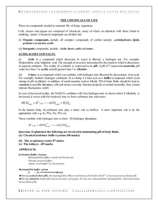
  • 1. 1 | C O R N E R S T O N E L E A D E R S H I P A C A D E M Y - B O Y S A - L E V E L B I O N O T E S 1 | E D I T E D b y K I N G S O Y E K W O k i n g s o y e 8 8 @ y a h o o . c o m 0 7 0 1 2 1 1 9 6 6 THE CHEMICALS OF LIFE These are compounds needed to maintain life of living organisms Cells, tissues and organs are composed of chemicals, many of which are identical with those found in nonliving matter. Chemical compounds are divided into (i) Organic compounds, include all complex compounds of carbon namely; carbohydrates, lipids, proteins and nucleic acids. (ii) Inorganic compounds, include; Acids,bases,salts and water. ACIDS, BASES AND SALTS. (i) Acid: Is a compound which dissociate in water to liberate a hydrogen ion. For example: Hydrochloric acid, Sulphuric acid. The strength of an acid is determined by the extentto which it dissociates in aqueous solutions. The acidity of a solution is expressed as its pH. A pH of 7 represents neutrality,pH value less than 7 is acidic and pH greater than 7 is Alkaline. (ii) A base:is a compound which can combine with hydrogen ions liberated by dissociation of an acid. For example; Sodium hydrogen carbonate. In so doing it a base acts as a buffer (compound which resist change in pH on dilution or addition of small amounts acid or Alkali). PH of body fluids should be kept as constant as possible because, cells and tissues can only function properly at around neutrality, they cannot tolerate fluctuations of pH. In case of increased acidity, the NaHCO3 combines with free hydrogen ions as shown above if alkalinity is increased, it reacts with free hydroxyl ions to form carbonate ions and water. ) ( 2 ) ( 2 ) ( ) ( 3 l aq aq aq O H CO H HCO       In the human body, bi-carbonate ions play a minor role as buffers. A more important role is by the appropriate salts e.g. K3 PO4,Na3 PO4 etc. These combine with hydrogen ions to form Di-hydrogen phosphate.       ) ( 4 2 2 ) ( 4 ) ( aq aq aq PO H HPO H Question. Explain howthe following are involved in maintaining pH of body fluids. (a) Chemical acid-base buffer systems (08 marks) (b) The respiratory centre 07 marks (c) The kidneys (05 marks APPROACH: (i) Protein Buffer System -Haemoglobin buffers inside red blood cells -Plasma protein buffers -Amino acid buffers by all proteins Haemoglobinbuffer system H+ + Hb HHb During acidosis(low pH), the haemoglobin (Hb) in red blood cells binds with H+ to increase intracellularpH; During alkalosis (high pH) and in presence of oxygen, H ions are released from haemoglobin, which decreases intracellularpH;
  • 2. 2 | C O R N E R S T O N E L E A D E R S H I P A C A D E M Y - B O Y S A - L E V E L B I O N O T E S 2 | E D I T E D b y K I N G S O Y E K W O k i n g s o y e 8 8 @ y a h o o . c o m 0 7 0 1 2 1 1 9 6 6 Plasma protein and Amino acid buffer systems During acidosis, exposed amino group of amino acids like histidine for plasma proteins like albumin accept H +To decrease acidity; During alkalosis,the exposed carboxyl group of amino acids can release H to increase acidity; (ii) Carbonic Acid - Bicarbonate Buffer System During acidosis(Low pH), NaHCO3 dissociates to form bicarbonate ions which combine with excess hydrogen ions to form carbonic acid; which dissociates into water and carbon dioxide,which can be eliminated by the respiratory system as pH is increased to the norm; During alkalosis(High pH), carbon dioxide reacts with water to form carbonic acid (H2CO3) which dissociate to form bicarbonate ionsand hydrogen ions;which lower the pH to the norm; OR (iii) Phosphate Buffer System When pH increases(alkalosis), dihydrogenphosphate dissociates into hydrogen ions and monohydrogen Phosphate ions; to lowerpH to the norm; When pH decreases (acidosis), hydrogenions react with monohydrogen phosphate ions to form dihydrogen phosphate; to increase pH to the norm; - (refer to proteins for more information) (iii) Salts: In the body, some of the common solutes found dissolved in water are mineral salts, compounds of a metal with nonmetal or non-metallic radical, e.g.sodium chloride. Some nutrients are need by the body in large amounts and are called Macronutrients while other nutrients are needed in small amounts thus called micronutrients. The functions of theses mineral salts and their relative derivatives are varied, but their roles can be summed up thus; (i) As constituents of various chemicals. (ii) As constituents of structures. (iii) As constituents of enzymes. (iv) As metabolic activators. (v) As constituents of certain pigments. (vi) As determinants of the anion – cation balance in cells. (vii) As determinants of osmotic pressure. Question: account for the different functions ofmineral salts in organisms General functions ofmineral salts (summary) - Formation of tissues such as a bone and teeth. - Essential for clothing of blood - Function as enzyme activators and components of enzymes. - Formation of hydrochloric acid in stomach.
  • 3. 3 | C O R N E R S T O N E L E A D E R S H I P A C A D E M Y - B O Y S A - L E V E L B I O N O T E S 3 | E D I T E D b y K I N G S O Y E K W O k i n g s o y e 8 8 @ y a h o o . c o m 0 7 0 1 2 1 1 9 6 6 - Maintenance of water balance - Maintenance of onion/cation balance - Components of haemoglobin, myoglobin and cytochromes. - Components of nuclei acids / DNA and RNA. - Involved in transfer of energy in ATP - Important in muscle and nuclear activity. - Needed for proper heart functioning - Important for amino acid formation and muscle growth. - Important for wound healing - Formation of hormones. - Preventing infection and body immunity. Inorganic ions and their importance in plants and animals. Macro element Function Notes and deficiency Nitrate NO3 - Ammonium NH4+ (i) Nitrogen is a component of amino acids, proteins, vitamins, coenzymes, nucleotides and chlorophyll: (ii) some hormones contain nitrogen e.g. Auxins in plants and insulin in animals In plants causes chlorosis (yellowing of leaves)and stunted growth. Phosphate PO43- Orthophosphate H2PO42- (i) A component of nucleotide, ATP and some proteins used in phosphorylation of sugars in respiration. (ii) Constituent of bones and teeth. (iii) Component of cell membranes in form of phospholipids. In plants leads to stunted growth, especially of roots, and formation of dull, dark green leaves: in animals, canresult into bone malformation called crickets. Sulphate SO42- Sulphur is a component of some proteins and certain coenzymes e.g. acetyl coenzyme A Sulphur forms important bridges between the polypeptide chains of some proteins, giving them their tertiary structure. Deficiency causes chlorosis and poor root development. Potassium K+ (i) Maintains electrical, osmotic and anion / cation balance across cell memb ranes. (ii) Involved in Potassium plays role in transmission of impulses. 1
  • 4. 4 | C O R N E R S T O N E L E A D E R S H I P A C A D E M Y - B O Y S A - L E V E L B I O N O T E S 4 | E D I T E D b y K I N G S O Y E K W O k i n g s o y e 8 8 @ y a h o o . c o m 0 7 0 1 2 1 1 9 6 6 active transport of certain materials across cell membrane. (iii) Co factor in photosynthesis and respiration. (iv) Constituent of sap vacuoles in plants, maintains turgidity. Deficiency leads to yellow edged leaves and premature death. Calcium Ca2+ In plants: (i) component of middle lamella. (ii) Aids translocation of carbohydrates and amino acids. In animals, (i) Constituent of bones, teeth and shells. (ii) Blood clotting. (iii) Muscle contraction. In plants, death of growing points. In animals causes rickets. Sodium Na+ (i) Maintains electrical, osmotic and anion / cation balance across membranes. (ii) Active transport across membranes. (iii) Constituent of sap vacuole. (i) Muscular clumps. (ii) Deficiency rare in plants. Chlorine Cl- (i) Maintains electrical, osmotic and anion / cation balance across membranes. (ii) Formation of hydrochloric acid in gastric juice. (iii) transport of carbon dioxide by blood (chloride shift) Causes muscular clumps in animals. Magnesium Mg2+ (i) Constituent of chlorophyll. (ii) Activator of some enzymes e.g. ATPase. (iii) Component of bone and teeth. Chlorosis in plants. Iron Fe2+ (i) Constituent of electron carriers e.g. cytochromes, needed in respiration and photosynthesis. (ii) A constituent of certain enzymes e.g. peroxidases, dehydrogenases, decarboxylases. (iii) Required in synthesis of chlorophyll. (iv) Forms part of haem group in respiratory pigments such as haemoglobin,,heamoerythrin, myoglobin. Chlorosis in plants and Aneamia in animals. Micronutrients / trace elements Magnesium Mn2+ (i) Activator of certain ennzymes eg phosphatases. (ii) Growth factor ion bone development Leaves with mottled with grey Bone deformation Copper Cu2+ (i) Constituent of some enzymes e.g. Cytochrome oxidase and tyrosinase. (ii) Component of haem ocyanin (a respiratory pigment) Young shoots die back at an early stage. Iodine I- Constituent of hormone thyroxin. Not required in higher plants. Causes cretinism in children and goiter in adults. Essential for metamorphic changes in some vertebrates. Cobalt Co2+ (i) Constituent of Vitamin B12, important in synthesis of RNA,nucleoprotein and red blood cells. Anemia
  • 5. 5 | C O R N E R S T O N E L E A D E R S H I P A C A D E M Y - B O Y S A - L E V E L B I O N O T E S 5 | E D I T E D b y K I N G S O Y E K W O k i n g s o y e 8 8 @ y a h o o . c o m 0 7 0 1 2 1 1 9 6 6 Zinc Zn2+ (i) Activator of certain enzymes, e.g. carbonic anhydrase. (ii) Required in plants for leaf formation, synthesis of auxins and alcoholic fermentation. Carbonic anhydrase is important in transportation of carbon dioxide in vertebrates’ blood. In plants, produces malformes and sometimes mottled leaves. Molybdenum Mo4+ Mo5+ (i) Reduction of nitrate to nitrite in formation of amino acids. Reduction in crop yields. Not vital in most animals. 2 Boron BO33+ B4O2+ (i) Required for uptake of Ca2+ by roots. (ii) Aids the germination of pollen grains and mitotic division in meristems. Not required by animals. Death of young shoots and abnormal growth. Fluorine F- Component of teeth and bones. Not required by most plants. Teeth decay QUESTION: OUTLINE THE ROLE OF MINERALS AND IONS IN BIOLOGICAL SYSTEMS. 1) They are components of smaller molecules e.g. phosphorus is contained in ATP and iodine is contained in thyroxin, etc. 2) They are constituents of large molecules e.g. proteins contain nitrogen and sulphur, phospholipids contain phosphorus, nucleic acids contain nitrogen and phosphorus, etc. 3) They are components of pigments e.g. haemoglobin and cytochromes which contain ion, chlorophyll contain magnesium, etc. 4) They are metabolic activators e.g. activates glucose before it is broken down in cell respiration, calcium ions activate ATPase enzyme during muscle contraction. 5) They determine the anion, cation balance e.g. Na+ ,K+ and Ca+ are important in transmission of impulses and muscle contraction. 6) They determine the osmotic pressure and water potential so that it does not fluctuate beyond narrow limits e.g. Na+ , K+ and Cl- are involved in water balance in the kidneys. 7) They are constituents of structures in cell membranes, cell walls, bones, enamel and shells. CHECK UP: By now you should be able to: (i) Describe properties of acids, bases and salts. (ii) Explain the role of acids, bases and salts in maintaining a stable internal environment for physiological processes. WATER Water is by far the most abundant component of organisms. Individual human cell contains about 80% water,and the whole body is made up of over 60%. Life originated in water and today numerous organisms make their home in it. Water provides the medium in which all biochemical reactions take place and has played a major role in the evolution of biological systems.
  • 6. 6 | C O R N E R S T O N E L E A D E R S H I P A C A D E M Y - B O Y S A - L E V E L B I O N O T E S 6 | E D I T E D b y K I N G S O Y E K W O k i n g s o y e 8 8 @ y a h o o . c o m 0 7 0 1 2 1 1 9 6 6 Some Biologically important functions of water. All organisms Plants Animals Structure- high water content of cells. Osmosis and turgidity, ( cell enlargement, guard cell mechanism, support) Transport in vascular system, lymphatic and excretory system. Solvent and medium for diffusion Reagent in photosynthesis. Osmoregulation. Reagent for hydrolysis Transpiratio n Cooling by evaporation, such as sweating and panting. Support for aquatic organisms. Translocation of inorganic ions and organic compounds. Lubrication, as in joints. Fertilization by swimming gametes Germination of seeds, swelling and breaking open of the testa and further development. Support, Hydrostaatic skeleton e.g. annelid worms. Dispersal of seeds, gametes and larval stages of aquatic organisms and seeds of some terrestrial species e.g. coconuts. Protection; e.g. mucus, tears Migration in ocean currents The importance of water as a medium for life comes from four of its properties: (a) Its solvent properties. Water’s properties as a solvent depend on the fact that it is a polar molecule (the distribution of electric charge is such that the centers of negative and positive charges are separated by a short distance). Because structure of a water molecule; instead of being arranged in a straight line, the hydrogen and oxygen atoms are situated asymmetrically (V – shape). The molecule as a whole shows polarity, Because Oxygen part has a net negative charge and hydrogen parts a net positive charge. Consider: if sodium chloride is placed in water, having both positive and negative charges,water attracts both ions of the molecule breaking it and clustering around it. Because of this:
  • 7. 7 | C O R N E R S T O N E L E A D E R S H I P A C A D E M Y - B O Y S A - L E V E L B I O N O T E S 7 | E D I T E D b y K I N G S O Y E K W O k i n g s o y e 8 8 @ y a h o o . c o m 0 7 0 1 2 1 1 9 6 6 (i) Water is a good solvent, ionic solids and polar molecules readily dissolve in it , this is of a very biological importance because all chemical reactions taking place in a cell occur in aqueous solution. (ii) Water molecules associate with each other (positive hydrogen atom of one molecule maybe attracted to negative oxygen atom of another) forming hydrogen bonds important in holding organic molecules together. (b) Thermal properties: Heat capacity, is the amount of heat required to raise the temperature of 1g by 1o C. Water has very high heat capacity compared with other liquids. Therefore a large increase in heat results into a comparatively small rise in temperature of water. This implies water is good at maintaining its temperature constant irrespective of changes in temperature of the surrounding environment. This is biologically important because; (i) Biochemical processes proceed in very narrow range of temperatures,and most organisms cannot tolerate wide variations of temperature (ii) High thermal capacity of water keeps the temperature constant, thus making it an ideal for plant and animal life. (iii) Water has remarkably high boiling point, thus hardly evaporate. (iv) It provides a sustainable habitat for aquatic organism the habitat does not easily get frozen in cold weather. (v) It maintains an effective transport medium of materials in the body since it maintains its liquid form even on the cold weather. (vi) Aquatic organisms can survive even in winter because the lower part of the water bodies hardly freezes
  • 8. 8 | C O R N E R S T O N E L E A D E R S H I P A C A D E M Y - B O Y S A - L E V E L B I O N O T E S 8 | E D I T E D b y K I N G S O Y E K W O k i n g s o y e 8 8 @ y a h o o . c o m 0 7 0 1 2 1 1 9 6 6 (c) Surface tension. This is the force that causes the surface of liquid to contact so that it occupies the least possible area. It is due to inward – acting cohesive forces between molecules at the surface being caused by polarity of water. Waterhashigh surface tension and molecules dissolved in watertend to lower its surface tension and collect at the interface between liquid and other phases. This is biologically important in: (i) Development of plasma membrane and movement of molecules across it. (ii) Movement of water up the capillary like vessels and tracheid in the stems of plants (iii) Enables the surface film to support and provide habitat for certain aquatic organisms. (d) Freezing properties: When water is cooled below a certain temperature, its volume increases and its density decreases. This means ice tends to float than to sink. When the temperature drops the coldest water is at the surface and being less dense than the slightly warmer water lower down, tends to remain at the surface. So ice forms at the surface fist, and bottom later. Organisms which live towards the bottom of fresh water lakes are protected from freezing increasing their survival. QUESTION: HOW DO THE PROPERTIES OF WATER RELATE TO ITS BIOLOGICAL ROLE? 1 Water is transparent and this allows light penetration in aquatic habitats to enable photosynthesis of aquatic autotrophs and visibility of aquatic animals. 2 Water has a low viscosity and this al lows for smooth flow of water and other dissolved substances in an aquatic medium for easy transport. 3 It has a high surface tension providing support to aquatic organisms and allowing movement of living organisms on water surface. 4 Has a high latent heat of vaporization hence a cooling effect on the body surface since evaporation of water from the body of an organism draws out excess heat. 5 It has a high boiling point thus provides a stable habitat and medium since a lot of heat which is not normally provided in the natural environment is needed to boil the water. 6 It has a high latent heat of fusion and hence a low freezing point thus providing a wide range of temperature for survival of aquatic organisms since it prevents freezing of cells and cellular components. 7 It has a high specific heat capacity which minimizes drastic temperature changes in biological systems and provides a constant external environment for many plant cells and aquatic organisms. 8 It has a maximum density at 4o C hence ice floats on top of water insulating the water below hence increasing the chances of survival of aquatic organisms below the ice. 9 Water is liquid at room temperature providing a liquid medium for living organisms and metabolic reactions and a medium of transport.
  • 9. 9 | C O R N E R S T O N E L E A D E R S H I P A C A D E M Y - B O Y S A - L E V E L B I O N O T E S 9 | E D I T E D b y K I N G S O Y E K W O k i n g s o y e 8 8 @ y a h o o . c o m 0 7 0 1 2 1 1 9 6 6 10 It has high adhesive and cohesive forces creating enough capillarity forces for transport in narrow tubes of biological systems. 11 It is a universal solvent hence providing a medium for biochemical reactions. 12 Water is a polar molecule allowing solubility of polar substances, ionization or dissociation of biochemical substances. 13 Water is incompressible thus providing support in hydrostatic skeleton and herbaceous stems. 14 Water is neutral hence does not alter the pH of cellular components on their environment. 15 A water molecule is relatively small for easy and fast transport across a membrane. CHECK UP: By now you should be able to: (i) Describe the molecular structure of water. (ii) State f unctions of water. (iii) Explain the importance of water as a solvent. (iv) Relate the water properties to its role in the life of organisms. A. CARBOHYDRATES These are food substances which contain elements; carbon, hydrogen and Oxygen. They have a general formula C x (H2O) y, where Xand Y are variable numbers. Their name Hydrate of carbon is derived from the fact that Hydrogen and oxygen are present in the same proportions as in water, namely two hydrogen atoms per oxygen atom. In addition: (i) All carbohydrates are either aldehydes or ketones. (ii) All contain severalhydroxyl groups. Question Howis carbon uniquely suited to its role as the main element in living organisms. APPROACH  Great strength of the carbon-to-carbon bond ensures that carbon-based molecules are very stable.  Great strength of the carbon-to-hydrogen bond ensures that the hydrocarbon skeleton, typical of carbon-based organic moleculesis very stable.  Ability of carbon to form repetitive bonds with itself/catenation leads to; enormously varying lengths of chains,enormously varying extent of branching, enormously varying length of branches, enormously varying forms of ringed compounds,ensuring that carbon-based molecules can form a variety of shapes.  Great stability of the +4 oxidation state of carbon owing to its very small atomic size, ensures high stability of carbon-based molecules formed in the +4 oxidation state.
  • 10. 10 | C O R N E R S T O N E L E A D E R S H I P A C A D E M Y - B O Y S A - L E V E L B I O N O T E S 10 | E D I T E D b y K I N G S O Y E K W O k i n g s o y e 8 8 @ y a h o o . c o m 0 7 0 1 2 1 1 9 6 6  Ability of carbon to formmultiple bondswith itself and other elements, leads to a wide variety of carbon-based molecules.  Ability of carbon to form stable covalent bonds with other elements, ensures that carbon-based molecules contain a variety of elements Carbohydrates are divided into three main classes namely; (i) Monosaccharaides (ii) Disaccharides (iii) polysaccharides. 1. Monosaccharaides; These are single sugar units. Their generalformula is (CH2O) n ., Where n is variable number. Properties:(i) sweet. (ii) Soluble in water. (iii) Crystalline. (iv) Low molecular weight Classification: They are class ified according to number (n) of carbon atoms. Example: (i) Trioses, 3 carbon atoms, e.g. Glyceraldehyde. (ii) Pentoses,5 carbon atoms, e.g.Ribose, Deoxyribose. (iii) Hexoses,6 carbonatoms e.g. Glucose, Fructose, Galactose. Aldoses and ketoses:In monosaccharaides,all carbon atoms except one having a hydroxyl group attached, the remaining carbon is either part of an aldehyde group, and the monosaccharide is called aldose or aldo sugar, or is part of a keto group, monosaccharide called ketose or keto sugar. Aldoses include: Glyceraldehyde, Ribose, and Glucose. And Ketoses include: Dihdroxyacetone, Ribulose, and fructose. Trioses ( C3H6O3 ) Pentoses ( C5H10O5) Hexoses ( C6H12O6 ) Aldoses ( - CHO) ( Aldo – sugar) Glyceraldehyde Ribose Glucose, Galactose Ketoses ( C=O ) (keto sugars) Dihdroxyacetone Ribulose Fructose Glyceraldehyde Dihydroxyacetone.
  • 11. 11 | C O R N E R S T O N E L E A D E R S H I P A C A D E M Y - B O Y S A - L E V E L B I O N O T E S 11 | E D I T E D b y K I N G S O Y E K W O k i n g s o y e 8 8 @ y a h o o . c o m 0 7 0 1 2 1 1 9 6 6 Open chain and ring forms. The open chain form can be straight, Because of bond angles between carbon atoms, hexoses and pentoses, b end to form a stable ring structures. In hexoses like glucose, First carbon atom combines with the oxygen on carbon atom five to give a six – membered ring. Note that oxygen is part of the ring and one carbon atom, carbon atom number 6, sticks up out of the ring. In Pentoses, the first carbon atom combines with oxygen atom on the fourth carbon atom forming a five – membered ring. These ring structures are the forms used to make disaccharides and polysaccharides. Ribose open chain Ribose with five – membered ring Glucose:Is a common monosaccharide, It a hexose sugar ( C6H12O6 ). Has open chain structure and ring structure. In Ring structure, has two isomers namely; Alpha and Beta isomers. The hydroxyl group on carbon atom 1 canproject below the ring forming Alpha glucose or above the ring forming the Beta glucose. Alpha glucose is used in making polysaccharide starch, Beta glucose is used in making polysaccharide cellulose. Open chain form  glucose  Glucose
  • 12. 12 | C O R N E R S T O N E L E A D E R S H I P A C A D E M Y - B O Y S A - L E V E L B I O N O T E S 12 | E D I T E D b y K I N G S O Y E K W O k i n g s o y e 8 8 @ y a h o o . c o m 0 7 0 1 2 1 1 9 6 6 Formation of a ring molecule of beta- D-glucose Functions of monosaccharaides: Monosaccharides Examples Functions Trioses Glyceraldehyde Dihdroxyacetone Intermediates in respiration (Glycolysis) , Photosynthesis (dark reactions) and other branches of carbohydrate metabolism. Pentoses Ribose Deoxyribose Ribulose (i) Synthesis of nucleic acids. (ii) Synthesis of some coenzymes (Ribose is used in synthesis of NAD and NADP). (iii) Synthesis of ATP requires ribose. (iv) Ribulose biphosphate is the CO2 acce[ptor in photosynthesis, made from ribulose. Hexoses Glucose Fructose Galactose (i) Source of energy when oxidized in respiration. (ii) Synthesis of disaccharides. (iii) Synthesis of polysaccharides
  • 13. 13 | C O R N E R S T O N E L E A D E R S H I P A C A D E M Y - B O Y S A - L E V E L B I O N O T E S 13 | E D I T E D b y K I N G S O Y E K W O k i n g s o y e 8 8 @ y a h o o . c o m 0 7 0 1 2 1 1 9 6 6 2. Disaccharides: These are double sugar units formed when two monosaccharaides usually hexoses combine by means of a chemical reaction called condensation. This means removal of water. The bond formed between two monosaccharaides as a result of condensation is called Glycosidic bond and it normally forms between carbon atom 1 and 4 of neighboring units ( 1,4 bond or 1,4 linkage ). The monosaccharide molecules are called residues once they have been linked. Maltose molecule contains two glucose residues. Common disaccharides are; (i) Maltose (Glucose + Glucose). (ii) Lactose (Galactose + Glucose). (iii) Sucrose (Fructose + Glucose). Formation of Maltose molecule (disaccharide) from Glucose molecules (monosaccharaides) Addition of water to a disaccharide under suitable conditions, splits into monosaccharaides and this called Hydrolysis (splitting by water). Likes monosaccharaides,disaccharides have the following characteristics;  They are small molecules with a low molecular mass  They are readily soluble in water  Have a sweet taste  They exist in crystalline form  They are made by combining two monosaccharides in a condensation reaction Formation ofa sucrose molecule
  • 14. 14 | C O R N E R S T O N E L E A D E R S H I P A C A D E M Y - B O Y S A - L E V E L B I O N O T E S 14 | E D I T E D b y K I N G S O Y E K W O k i n g s o y e 8 8 @ y a h o o . c o m 0 7 0 1 2 1 1 9 6 6 Formation ofa maltose molecule + H2O Reducing sugars: All monosaccharaides and some disaccharides (Maltose and Lactose) are reducing sugars, meaning they can carry out a type of chemical reaction called Reduction. Sucrose is the only common non reducing sugar.Two common testsfor reducing sugars; (i)Benedict’stest.(ii)Fehling’stest, make use of the ability of these sugars to reduce copper from a valence of 2 to a valence of 1. Both tests involve use of alkaline blue solution of copper (II) sulphate which is reduced to insoluble brick-red precipitate copper (I) oxide. Equation showing reduction reaction involving a reducing sugar and test reagent FUNCTIONS OF DISSACCHARIDES i. They are food reserves in organisms and when they are hydrolysed to monosaccharide and used in cell metabolism. ii. They are the main forms of transport of organic substances in the phloem. iii. Storage materials in some plants like sugar canes. iv. They are energy reserves. NB: Starch is converted in plants to maltose so as;-
  • 15. 15 | C O R N E R S T O N E L E A D E R S H I P A C A D E M Y - B O Y S A - L E V E L B I O N O T E S 15 | E D I T E D b y K I N G S O Y E K W O k i n g s o y e 8 8 @ y a h o o . c o m 0 7 0 1 2 1 1 9 6 6 i. To be transported easily because it’s soluble in water. ii. Maltose is less reactive hence won’t be used. 3. Polysaccharides: Polysaccharides are formed when monosaccharaides polymerize in a condensation reaction between two hydroxyl groups, resulting in a covalent interaction called a glycosidic linkage. They are normally used for food storage because;- i. They can be hydrolyzed to sugars when required for production. ii. They fold into compact shapes which cannot diffuse out of the cells. iii. They exert no osmotic or chemical influence on the cell. iv. They have large sizes that make them insoluble in water. v. They are non-diffusible i.e. they don’t leave sites of storage vi. Making structures compact e.g. cellulose. The monosaccharide polymer may be branched, unbranched or coiled. In cells they exists as granules or grains. Examples ofpolysaccharides include: (a) Starch: [A storage polysaccharides in plants] It is a main food storage form of plants and it is a polymer of  glucose it exists as grains in the cells .it is mainly found in seeds, leaves and tubers. It comprises of amylose 20%,Amylopectin 79% together with other substances(1%)e.g.phosphate..Starch has two components: (i) Amylose, this has straight chain structure consisting of several thousand α-glucose residues joined by α (1-4) glycosidic bonds. Soluble starches are mainly made up of amylose. They do not dissolve, but form colloidal suspensions. Amylose is a polymer of α-glucose molecules that form unbranched molecule wound in a helix. The bore of the helix is large enough to trap iodine molecules and produce the blue complex during iodine tests for starch using iodine solution in potassium iodide,
  • 16. 16 | C O R N E R S T O N E L E A D E R S H I P A C A D E M Y - B O Y S A - L E V E L B I O N O T E S 16 | E D I T E D b y K I N G S O Y E K W O k i n g s o y e 8 8 @ y a h o o . c o m 0 7 0 1 2 1 1 9 6 6 Amylose helix (iii) Amylopectin, is compact with many branches, formed by 1, 6 glycosidic bonds. It has up to twice as many glucose molecules as amylose. Just like in amylose, the unbranched chain of amylopectin has molecules of α-glucose held together by 1-4 glycosidic bonds, but the side branches are held together by α(1-6) glycosidic bonds. Amylopectin also forms colloidal suspension in water. The two components of starch fit together into a compact 3-D structure in which the branches of amylopectin are entangled by the helices of amylose In iodine (potassium iodide solution); (i) Amylose suspension gives a blue black colour. (ii) Amylopectin Suspension gives a red – violet colour. Starchmolecules accumulate to form starch grains found in; many plant cells (chloroplast of leaves), Storage organs (tubers), seeds of cereals and legumes. Formation of one branch in amylopectin
  • 17. 17 | C O R N E R S T O N E L E A D E R S H I P A C A D E M Y - B O Y S A - L E V E L B I O N O T E S 17 | E D I T E D b y K I N G S O Y E K W O k i n g s o y e 8 8 @ y a h o o . c o m 0 7 0 1 2 1 1 9 6 6 Differences between amylose and amylopectin Amylose Amylopectin Stains blue with iodine Stains red-purple with iodine Has a low RMM, maximum 500000 Has a high RMM up to 500000 Has between 200 to 500 glucose units Has between 5000 to 100000 glucose units Has unbranched helical chain Has branched chain Has 1,4 gylcosidic bonds Has both 1,4 and 1,6 gylcosidic bonds Has 20% composition in starch molecule Has 79% composition in starch molecule Adaptations of starch as a storage compound: - It is in soluble in water, therefore it exerts no osmotic influence in the cell. - It exerts no chemical influence in the cells due to its insoluble chemical nature. - It can fold into compact shape, thus minimizing the space occupied. - It can easily be converted back to glucose by hydrolysis. The glucose can be used in respiration. (b) Glycogen:[Highly branched storage polysaccharides in animals and also fungi] This is an animal equivalent of starch, being a storage polysaccharide made from α-glucose, many fungi also store it. In invertebrates, it’s stored mainly in liver and muscles, both centers of high metabolic activity, where it provides a useful energy reserve. Similar to amylopectin, but shows more branching.
  • 18. 18 | C O R N E R S T O N E L E A D E R S H I P A C A D E M Y - B O Y S A - L E V E L B I O N O T E S 18 | E D I T E D b y K I N G S O Y E K W O k i n g s o y e 8 8 @ y a h o o . c o m 0 7 0 1 2 1 1 9 6 6 A molecule ofglycogen (c) Cellulose:[A structural polysaccharide in plants] Cellulose is a polymer of β-glucose monomers, joined by β-1,4glycosidic linkages.In plants, cellulose is the major component of the cell wall. The geometry of the linkage is such that each glucose monomer in the chain is flipped in relation to the adjacent monomer. The flipped orientation is important because (i) It generates a linear molecule, rather than the helix seen in starch; and (ii) It permits multiple hydrogen bonds to form between adjacent, parallel strands of cellulose. As a result, cellulose forms long, parallel strandsthat are joined by hydrogen bonds. The linked cellulose fibers are strong and give the cell structural support. Structure It is a polymer with straight chains of β-glucose units held by glycosidic bonds with the OHgroup projecting out wards from each chain forming cross linkages of hydrogen bonds with adjacent chains. The cross linking binds the chains together which associate to form micro fibrils that are arranged in larger bundles to form macro fibrils. Note: Starch lacks the structural properties possessed by cellulose because it lacks cross linkages. The stability of cellulose makes it difficult to digest and therefore not a good source of food to animals except those which have cellulase producing microorganisms which live in them in a symbiotic way e.g. in the rumen of cattle, goats, sheep, etc. Formation of Cellulose from Beta glucose. Question: What properties of glycogen makes it a betterstoragecarbohydrate in animals than starch
  • 19. 19 | C O R N E R S T O N E L E A D E R S H I P A C A D E M Y - B O Y S A - L E V E L B I O N O T E S 19 | E D I T E D b y K I N G S O Y E K W O k i n g s o y e 8 8 @ y a h o o . c o m 0 7 0 1 2 1 1 9 6 6 The plant cell wall is permeable to water because the matrix has minute water channels through which diffusion occurs. Salts, sugars, and other polar molar molecules can easily disuse through these minute channels. Cellulose is hydrophilic and this makes the cell walls always saturated with water. Structure of cellulose showing Hydrogen bonds. Question: Explain why cellulose formsa structural carbohydrate while starch does not (07 marks) Approach Cellulose is made up long chains of glucose linked by β-1–4 glycosidic bonds; This results in the glucose molecules being alternately one way up, then the other; This means that chains lying side
  • 20. 20 | C O R N E R S T O N E L E A D E R S H I P A C A D E M Y - B O Y S A - L E V E L B I O N O T E S 20 | E D I T E D b y K I N G S O Y E K W O k i n g s o y e 8 8 @ y a h o o . c o m 0 7 0 1 2 1 1 9 6 6 by side can form hydrogen bonds with each other, making microfibrils; Starch, on the other hand, is made up of long chains of glucose linked by α 1–4 glycosidic bonds; It twists into a spiral, and one starch molecule does not form bonds with others; so no fibrils are formed; Moreover, it is difficult to break down β 1–4 glycosidic bonds;so not many organisms can digest cell walls; Uses of cellulose  Rayon produced form cellulose extracted from wood is used in the manufacture of tyre cords.  Cotton is used in the manufacture of fibres and closes.  Cellophane used in packaging is produced from cellulose.  Paper is a product of cellulose.  Celluloid used in photographic films is also a derivative of cellulose. DIFFERENCES BETWEENCELLULOSE ANDGLYCOGEN CELLULOSE GLYCOGEN Polymer of ß-glucose Polymer of ∞-glucose Monomers rote alternately at1800 to eachother No rotation of adjacent monomers Straight chains, with cross-linking due to hydrogen bonds Branched chains ß-1,4 linkages ∞-1,4 and 1,6 linkages forms micro fibrils Does not form micro fibrils hydroxyl groups project outwards Hydroxyl groups project inwards performs structural role not for storage Performs storage role, no structural role found in plants Found in animals & fungi feworganisms can digest cellulose/ Difficult to digest Many organisms can digest cellulose/easily digested HOW DOES THE STRUCTURE OF CELLULOSE RELATE TO ITS ROLES ® The cross linking binds the chains together offering much tensile strength. ® The micro fibrils in cell walls are arranged in several layers offering protection to the plant cell preventing it from bursting when water enters by osmosis. ® The arrangement of micro fibrils in cell walls contributes to turgidity hence offering support. ® The parallel layers of cellulose are fully permeable to water and solutes. ® The arrangement of micro fibrils determines the shape of the cells and hence plant organs since it determines the direction in which cells expand as they grow.
  • 21. 21 | C O R N E R S T O N E L E A D E R S H I P A C A D E M Y - B O Y S A - L E V E L B I O N O T E S 21 | E D I T E D b y K I N G S O Y E K W O k i n g s o y e 8 8 @ y a h o o . c o m 0 7 0 1 2 1 1 9 6 6 ® The glycosidic bonds holding the β-glucose units in cellulose can be broken down in presence of enzyme cellulase so that a free glucose molecule can be respired. (d) Chitin: [A structural polysaccharide in fungi and animals]. Chitin is similar to cellulose, but instead of consisting of glucose monomers, the monosaccharide involved is one called N-acetylglucosamine (NAG). NAGhas an aminoacyl group (NHOCCH3) at Carbon 2 of the glucose molecule, therefore cellulose is very similar to chitin. These NAG monomers are joined by β-1, 4-glycosidic linkages. As in cellulose, the geometry of these bonds results in every other residue being flipped in orientation. Like the glucose monomers in cellulose, the NAG subunits in chitin form hydrogen bonds between adjacent strands. The result is a tough sheet animals. It is, for example, the most important that provides stiffness and protection. Chitin is a polysaccharide that stiffens the cell walls of fungi. It is also found in a few types of protists and in many component of the external skeletons of insects and crustaceans. (e) Peptidoglycan:[A Structural Polysaccharide in Bacteria] Peptidoglycan is the most complex of the polysaccharides discussed thus far. It has a long backbone formed by two types of monosaccharaides that alternate with each other and are linked by β-1,4-glycosidic linkages. Most bacteria,like all plants, have cell walls. But unlike plants, in bacteria the ability to produce cellulose is extremely rare. Instead,peptidoglycan gives bacterial cell walls strength and firmness. (f) INULUN(α-fructose ) This is a major storage form of carbohydrates in plants like Dahlia (Jerusalem artichoke). It is a polymer of fructose molecules .It comprises 1, 2 glycosidic bonds and has unbranched chain of fructose molecules. Question: Describe the Structural features of carbohydrates accounting for wide variety of polysaccharides Approach ® Both pentoses and hexoses can be used to make polysaccharides ® Two types of linkage 1,4 and 1,6 glycosidic bonds occur between sugar units leading to branching. ® Lengths of chains vary enormously ® Lengths of branches vary enormously ® Extent of branching varies enormously ® Monosaccharides have alpha and beta isomeric ring forms.
  • 22. 22 | C O R N E R S T O N E L E A D E R S H I P A C A D E M Y - B O Y S A - L E V E L B I O N O T E S 22 | E D I T E D b y K I N G S O Y E K W O k i n g s o y e 8 8 @ y a h o o . c o m 0 7 0 1 2 1 1 9 6 6 ® Sugars may be ketoses or aldoses ® Aldehyde, ketone,and hydroxyl groups of sugars, makethem highly reactive molecules. (g) Mucopolysaccharides: These are carbohydrate derivatives derived from a combination of sugar molecules and amino acids in a condensation reaction. This group includes hyaruronic acid which forms part of the matrix of vertebrae connecting tissue. Heparin, an anti-coagulant also contains muco- polysaccharides. They contain amino sugars and are found in; (i) The basement membrane of epithelium (ii) The matrix of connective tissues (iii) Synovial fluid in joints of vertebrates (iv) Matrix of bone and cartilage. (v) Vitreous humor of the eye LIPIDS Lipids include Fats (solids at room temperature) and Oils (Liquids at room temperature). Like carbohydrates, lipids contain Carbon, Hydrogen and oxygen BUT a lipid molecule contain smaller proportion of oxygen than carbohydrate molecule. Constituents oflipids. Lipids are made up of; (a) Fatty acids These contain the acidic group –COOH (Carboxyl group). Named so because larger molecules in the series occur in fats. General formula of R.COOH where R is a hydrogen or group (-CH3 , -C2H5 etc increasing by –CH2 for each subsequent member of the series ). Fatty acids have a long chain of long chain of carbon and hydrogen atoms forming a hydrocarbon tail. The tail determines many properties of lipids including solubility in water. The tails are Hydrophobic (Hydro,water; Phabos,fear), water hating. Fatty acids can be: (i) Saturated, lack double bonds. (ii) Unsaturated,Contain one or more double bonds (C=C ). (b) Glycerol: The formula of glycerol is C3H8O3,This shows no variation and is the same in all lipids. Glycerol has three hydroxyl groups of which can condense with a fatty acid. Usually all the three undergo condensation and the lipid formed is called a triglyceride. Formation oftriglycerides Lipids are made of fatty acids and glycerol. Glycerol has 3 OH groups and each combines with a separate fatty acid to form a lipid chemically known as a triglyceride. This is a condensation reaction that leads to liberation of 3 water molecules.
  • 23. 23 | C O R N E R S T O N E L E A D E R S H I P A C A D E M Y - B O Y S A - L E V E L B I O N O T E S 23 | E D I T E D b y K I N G S O Y E K W O k i n g s o y e 8 8 @ y a h o o . c o m 0 7 0 1 2 1 1 9 6 6 Metabolic water is formed and an ester bond is formed between the glycerol and fatty acid. Since the glycerol possesses 3 hydroxyl groups to which 3 fatty acids attach themselves, 3 water molecules are formed. In this reaction, the fatty acids may all be the same or different, saturated or unsaturated. Formation of Triglyceride from glycerol and fatty acids. Characteristicsof lipids. 1. They are made up of carbon, hydrogen and oxygen. They are composed of 3 fatty acids (glycerol alcohol), 3 CH groups. The general formula of glycerol is C3H8O2. The fatty acids contain the carboxylic acid/carboxyl group (-COOH) and a hydrocarbon group.
  • 24. 24 | C O R N E R S T O N E L E A D E R S H I P A C A D E M Y - B O Y S A - L E V E L B I O N O T E S 24 | E D I T E D b y K I N G S O Y E K W O k i n g s o y e 8 8 @ y a h o o . c o m 0 7 0 1 2 1 1 9 6 6 2. They have a high proportion of hydrocarbon groups in their molecules. 3. They are insoluble in H2O but soluble in organic solvents e.g. benzene and chloroform alcohol. This is because lipids have a low O2 and the numbers of polar hydroxyl groups are few and this decreases their solubility in H2O. 4. They are either solids (fats) or liquids (oils) at room temperature. 5. The higher the proportion of unsaturated fatty acids, the lower will be their melting points. 6. They are non-polar and this makes them effective storage materials. 7. They are less dense than H2O and this helps the aquatic organism to float on H2O. 8. They have a function group COOH composed of fatty acids and glycerol. FATTY ACIDS Fatty acids are long molecules with a polar, hydrophilic end and a non-polar, hydrophobic "tail". The hydrocarbon chain can be from 14 to 22 CH2 units long. The hydrocarbon chain is sometimes called an R group, so the formula of a fatty acid can be written as R-COOH. All occurring fatty acids can be saturated or unsaturated Unsaturatedfatty acids are ones where the hydrocarbon part contains one or more double bonds e.g. linoleic acid and oleic acid .The saturated don’t contain double bonds. The saturated fatty acids have a high boiling or melting points compared to the unsaturated of the same molecular mass. Fatty acids Formula Unsaturated Sources Butyric acid C3H17COOH Unsaturated Butter Linolenic acid C17H31COOH Saturated Lin seeds Oleic acid C17H33COOH Unsaturated All fat Palmitic acid C15H31COOH Unsaturated Palm oils
  • 25. 25 | C O R N E R S T O N E L E A D E R S H I P A C A D E M Y - B O Y S A - L E V E L B I O N O T E S 25 | E D I T E D b y K I N G S O Y E K W O k i n g s o y e 8 8 @ y a h o o . c o m 0 7 0 1 2 1 1 9 6 6 Stearic acid C17H35COOH Saturated Adipose fats Arachidonic acid C19H39COOH Saturated Peanut oils Cerotic C25H51COOH Saturated Wool oil. Illustration of different fatty acids NB: There are some fatty acids which cannot be synthesized in the body. Therefore, they have to be obtained from the diet and these are called essential fatty acids. Their deficiency results into retarded growth, reproductive deficiency and kidney failure. The body synthesizes some fatty acids and they are synthesized from compounds of proteins and carbohydrate metabolism. FUNCTIONS OF LIPIDS. i) They serve as energy sources or stores and yield much more energy than carbohydrates i.e. 1g of lipids yields 38kJ or 9k calon oxidation while that the carbohydrates of 1g of carbohydrates yields 17kJ OR 4k Cal. ii) In animals, it is found below the skin and acts as an insulator since it’s a poor conductor of heat. It is most found in animals living in cold climates and aquatic mammals have very thick subcutaneous fat called BLUBBER. iii) Fats provide H2O to desert animals when oxidized e.g. on camels where they are heavily deposited in the hump.
  • 26. 26 | C O R N E R S T O N E L E A D E R S H I P A C A D E M Y - B O Y S A - L E V E L B I O N O T E S 26 | E D I T E D b y K I N G S O Y E K W O k i n g s o y e 8 8 @ y a h o o . c o m 0 7 0 1 2 1 1 9 6 6 iv) They are constituents of the plasma membrane when they combine with phospholic acids to form phospholipids. They are constituents of the waxy cuticle of plants and insects giving water repelling properties making them cuticle waterproof Since they are insoluble in water v) They are used to protect delicate organs of the body by offering cushion vi) Buoyancy function in that they help the aquatic organism to float and the oils on bird feathers help the aquatic varieties to float. vii) Normal functioning of the brain. SUITABILLITYOF LIPIDS OVER GLYCOGEN FOR STORAGE - Lipids yield more metabolic water on oxidation important for survival of desert animals - Fat insulates the body against heat loss whereas glycogen does not. - Fats stored around delicate organs protect them from mechanical/physical damage. - Fats store fat-soluble vitamins A, D, E and K - Fats are more compact than glycogen and so occupy less space - Lipids are lighter than glycogen hence contribute less weight to organism and structures - Lipids are store and yield more energy than glycogen - Lipids are less dense than water and so aid buoyancy of aquatic animals QUESTION: Discuss the structural and physiological functions oflipids (10 marks) Structural: i) They are components of the plasma/cell membrane. ii) They form subcutaneous fat in the dermis of the skin hence insulating the body since they are poor conductors of heat. iii) They are components of the waxy cuticle in plants and insects there by preventing water loss (desiccation). iv) They form a component of the myelin sheath of nerves hence playing a role in the transmission of impulses. iv) They protect delicate organs e.g. the heart and kidney from injury. v) They coat on fur of animals enabling it to repel water which would otherwise wet the organism. vii) They are component of adipose tissue. Physiological: i) They provide energy through oxidation. ii) They are solvents for fat soluble vitamins (ADEK). iii) They are a good source of metabolic water to desert animals, young birds and reptiles while still in their shells. iv) They are a constituent of the brown adipose tissue which provides heat for temperature regulation (thermogenesis). Other functions: i) Some lipids provide a scent in plants which attracts insects for pollination. ii) Wax is used by bees to construct honey combs. iii) Wax from bees is used in the manufacture of candles. LIPID DERIVATIVES
  • 27. 27 | C O R N E R S T O N E L E A D E R S H I P A C A D E M Y - B O Y S A - L E V E L B I O N O T E S 27 | E D I T E D b y K I N G S O Y E K W O k i n g s o y e 8 8 @ y a h o o . c o m 0 7 0 1 2 1 1 9 6 6 Phospholipids Phospholipids consist of a glycerol that is linked to a phosphate group and two hydrocarbon chains of fatty acids. Has phosphate head which is Hydrophilic (water loving) and water hating tails. This is important in formation of cell membranes. The fatty acid chains have no electrical charge and so are not attracted to the dipoles of water molecules They are said to be hydrophobic. The phosphate group has an electrical charge and is attracted to water molecules. It is hydrophilic. In water, a group of phospholipid molecules therefore arranges itself into a bilayer, with the hydrophilic heads facing outwards into the water and the hydrophobic tails facing inwards, therefore avoiding contact with water Formation of a phospholipid. How the phospholipids arrange themselvesin water environment
  • 28. 28 | C O R N E R S T O N E L E A D E R S H I P A C A D E M Y - B O Y S A - L E V E L B I O N O T E S 28 | E D I T E D b y K I N G S O Y E K W O k i n g s o y e 8 8 @ y a h o o . c o m 0 7 0 1 2 1 1 9 6 6 Glycolipid: Are associations of lipids with carbohydrates. The carbohydrate forms a polar head to the molecule. Glycolipids like phospholipids are found in membranes. Waxes: Waxes are formed by combination of fatty acids with alcohol other than glycerol. Alcohol is much larger than glycerol therefore waxes have more complex chemical structure. Waxes are important for; (i) Waterproofing in plants and animals. (ii) Form Storage compounds in few organisms like castor oil and in fish. Steroids: Steroids are a family of lipids distinguished by the bulky, four-ring structure. The various steroids differ from one another by the functional groups or side groups attachedto different carbons in those hydrophobic rings. Cholesterol is common steroid in animals. It’s important for: i) Limit uncontrolled leakage of small molecules (ions and water) in and out of the plasma membranes. ii) Constituent of myelin, facilitates movement of impulse along neuron. iii) Formation of steroid hormones and bile salts. How cholesterol causes arthesclerosis Cholesterol is produced by the liver and is used as the starting point for the synthesis of other steroid molecules. The major source of cholesterol is diet and many dairy products are rich in cholesterol or fatty acids from which cholesterol can be synthesized. Thyroxin stimulates cholesterol production in the liver and also increases the rate of excretion of bile. Excessive amounts of cholesterol in blood can be harmful. It can be deposited in walls of arteries leading to arthesclerosis and increased risk of formation of a blood clot which may block blood vessels, a condition known as thrombosis. This is often fatal if it occurs in the coronary artery in the wall of the heart (coronary thrombosis or heart attack) or brain (cerebralthrombosis). Although cholesterol is harmful in excess,it is essential to have some in the diet for reasons stated B. PROTEINS Proteins contains Carbon, hydrogen, oxygen and Nitrogen but sometimes sulphur and phosphorus. Proteins are built up from amino acids. AMINO ACIDS These are groups of many chemicals of which around 20 occur in proteins. They contain an amino group (NH2) and a carboxyl group (COOH). Most amino acids have one of each and are therefore neutral but a few have more amino groups than carboxyl making them alkaline or may have more carboxyl than amino groups making them acidic.
  • 29. 29 | C O R N E R S T O N E L E A D E R S H I P A C A D E M Y - B O Y S A - L E V E L B I O N O T E S 29 | E D I T E D b y K I N G S O Y E K W O k i n g s o y e 8 8 @ y a h o o . c o m 0 7 0 1 2 1 1 9 6 6 Structure of amino acids Where R is a variable Amino acids are soluble in water and ionize to form ions. The carboxyl end of the amino acid is acidic in nature. It will ionize in water to give H+. This will make the COOH group negatively charged. The amino end (NH2) is basic in nature. It attracts the H+ in solution making it positively charged. The ion is now dipolar i.e. having a negative and a positive pole. Such ions are called zwitter ions i.e. the negative and positive charges exactly balance and the amino acid ion has no overall charge i.e. An Illustration of the Amphoteric Nature of Amino Acids Zwitter ion (no overall charge) Therefore in acidic solutions, an amino acid acts like a base and in alkaline solutions, it acts as an acid. In neutral conditions found in the cytoplasm of most living organisms, the amino acid acts as both. Amino acids therefore show both acidic and basic properties i.e. they are amphoteric. The overall charge of the amino acid depends on the pH of the solution. At some characteristic pH, the amino acid has no overall electric charge i.e. it exists as a zwitter ion. This pH is called the isoelectric point of an amino acid. If the pH falls below the iso-electric point i.e. the solution becomes more acidic, H+ are taken up by the carboxyl ion. This reduces the concentration of the H+ in solution making the solution less acidic and the amino acid gains an overall positive charge. If the pH rises above the iso-electric point i.e. it becomes less acidic or more alkaline, hydrogen ions are lost by the amino group. This increases the concentration of free H+ in the solution making it more acidic and the amino acid gains an overall negative charge. Therefore being amphoteric, amino acids are buffers. NOTE:a buffer solution is one which resists the tendency to alter its pH even when small amounts of acid or base are added to it.
  • 30. 30 | C O R N E R S T O N E L E A D E R S H I P A C A D E M Y - B O Y S A - L E V E L B I O N O T E S 30 | E D I T E D b y K I N G S O Y E K W O k i n g s o y e 8 8 @ y a h o o . c o m 0 7 0 1 2 1 1 9 6 6 All amino acids have; (i) Central carbon atom. (ii) Amino group (NH2). (iii) Hydrogen atom. (iv) Carboxyl group (COOH). (v) R group, this varies from one amino acid to another. The simplest amino acid is glycine in which R is a hydrogen atom. Neutral zwitterion form of an amino acid -NH2, being a base, possess a high affinity for H+ ions. The acidic COOH dissociates, releasing H+ which can attach to the basic amino group, giving it a positive charge. Amino acids polymerize when a bond forms between the carboxyl group of one amino acid and the amino group of another. The C-N covalent bond that results from this condensation reaction is called a peptide bond. When a water molecule is removed in the condensation reaction, the carboxyl group is converted to a carbonyl functional group (C=O) in the resulting polymer, and the amino group is reduced to an N-H. Peptide bonds are stable because a pair of valence electrons on the nitrogen is partially shared in the C-N bond. When fewer than 50 amino acids are linked polymer is called an oligopeptide (“few peptides”) ora peptide.Polymersthat contain 50 or more amino acids are called polypeptides (“manypeptides”). Protein is used to describe any chain of amino acid residues, complete, functional form of the molecule. Formation of a dipeptide by a condensation reaction between amino acids.
  • 31. 31 | C O R N E R S T O N E L E A D E R S H I P A C A D E M Y - B O Y S A - L E V E L B I O N O T E S 31 | E D I T E D b y K I N G S O Y E K W O k i n g s o y e 8 8 @ y a h o o . c o m 0 7 0 1 2 1 1 9 6 6 There are three key points to note about the peptide bonded backbone: i) R-group orientation; the side chains in each residue extend out from the backbone, making it possible for them to interact with each other and with water. ii) DirectionalityThere is an amino group (NH3+ )on one end of the backbone (N-terminus,oramino- terminus) and a carboxyl group (COO−) on the other (C-terminus, or carboxylterminus. ). iii) Flexibility although the peptide bond itself cannot rotate because of its double-bond nature, the single bonds on either side of the peptide bond can rotate . Questions: How do amino acids act as buffer solutions? Amino acids make the proteins serve as buffers; The amphoteric nature of amino acids is useful biologically asitmeansthatthey serve asbuffersin solution resistingchangesin PH;The bufferaction dependson concentrationof hydrogenions;Whenan acidisi.e more hydrogenions(H+),the hydrogen ions(H+) are accepted by the amino acids making thempositively charged; hence reducing the H+ concentration of the solution; When a base is added, or a decrease in H+ concentration of the solution, the amino acid releases H+ to the solution; Properties: (a) Functional Groups Affect Reactivity: Severalof the side chains found in amino acids contain carboxyl, sulfhydryl, hydroxyl, or amino functional groups. These participate in chemical reactions. For example, amino acids with a sulfhydryl group (SH) in their side chains can form disulfide (S-S) bonds that help link different parts of large proteins. Such bonds naturally form between the proteins in your hair; curly hair contains many cross-links and straight hair far fewer. Some amino acids contain side chains that rarely participate in chemical reactions. (b) The Polarity of Side Chains Affects Solubility: The nature of its R-group affects the polarity, and thus the solubility, of an amino acid in water. Nonpolar side chains lack charged or highly electronegative atoms capable of forming hydrogen bonds with water. These R-groups are hydrophobic. Polar or charged side chains interact readily with water and are hydrophilic. Amino acid side chains distinguish the different amino acids and can be grouped into four general types: (i) Acidic,side chain has a negative charge,has lost a proton. (ii) Basic, has a positive charge, has taken on a proton. (iii) Uncharged polar, side chain uncharged, has an oxygen atom, the highly electronegative oxygen will result in a polar covalent bond and thus is uncharged polar. (iv) Nonpolar. Side chain has no charge and has no oxygen atom. Structure of proteins. Each protein contain a characteristic three dimensional shape, its conformation. There are four separate levels of structure and organization as shown.
  • 32. 32 | C O R N E R S T O N E L E A D E R S H I P A C A D E M Y - B O Y S A - L E V E L B I O N O T E S 32 | E D I T E D b y K I N G S O Y E K W O k i n g s o y e 8 8 @ y a h o o . c o m 0 7 0 1 2 1 1 9 6 6 (a) Primary structure. This is the sequence ofamino acids in polypeptide chain. The sequence of amino acids of a protein dictates its biological function. Primary structure is also fundamental to the higher levels of protein structure: secondary, tertiary, and quaternary. An illustration of a chain of amino acids in a polypeptide (c) Secondary structure. This involves folding or twisting of the polypeptide chains into a spiral shape or beta-pleated shape. It is maintained by many ionic bonds which are formed between neighbouring COO- and NH3+ groups. i) Spiral shape; proteins in this shape take the form of an alpha helix. They are hard but stretchable. The etc. keratin is helical structure is maintained by hydrogen bonds. Examples of such proteins include; keratin, collagen, found in hair, beaks,nails, feathers,claws, horns, etc. ii) Beta pleated sheets: These result from the folding of two or more adjacently joined parallel polypeptide chains which are stabilized by hydrogen bonds. The segments of a peptide chain bend 180° and then fold in the same plane. They then get arranged into sheets. Proteins with this structure are very stable and rigid (unstretchable) e.gfibroin protein found in silk. Beta pleated sheetshave a flat zig-zag structure. They commonly occur in insoluble structuralproteins but also in parts of some soluble proteins Secondary structure is created in part by hydrogen bonding between components of the peptide-bonded backbone. They are distinctively shaped sections of proteins that are stabilized largely by hydrogen bonding that occurs between the oxygen on the C=O group of one amino acid residue (oxygen has a partial negative charge due to its high electronegativity) and the hydrogen on the N-H groups of another (has a partial positive charge because it is bonded to nitrogen, which has high electronegativity). An α-helix A β-pleated sheet
  • 33. 33 | C O R N E R S T O N E L E A D E R S H I P A C A D E M Y - B O Y S A - L E V E L B I O N O T E S 33 | E D I T E D b y K I N G S O Y E K W O k i n g s o y e 8 8 @ y a h o o . c o m 0 7 0 1 2 1 1 9 6 6 (d) Tertiary structure. TERTIARY STRUCTURE This is the 3 dimensional structure formed by the folding up of a whole polypeptide chain. Every protein has a unique tertiary structure, which is responsible for its properties and function. For example the shape of the active site in an enzyme is due to its tertiary structure. The tertiary structure is held together by bonds between the R groups of the amino acids in the protein, and so depends on what the sequence of amino acids is. Tertiary (Overall) structure, is as result from interactions between R-groups or between R-groups and the backbone. These side chains can be involved in a wide variety of bonds and interactions. In addition, the amino acid residues that interact with one another are often far apart in the linear sequence. Because each contact between R-groups causes the peptide-bonded backbone to bend and fold, each contributes to the distinctive three dimensional shape of a polypeptide. Five types of interactions involving side chains are particularly important: (i) Hydrogen bonding: Hydrogen bonds form between polar R-groups and opposite partial charges either in the peptide backbone or other R-groups. (ii) Hydrophobic interactions: In an aqueous solution, water molecules interact with the hydrophilic polar side chains of a polypeptide and force the hydrophobic nonpolar side chains to coalesce into globular masses. When these nonpolar R-groups come together, the surrounding water molecules form more hydrogen bonds with each other, increasing the stability of their own interactions. (iii) Van der Waals interactions: Once hydrophobic side chains are close to one another, their association is further stabilized by electrical attractions known as van der Waals interactions because the constant motion of electrons gives molecules a tiny asymmetry in charge that changeswith time. If nonpolar molecules get extremely close to each other,the minute partial charge on one molecule induces an opposite partial charge in the nearby molecule and causesanattraction.Although the interaction is very weakrelative to covalent bonds or even hydrogen bonds, a large number of van der Waals attractions can significantly increase the stability of the structure. (iv) Covalent bonding:Covalent bonds can form between the side chains of two cysteines through a reaction between the sulfhydryl groups. These disulfide (“two-sulfur”) bonds are frequently referred to as bridges, because they create strong links between distinct regions of the same polypeptide or two separate polypeptides. (v) Ionic bonding: Ionic bonds form between groups that have full and opposing charges,such as the ionized acidic and basic side chains. The overall shape of many proteins depends in part on the presence of secondary structures like α-helices and β-pleated sheets. Thus, tertiary structure depends on both primary and secondary structures.
  • 34. 34 | C O R N E R S T O N E L E A D E R S H I P A C A D E M Y - B O Y S A - L E V E L B I O N O T E S 34 | E D I T E D b y K I N G S O Y E K W O k i n g s o y e 8 8 @ y a h o o . c o m 0 7 0 1 2 1 1 9 6 6 An illustration showing the forces that stabilize the tertiary structure of a protein (d) Quaternary structure: The combination of multiple polypeptides, referred to as subunits, gives a protein quaternary structure. The individual polypeptides are held together by the same types of bonds and interactions found in the tertiary level of structure. The key thing to note is that protein structure is hierarchical. Quaternary structure is basedon tertiary structure,which is based in part on secondarywhich is basedon primary. The quartenary structure refers to the way in which the polypeptides in secondary structure clump together. E.g haemoglobin has four polypeptide chains of two different types. Tertiary structure Quaternary structure
  • 35. 35 | C O R N E R S T O N E L E A D E R S H I P A C A D E M Y - B O Y S A - L E V E L B I O N O T E S 35 | E D I T E D b y K I N G S O Y E K W O k i n g s o y e 8 8 @ y a h o o . c o m 0 7 0 1 2 1 1 9 6 6 Question: Give an account of the structure of proteins. (12 marks) Approach: There are 3 main protein structures i.e. primary structure, secondary structure and tertiary structure. Primary structure: It is a sequence of amino acids in a polypeptide chain. It is made up of 2 polypeptide chains held together by di-sulphide bridges. The sequences of amino acids of a protein dictate its biological functions. Examples of primary structures are insulin and lysosomes. Secondary structure: This involves folding or twisting of the polypeptide chains into a spiral shape or beta-pleated shape. It is maintained bymany ionic bonds which are formed between neighbouring COO- and NH2 groups. (i) Spiral shape; proteins in this shape take the form of an alpha helix. They are hard but stretchable. The helical structure is maintained by hydrogen bonds. Examples of such proteins include; keratin, collagen, etc. keratin is found in hair,beaks, nails, feathers, claws, horns, etc. (i) Beta pleated sheets: These result from the folding of two or more adjacently joined parallel polypeptide chains which are stabilized by hydrogen bonds. They then get arranged into sheets. Proteins with this structure are very stable and rigid (unstretchable) e.g fibroin protein found in silk. Beta pleated sheets have a flat zig zag structure. They commonly occur in insoluble structural proteins but also in parts of some soluble proteins. Tertiary structures: The polypeptide chain coils extensively forming a compact globular shape. This structure is maintained by interaction of the four types of bonds i.e. ionic, hydrogen, di sulphide bonds and hydrophobic interactions. The hydrophobic interactions are quantitatively the most important and occur when a protein folds to shield the hydrophobic side groups from the aqueous surrounding and at the same time exposing hydrophobic side chains. Quaternary structure: It is a combination of several polypeptide chains clumped together and associate with non-protein parts to form complex proteins e.g. in haemoglobin. Classification ofproteins Because ofthe complexity of protein molecules, and their diversity of function, it is very difficult to classify them into a single, well defined fashion. Three alternative methods include: (a) Classification according to structure. Type Nature Function
  • 36. 36 | C O R N E R S T O N E L E A D E R S H I P A C A D E M Y - B O Y S A - L E V E L B I O N O T E S 36 | E D I T E D b y K I N G S O Y E K W O k i n g s o y e 8 8 @ y a h o o . c o m 0 7 0 1 2 1 1 9 6 6 Fibrous (i) Secondary structure most important, little/no tertiary structure. (ii) Insoluble in water. (iii) Physically tough. (iv) Long parallel polypeptide chains cross linked at intervals forming long fibres or sheets. Structural functions in cells and organisms: (i) Collagen (tendons, bone, connective tissue). (ii) Myosin in muscle. (iii) Silk in spider’s web. (iv) Keratin (hairs, nails, feathers,horn) Globular (i) Tertiary structure most important. (ii) Polypeptide chains tightly folded to form spherical shape. (iii) Easily soluble Form enzymes, antibodies and some hormones. Intermediate Fibrous but soluble Fibrinogen (forms insoluble fibrin when blood clots) (b) Classification according to composition. They can be: i) Simple, Only amino acids form their structure. ii) Conjugated, complex compounds consisting of globular proteins and tightly bound non – protein material (prosthetic group). Conjugated protein Prosthetic group Location Phosphoprotein Phospheric acid Casein in milk. Vitellin of egg yolk Glycoprotein Carbohydrate Membrane structure, mucin in saliva Nucleoprotein Nucleic acid Component of viruses, chromosomes Chromoprotein Pigment Heamoglobin, phytochrome, chytochrome Lipoprotein Lipid Membrane structure Flavoprotein FAD (flavin adenine dinucleotide) Electron transport chain in respiration. Metalprotein Metal E.g. Nitrate reductase. (c) Protein classification according to function. Type Examples,Occurrence and function. Structural (i) Collagen,component of connective tissue, bone, tendon, cartilage. (ii) Keratin: Skin, feathers, hairs, nails, horns. (iii) Elastin: Elastic connective tissue (ligaments). (iv) Viral coat proteins,wraps up nucleic acid of virus. Enzymes (i) Ribulose bisphosphate carboxylase, catalyses carboxylation. (ii) Glutamine Synthetase, catalyses synthesis of glutamine from glutamic acid + ammonia. (iii) Trypsin , catalyses hydrolysis of proteins. Hormones (i) Insulin and Glucagon, Regulate sugar metabolism. (ii) ACTH, stimulates growth and activity of adrenalcortex. Pigment (i) Haemoglobin, transports oxygen in blood. (ii) Myoglobin, stores oxygen in muscles. Transport Serum albumin, Transport of fatty acids and lipids in blood. Protective (i) Antibodies, complexes with foreign proteins. (ii) Fibrinogen, forms fibrin in blood clotting. (iii) Thrombin, involved in blood clotting.
  • 37. 37 | C O R N E R S T O N E L E A D E R S H I P A C A D E M Y - B O Y S A - L E V E L B I O N O T E S 37 | E D I T E D b y K I N G S O Y E K W O k i n g s o y e 8 8 @ y a h o o . c o m 0 7 0 1 2 1 1 9 6 6 Contractile (i) Myosin, moving filaments in myofibrils of muscles. (ii) Actin, Stationary filaments in myofibrils of muscles. Storage (i) Ovalbumin, Egg white protein. (ii) Casein, Milk protein. Toxins (i) Snake venon, enzymes. (ii) Diphtheria toxin, made by diphtheria bacteria. Denaturation and renaturation ofproteins. Denaturation is the loss of specific three dimensional shape of a protein molecule. This is due to the breaking of the weak hydrogen and ionic bonds that support it. The peptide and disulphide bonds remain intact but the strong  3 dimensional structure is altered which is important in the function of the protein i.e. It can no longer perform its biological function when a protein refolds into its original 3 dimensional structure after denaturation, it is said to be renatured and this happens when suitable conditions are availed. This proves that tertiary structures are determined purely by primary structures. AGENTS THAT CAUSE DENATURINGOF PROTEINS. Factor Explanation Example Heat or radiation Causes the atoms of the proteins to vibrate to more (Increased kinetic energy) Thus breaking hydrogen and ionic bonds Coagulation of albumen (boiling eggs makes the white more fibrous and hence insoluble) Acid Addition H+ in acid which combine with COO-groups on amino acids and form COOH. Ionic bonds are broken. The souring of milk by acid e.g. lactobacillus bacterium produces lactic acid. Bases Reduces no of H+ ions causes NH  3 groups to lose H+ ions and from NH2. Ionic bonds are hence broken. Inorganic chemicals The ions of heavy metals such as Hg and silver are highly electropositive. They combine with COO-groups and disrupt ionic bonds. Similarly, highly electronegative ions e.g. cyanide (CN- ) combine with NH  3 groups and disrupt ionic bonds. Many enzymes are inhibited being denatured in presence of certain ions e.g. cytochrome oxidase (respiratory enzymes) is inhibited by cyanide (CN- ) Salts cuddling milk. Organic chemicals Organic solvents alter hydrogen bonding with in a protein Alcohol denatures certain bacteria proteins. This is what makes it useful for sterilization Mechanical force Physical movement may break hydrogen bonds. Stretching the hair breaks the H2 bonds in the Keratin helix. The helix is extended and hair stretches. It releases, the hair returns to the normal length. If
  • 38. 38 | C O R N E R S T O N E L E A D E R S H I P A C A D E M Y - B O Y S A - L E V E L B I O N O T E S 38 | E D I T E D b y K I N G S O Y E K W O k i n g s o y e 8 8 @ y a h o o . c o m 0 7 0 1 2 1 1 9 6 6 however wetted and dried under tension, it keeps its new length. Heavy metals Cations (Positively charged ions) of heavy metals form strong bond with carboxyl groups (negatively charged) of R-groups and disrupt the ionic bonds. Reduce protein’s polarity and increases its insolubility. This causes the protein to participate out of solution. Renaturation: This is the spontaneous refolding of the protein into its original structure after denaturation under suitable conditions. Illustration of the denaturation of proteins Differences between Globular and Fibrous proteins. Fibrous Globular Repetitive regular sequences of amino acids Irregular amino sequence. Actual sequences may vary slightly between 2 examples of the some proteins. Never varies between 2 examples of the same protein Stable structure to heat. Relatively unstable to heat structure. . Insoluble. Soluble forms colloidal suspensions. Support and structural functions keratin, collagen , myosin ,actin Metabolic function. Example includes collagen and keratin. Examples all enzymes, some hormones e.g. insulin and haemoglobin Enzymes, hormones, antibodies. Secondary structures is vital Tertiary structure is vital Arranged in parallel chains. Arranged in coiled forms. Chain length varies. Chain length is specific
  • 39. 39 | C O R N E R S T O N E L E A D E R S H I P A C A D E M Y - B O Y S A - L E V E L B I O N O T E S 39 | E D I T E D b y K I N G S O Y E K W O k i n g s o y e 8 8 @ y a h o o . c o m 0 7 0 1 2 1 1 9 6 6 QUESTION: HOW DOES THE MOLECULAR STRUCTURE OF PROTEINS RELATE TO THEIR ROLES? i) Some proteins have a structural function, these are fibrous proteins with a secondary structure insoluble in water and physically tough e.g. collagen in connective tissues, bone, tendons and cartilage. Other structural proteins include keratin in feathers,nails, hair, horns, beaks and skin. ii) Some proteins function as enzymes. These have a globular structure and are soluble in water e.g. digestive enzymes like pepsin, respiratory and photosynthetic enzymes. iii) Some proteins function ashormones regulating metabolic processes.These are globular and soluble in water e.g. insulin which regulates metabolic activity. iv) Some proteins functions as respiratory pigment. These are globular proteins with a quaternary structure that increases their surface area for transport or storage of respiratory gases e.g. haemoglobin which transports oxygen in blood and myoglobin that stores oxygen in muscles. v) Some proteins are involved in transport and are globular with primary or tertiary structures e.g. serum albumen that transports fatty acids and lipids in blood. vi) Some proteins are involved in immunological responses hence protecting the body. These are globular e.g. antibodies, fibrinogen and thrombin. vii) Some proteins are contractile e.g. they are fibrous with a secondary structure e.g. myosin and actin filaments in muscles. viii)Storage proteins are toxins and soluble and water with a globular structure e.g. snake venom, bacteria toxins, etc. ix) Some proteins are insoluble in water e.g. ovalbumin that occurs in egg white, casein in milk, etc. x) Globular proteins form colloidal suspensions that hold molecules in position within cells e.g. proteins in the cytoplasm of most cells where they are soluble in water and have a large surface area. xi) Globular proteins in blood are buffers since they are soluble in water. ENZYMES These are biological catalysts, protein in nature which alter the speed of chemical reactions in living cells but remain unchanged at the end of the reaction. NOTE Most enzymes speed up chemical reactions. Most enzymes are important because in their absence reactions in the cell would be too slow to sustain life. CHARACTERISTICS OF ENZYMES ® All are globular proteins ® They are very efficient i.e. a very small amount of catalystsbring about the change of large amount of substrates . ® Enzymes lower the activation energy of the reactions they catalyze ® They are specific in nature i.e. an enzyme will catalyze only a single reaction e.g. Catalase will only catalyze the decomposition of hydrogen peroxide ® The catalysed reactions are reversible ® Their activity is affected by pH i.e. they work in a narrow range of pH ® They are denatured by high temperatures ® They lower the activation energy of the reactions they catalyze ® They possess active sites where the reactions take place. These sites have specific shape
  • 40. 40 | C O R N E R S T O N E L E A D E R S H I P A C A D E M Y - B O Y S A - L E V E L B I O N O T E S 40 | E D I T E D b y K I N G S O Y E K W O k i n g s o y e 8 8 @ y a h o o . c o m 0 7 0 1 2 1 1 9 6 6 MOMENCLATURE OF ENZYMES Normally an enzyme is named by attaching the suffix “ase” to the name of the substrate on which it acts for example i. Proteins - protease. ii. Lipids - lipase iii. Maltose - maltase iv. Sucrose - sucrase. CLASSIFICATION OF ENZYMES (UNEB 2016 P2) Enzymes group Type of RxN (Role) Enzyme example Oxi-reductases They catalyse biological oxidation and reduction reaction. Dehydrogenase, oxidase Transferases Transfer of a chemical group from one substance to another. Transaminases Phosphorylases Hydrolases Catalyse reactions involving addition of water (hydrolysis) and loss of water molecules (condensation) Proteases,Lipases Lyases These catalyse breakdown of chemical bonds without addition of water or a new group to the free bonds Decarboxylase Carboxylase Isomerases These catalyse the rearrangement of groups within a molecule. Isomerases Phosphoglumutase Ligases Formation of bonds between two molecules using energy derived from the breakdown of ATP Phosphokinases. ENZYME AND ACTIVATION ENERGY Before a reaction can take place, it must overcome an energy barrier by exceeding its activation energy. Enzymes work by lowering the activation energy and thus permit the reaction to occur more readily. As heat is often the source of activation energy, enzymes often dispense with the need for this heat and so allow reaction to take place at lower temperatures. Many reactions which would not ordinarily occur at the temperature of an organism do so readily in the presence of enzymes MECHANISM OF ENZYME ACTION
  • 41. 41 | C O R N E R S T O N E L E A D E R S H I P A C A D E M Y - B O Y S A - L E V E L B I O N O T E S 41 | E D I T E D b y K I N G S O Y E K W O k i n g s o y e 8 8 @ y a h o o . c o m 0 7 0 1 2 1 1 9 6 6 Enzymes generally work by lowering the activation energy needed to initiate the reaction. They make it easy for a reaction to take place than it would without them. They lower the activation energy through a number of mechanisms described below. LOCK ANDKEY HYPOTHESIS This hypothesis is based on the fact that all enzymes have a complex globular shape with a specific surface configuration consisting of a specific site known as active sites which substrates of similar complementary configuration can fit just as a key fits into a lock. In this hypothesis, an analogy is made where the lock represents the enzyme and the key represents the substrate. It’s assumedthat once in the active site, reactions occur which results in the formation of enzyme substrate- complex and later the enzyme product complex after which the products are released and active sites set free for other reactions. An illustration of the lock and key hypothesis of enzyme action ADVANTAGES OF LOCK AND KEY HYPOTHESIS It explains the following; a) The specificity of enzymes. Only substrates with complementary shapes to the active site can fit in the active site of the enzyme. b) Why enzymes can be used over and over again. This is because after the enzyme product complex the active site of the enzymes is left free. c) Why to some extent the role of enzyme controlled reactions is limited by increasing the concentration of substrates. This is because all the active sites of the enzyme being used. d) Why and how enzyme can be inhibited (see enzyme inhibition) INDUCED FIT HYPOTHESIS It’s an alternative mechanisms proposed in line with more recentevidence that the lock and key are actually not static but may change their shapes during combination in that the two fit each other properly. The enzyme initially has a binding configuration which attracts the substrate on binding to the enzyme, the substrate disturbs the shape of the enzyme and causes it to assume a new configuration that is catalytically active and affect the shape of the enzyme, thus lowering its activation energy. Illustration of the induced fit hypothesis of enzyme action
  • 42. 42 | C O R N E R S T O N E L E A D E R S H I P A C A D E M Y - B O Y S A - L E V E L B I O N O T E S 42 | E D I T E D b y K I N G S O Y E K W O k i n g s o y e 8 8 @ y a h o o . c o m 0 7 0 1 2 1 1 9 6 6 FACTORS WHICH AFFECT ENYME ACTIVITY /REACTION ENZYME CONCETRATION Provided that the substrate concentration is maintained at a high level and other conditions e.g. pH and temperature are kept constant, the rate of the reaction is proportional to the enzyme concentration. As the enzyme concentration increases so will the rate of enzymatic reaction. Comparison of rate of reaction between enzyme catalyzed and uncatalyzed reaction.  Catalysed reaction  Uncatalysed reaction  Maximum rate is high  Maximum rate is low  Rate of product formation remains constant after a given substrate concentration.  Rate of product formation continues to increase  Rate of product formation is higher at all substrate concentrations  Rate of product formation is lower at all substrate concentrations  Highest/maximum rate achieved at a lower substrate concentration  Highest/maximum rate achieved at a higher substrate concentration  Rate of product formation initially increases rapidly.  Rate of product formation initially increases gradually. SUBSTRATE CONCENTRATION For a given enzyme concentration the rate of an enzymatic reaction increase with increasing substrate concentration. The active sites of the enzyme molecule at any given moment are virtually saturated with the substrate. Therefore in such circumstances any extra substrate have to wait until the enzyme substrate complex has dissociated into products and feed enzymes therefore at high substrate levels, both enzyme concentration and the time it takes for dissociation of the enzyme concentration and the time it takes for dissociation of the enzyme substrate molecules limit the ratio of the reaction.
  • 43. 43 | C O R N E R S T O N E L E A D E R S H I P A C A D E M Y - B O Y S A - L E V E L B I O N O T E S 43 | E D I T E D b y K I N G S O Y E K W O k i n g s o y e 8 8 @ y a h o o . c o m 0 7 0 1 2 1 1 9 6 6 TEMPERATURE An increase in temperature affects the rate of enzyme controlled reaction in two ways As the temperature increases, the kinetic energy of the substrate and enzymes increases and so they move faster. The faster these molecules move, the more often they collide with one another and the greater the rate of reaction. The temp that promotes maximum activity is known as optimum temperature. If the temperature increases above this level then a decrease in the rate of reaction occurs despite the increasing frequency of collisions. This is because the secondary and tertiary structure of enzyme has been disrupted and the enzyme is said to be denatured. In effect,the enzyme unfolds and the active site precise structure is lost as a result of breaking the bonds which are most sensitive to temperature like hydrogen bonds and hydrophobic interactions. If the temperature is reduced to near or below the optimum range, enzymes are inactivated. They will regain their catalytic influence when higher temperature are restored The temperature that promotes maximum activities is referred to as optimum temperature. If temperature is moved as above this level thus decrease in the rate of reaction occurs despite the increasing frequency of collision.
  • 44. 44 | C O R N E R S T O N E L E A D E R S H I P A C A D E M Y - B O Y S A - L E V E L B I O N O T E S 44 | E D I T E D b y K I N G S O Y E K W O k i n g s o y e 8 8 @ y a h o o . c o m 0 7 0 1 2 1 1 9 6 6 PH Under conditions of constant temperature every enzyme function must be efficient over a narrow pH range. The optimum pH is that at which the maximum rate of reaction occurs. As pH decreases, acidity and concentration of H+ ions increases .This increases the positive charge in the medium .The change in pH alter the ionic charge of the acidic or basic groups therefore disrupting ionic bond which maintains the specific shape of the enzyme including its active site. If extremes of pH are encountered by an enzyme, then it will be denatured. NB: Enzymes work best on specific pH ranges however most of them work best in neutral pH values. Consider the enzymes below and their corresponding pH values. Enzyme Optimum pH Pepsin 2.0 Sucrase 4.5 Enterokinase 5.5 Salivary amylase 5.8 Catalase 7.6 Pancreatic lipase 9.0 Arginase 9.1 ENZYME INHIBITORS Inhibitors slow down or stop the enzyme catalysed reactions. Inhibitors can be competitive or noncompetitive. Noncompetitive inhibitors may be reversible or irreversible. COMPETITIVE INBIBITORS
  • 45. 45 | C O R N E R S T O N E L E A D E R S H I P A C A D E M Y - B O Y S A - L E V E L B I O N O T E S 45 | E D I T E D b y K I N G S O Y E K W O k i n g s o y e 8 8 @ y a h o o . c o m 0 7 0 1 2 1 1 9 6 6 A competitive inhibitor is usually a substrate closely related to the normal substrate in both the chemical composition and structure. When both the substrate and inhibitor molecules are present,they compete for the active site of the enzyme thereby slowing down the rate of reaction. For this reason, the degree of inhibition depends on the relative concentration of both the substrate and the inhibitor and if the concentration of the substrate is high there is no much problems and this offers a means of reducing the inhibitors. If the concentration of the inhibitors is high, the reaction is slowed down, competitive inhibitors is not permanent and can be reversed. An example is in the oxidation of succinic acid. The enzyme catalyzing this reaction is succinic dehydrogenase. However, malonic acid has a molecular configuration similar to that of succinic acid if it’s added to the system; the rate of reaction is reduced. This is because malonic acid is so similar to succinic acid that it fits in the active site of the enzyme. Thereby competing with the normal substrate for the active site. When it is attached by the enzyme, it prevents the normal substrate from doing so. This is known as competitive inhibition. NB: The Degree of Inhibition by a competitive inhibitor is less if the ratio of enzyme-to-inhibitor is high as shown below. NON –COMPETITIVE INBIBITORS This is the type of inhibition in which inhibitors attach themselves to the enzyme molecule outside the active site thereby preventing the enzyme from carrying out its normal catalytic action. It brings about inhibition by causing the enzyme to change the shape in some way i.e. there is a conformational change or an allosteric effect thereby preventing the active site from fitting the substrate. Examples of noncompetitive inhibitors.
  • 46. 46 | C O R N E R S T O N E L E A D E R S H I P A C A D E M Y - B O Y S A - L E V E L B I O N O T E S 46 | E D I T E D b y K I N G S O Y E K W O k i n g s o y e 8 8 @ y a h o o . c o m 0 7 0 1 2 1 1 9 6 6  Cyanide, arsenic nerve gases,and heavy metals like Zinc, mercury and silver, etc. Unlike competitive inhibition, the degree of inhibition by non-competitive inhibitors is never reduce by increasing the number of substrate molecules. Non-competitive inhibitors can be divided into major categories i.e. non-competitive reversible inhibitor. a). Non-Competitive Reversible Inhibition This type of inhibitor has no structural similarity to the substrate and combines with an enzyme at a point other than the active site. It does not affect the ability of the substrate to bind to the enzyme but it makes it impossible for catalysis to take place. The rate of reaction decreases with increasing inhibitor concentration and when the inhibitor saturation is reached,the rate of reaction will be almost nil b). Non-Competitive Irreversible Inbibition This is where once the inhibitor molecule has combined with the enzyme it leaves it permanently damaged and unable to carry out its catalytic function .they cannot be detached even if the enzyme were to break onto fragments. c) End product inhibition This takes place in some metabolic pathways when the end product combines with the first enzyme controlling the first step of the pathway for its production so that further formation of the end product is slowed or stopped. This is an example of negative feedback which is important in regulation of body processes. Explain the changes in the graph shown below
  • 47. 47 | C O R N E R S T O N E L E A D E R S H I P A C A D E M Y - B O Y S A - L E V E L B I O N O T E S 47 | E D I T E D b y K I N G S O Y E K W O k i n g s o y e 8 8 @ y a h o o . c o m 0 7 0 1 2 1 1 9 6 6 Question Using examples, distinguish between competitive and non-competitive inhibitors (6 marks) Competitive Non competitive Inhibitor is similar in structure to the substrate Different in structure Attach at the active site. Attaches at another site, not the active site. Rate increases with increase in substrate concentration. Substrate concentration has no effect on rate of reaction Compete with the substrate for the active site Does not compete with the substrate for the active site. Does not alter shape of the active site Alters the shape of the active site For example, malonic acid and succinate For example cyanide in the respiratory chain for cytochrome oxidase @ 1 mark, total 6 marks Application of enzyme inhibitors  As antibiotics eg sulphonamides  In organophosphate insecticides  Chemotherapy ALLOSTERIC ENZYMES
  • 48. 48 | C O R N E R S T O N E L E A D E R S H I P A C A D E M Y - B O Y S A - L E V E L B I O N O T E S 48 | E D I T E D b y K I N G S O Y E K W O k i n g s o y e 8 8 @ y a h o o . c o m 0 7 0 1 2 1 1 9 6 6 One of the commonest ways of regulating metabolic pathways in the body cells is by means of allosteric enzymes .These are enzymes which are designed to change shape they are regulated by enzymes which act as noncompetitive inhibitors . These compounds bind to the enzyme at specific sites well away from the active site. They modify the enzyme activity by causing a reversible change in the structure of the enzymes active site. The word allosteric means different shapes and its characteristics of such enzymes that exist in 2 forms i.e. and active form and an inactive form. The inactive form of an enzyme is shaped in such a way that the substrate will not bind into the active site. For the enzyme to work it must be transformed into the active form and this means/involves changing its shape to that the substrate will fit into the active site. NB: Allosteric enzymes can be inhibited by certain molecules which combine with another part of the enzyme other than the active sites such inhibits prevents the enzyme form changing into the active form. . Question: Explain how allosteric inhibition occurs (3 marks) Inhibitor binds at a site away from the active site; and cause a reversible change in the structure of enzyme’s active site; which affects the ability of substrate to bind with the enzyme; @ 1 mark, total 3 marks c) State one way in which enzyme inhibition has been put to use in man. (1 mark)  As antibiotics eg sulphonamides  In organophosphate insecticides  Chemotherapy IMPORTANCE OF ENZYME INHIBITORS i. They provide important information about shapes and properties of the active sites of enzymes. ii. They can be used to block particular reaction thereby enabling bio chemists to construct metabolic pathways. iii. Can be used in medicine and agriculture for drugs and pesticides respectively. iv. They control metabolic pathways by regulating the stages e.g. end point inhibition and allosteric inhibition. ENZYME CO-FACTORS. These are non-protein substancesrequired by enzymes for their efficient activity. Co-factorsmay vary from simple inorganic ions to complex organic molecules and may either remain unchanged at the end of the reaction or be regenerated by a later process The co-factors fall under the following groups. i. Prosthetic group. ii. Co-enzymes iii. Activators. PROSTHETIC GROUP These are non-protein groups that are permanently attachedon to the enzyme. They function by transferring atoms from the active site of enzymes to other sites e.g.FAD( Flavine Adenine dinucleotide) thiscontains
  • 49. 49 | C O R N E R S T O N E L E A D E R S H I P A C A D E M Y - B O Y S A - L E V E L B I O N O T E S 49 | E D I T E D b y K I N G S O Y E K W O k i n g s o y e 8 8 @ y a h o o . c o m 0 7 0 1 2 1 1 9 6 6 riboflavin(vit B2) the function of which is to accept hydrogen .FAD is concerned with cell oxidation pathways and is part of the respiratory chain in respiration . ACTIVATORS (INORGANIC IONS) These are inorganic chemicals like metal ions e.g. Zn2+, Cu2+, Mg2+ Cl- etc. which mould the enzyme or substrate into a shape that an enzyme substrate complex canbe formed easily thereby increasing the chances of a chemical reaction e.g.  The activity of salivary amylase is increased by the presence of chloride ions. Enzyme thrombokinase which converts  Prothrombin to thrombin during blood clotting is activated by calcium ions Co enzymes These are non-protein organic molecules which act as co-factors but donot remain attached to the enzyme during the reaction. All co enzymes are derived from vitamins e.g. NAD is derived from nicotinic acid, a member of vitamin B complex, NADP, Co enzyme A. NAD acts as a co enzyme to dehydrogenase by acting as a hydrogen acceptor. It can exist in both reduced and oxidized form. NUCLEIC ACIDS Examples:1. Deoxyribonucleic acid (DNA) 2. Ribonucleic acid (RNA) GENERAL STRUCTURE OF NUCLEIC ACIDS – (Describe the structure of nucleic acids /a nucleic acid) Nucleic acids are polymers made of subunits called nucleotides. A nucleotide is made up of three molecules: Phosphate group Pentose sugar – either Deoxyribose (in DNA) or Ribose (in RNA) Nitrogen base – any purine (Adenine, Guanine) or pyrimidine (Cytosine and either Thymine in DNA or Uracil in RNA) Nucleoside forms when a pentose sugar joins an organic base by condensation reaction (a water molecule is lost). Nucleotide forms when a nucleoside (pentose sugar + organic base) joins a phosphate by loss of second water molecule. The sugar-phosphate-sugar backbone is formed when the 3’ carbon on one sugar joins to the 5’ carbon on the next sugar by phosphodiester bonds repeatedly to form a polynucleotide (long chain of nucleotides) with organic bases protruding sideways from sugars. COMPONENTS OF NUCLEOTIDES MOLECULE DNA RNA
  • 50. 50 | C O R N E R S T O N E L E A D E R S H I P A C A D E M Y - B O Y S A - L E V E L B I O N O T E S 50 | E D I T E D b y K I N G S O Y E K W O k i n g s o y e 8 8 @ y a h o o . c o m 0 7 0 1 2 1 1 9 6 6 Pentose sugar Phosphoric acid Purines (Double ringed organic bases) Pyrimidines (Single ringed organic bases)
  • 51. 51 | C O R N E R S T O N E L E A D E R S H I P A C A D E M Y - B O Y S A - L E V E L B I O N O T E S 51 | E D I T E D b y K I N G S O Y E K W O k i n g s o y e 8 8 @ y a h o o . c o m 0 7 0 1 2 1 1 9 6 6 DNA STRUCTURE ACCORDING TO WATSON AND CRICK  DNA is a very large polymer of nucleotides.  A DNA nucleotide is made up of three molecules: 1. Phosphate group 2. Deoxyribose sugar 3. Nitrogen base - either adenine (A), guanine (G), thymine (T) or cytosine (C).  The polynucleotide stands are antiparallel (face in opposite directions) i.e. one runs from 3’ to 5’ direction while another runs from 5’ to 3’ direction.  Untwisted DNA is ladder-like, in which the sugar-phosphate backbones represent the handrails while the nitrogen base pairs represent the rungs.  Twisted DNA forms a double helix of major and minor grooves.  The sugar-phosphate-sugar backbone is held by covalent phosphodiester bonds, while the nitrogen bases from the two strands form weak hydrogen bonds by complimentary base pairing i.e. A with T, C with G.  A section of the DNA helix with few nitrogenous base(less than 10 bases) forms a minor groove, while the section of the helix with ten or more nitrogenous bases is called the major groove.  The major groove is about 34A wide, while the minor groove is 3.4A wide.
  • 52. 52 | C O R N E R S T O N E L E A D E R S H I P A C A D E M Y - B O Y S A - L E V E L B I O N O T E S 52 | E D I T E D b y K I N G S O Y E K W O k i n g s o y e 8 8 @ y a h o o . c o m 0 7 0 1 2 1 1 9 6 6 Characteristics ofDNA as a genetic material  Consistency of DNAcontent in the nucleus. Diploid nuclei from cells in any speciesand atdifferent stages of mitosis all contain the same quantity of DNA.  The gamete nuclei contain half the quantity as expected.  Unlike other cell components, DNA remains stable and intact as a large molecule.  DNA is not metabolized at any stage.  DNA has the capacity to mutate. Mutagens like U.V. light bring about changes in the DNA molecule which acts as a basis for new material of inheritance. Mutation is however limited and does not change the whole organism.  Presence of DNA in chromosomes which are the materials of hereditary.  Ability of DNA to replicate. ADAPTATIONS OF DNAFOR ITS FUNCTIONS  Sugar-phosphate backbone is held together by strong covalent phosphodiester bonds to provide stability.  The two sugar-phosphate backbones are antiparallel which enables purine and pyrimidine nitrogen bases to project towards each other for complimentary pairing.  Sugar-phosphate backbones are two / it is double stranded to provide stability.  The two sugar-phosphate backbones form a double helix to protect bases/hydrogen bonds.  Long/large molecule for storage of much information.  Double helical structure makes the molecule compact to fit in the nucleus.  Base sequence allows information to be stored.  Double stranded for replication to occur semiconservatively/ strands can act as templates.  There is complementary base pairing / A-T and G-C for accurate replication/identical copies can be made;  Weak hydrogen bonds enable unzipping/separation of strands to occur readily.  There are many hydrogen bonds which increase stability of DNA molecule. THEORIES OF DNA REPLICATION DNA replication is the process by which the parent DNA molecule makes another copy of itself. 1. Fragmentation hypothesis (Dispersive hypothesis) The parent DNA molecule breaks into segments and new nucleotides fill in the gaps precisely.