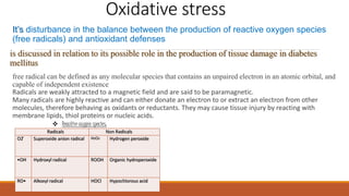
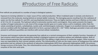
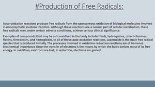
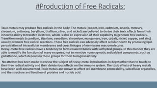
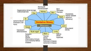
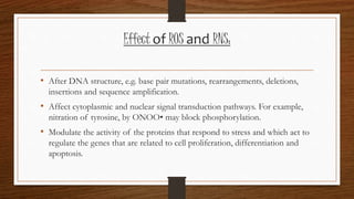
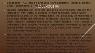
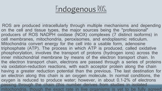
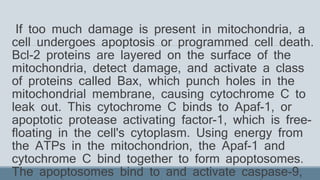
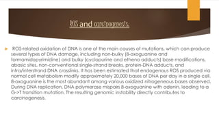
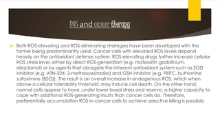
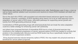
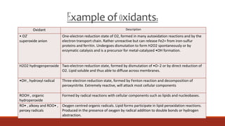
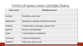
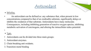
2. Oxidative stress
It’s disturbance in the balance between the production of reactive oxygen species
(free radicals) and antioxidant defenses
is discussed in relation to its possible role in the production of tissue damage in diabetes
mellitus
free radical can be defined as any molecular species that contains an unpaired electron in an atomic orbital, and
capable of independent existence
Radicals are weakly attracted to a magnetic field and are said to be paramagnetic.
Many radicals are highly reactive and can either donate an electron to or extract an electron from other
molecules, therefore behaving as oxidants or reductants. They may cause tissue injury by reacting with
membrane lipids, thiol proteins or nucleic acids.
Radicals Non Radicals
O2 ̄ Superoxide anion radical H2O2 Hydrogen peroxide
•OH Hydroxyl radical ROOH Organic hydroperoxide
RO• Alkoxyl radical HOCl Hypochlorous acid
Reactive oxygen species:
3. #Production of Free Radicals:
Free radicals are produced in a number of ways in biological systems :
Exposure to ionizing radiation is a major cause of free radical production. When irradiated water is ionized, and electron is
removed from the molecule, leaving behind an ionized water molecule. The damaging species resulting from the radiolysis of
water are the free radicals H× and OH× and eaq (hydrated electrons). They are highly reactive and have a lifetime on the order
of 10 -9 to 10 -11 seconds. The hydroxyl radical is extremely reactive and is carcinogenic. Since water presents the largest
number of target molecules in a cell, most of the energy transfer goes on in water when a cell is irradiated, rather then the
solute consisting of protein, carbohydrate, nucleic acid, and bioinorganic molecules. Oxygen is an excellent electron acceptor
and can combine with the hydrogen radical (H×) to form a peroxyl radical (H× + O2 ® HO2). Hydrogen peroxide is toxic and when
present in sufficient quantities can interfere with normal cellular metabolism.
Enzymes and transport molecules also generate free radicals as a normal consequence of their catalytic function. Examples of
two enzymes which have been extensively studied in biological systems are xanthine oxidase and aldahyde oxidase. Both of
these enzymes generate the superoxide anion radical (O2×) by adding a single electron to molecular oxygen. Other enzymes
may use superoxide for their normal catalytic activity. The mitochondria of cells are the major source of endogenous free radical
generation and are utilized in the synthesis of adenosine triphosphate (ATP) from adenosine diphosphate (ADP), the primary
energy currency of the body. Thus, the mitochondrion serves as the powerhouse of the cell and contains most of the respiratory
enzymes of the citric acid cycle.
4. #Production of Free Radicals:
Auto-oxidation reactions produce free radicals from the spontaneous oxidation of biological molecules involved
in nonenzymatic electron transfers. Although these reactions are a normal part of cellular metabolism, these
free radicals may, under certain adverse conditions, achieve serious clinical significance.
Examples of compounds that may be auto-oxidised in the body include thiols, hydroquines, catecholamines,
flavins, ferredoxins, and hemoglobin. In all of these auto-oxidation reactions, superoxide is the main free radical
species that is produced initially. The processes involved in oxidation-reduction reactions are of immense
biochemical importance since the transfer of electrons is the means by which the body derives most of its free
energy. In oxidation, electrons are lost; in reduction, electrons are gained.
5. #Production of Free Radicals:
Toxic metals may produce free radicals in the body. The metals (copper, iron, cadmium, arsenic, mercury,
chromium, antimony, beryllium, thallium, silver, and nickel) are believed to derive their toxic effects from their
inherent ability to transfer electrons, which is also an expression of their capability to generate free radicals.
Transition metals (scandium, titanium, vanadium, chromium, manganese, iron, cobalt, nickel, copper, and zinc)
usually promote free radical reactions. These free radicals can adversely affect cellular health by producing lipid
peroxidation of intracellular membranes and cross linkages of membrane macromolecules.
Heavy-metal free radicals have a tendency to form covalent bonds with sulfhydral groups. In this manner they are
able to modify the functions of many enzymes, not to mention nonenzymatic antioxidant compounds, such as
glutathione, which depend on these groups for their biological activity.
No attempt has been made to review the subject of heavy-metal intoxications in depth other than to touch on
their free radical activity and their deleterious effects on the immune system. The toxic effects of heavy metals
have been well-documented .Toxic metals are known to affect cell membrane permeability, subcellular organelles,
and the structure and function of proteins and nucleic acid.
7. Effect of ROS and RNS:
• After DNA structure, e.g. base pair mutations, rearrangements, deletions,
insertions and sequence amplification.
• Affect cytoplasmic and nuclear signal transduction pathways. For example,
nitration of tyrosine, by ONOO• may block phosphorylation.
• Modulate the activity of the proteins that respond to stress and which act to
regulate the genes that are related to cell proliferation, differentiation and
apoptosis.
8. EXOGENOUS ROS:
Exogenous ROS can be produced from pollutants, tobacco, smoke,
drugs, xenobiotics, or radiation.
Ionizing radiation can generate damaging intermediates through the
interaction with water, a process termed radiolysis. Since water
comprises 55–60% of the human body, the probability of radiolysis is
quite high under the presence of ionizing radiation. In the process,
water loses an electron and become highly reactive. Then through a
three-step chain reaction, water is sequentially converted to hydroxyl
radical (-OH), hydrogen peroxide (H2O2), superoxide radical (O2-)
and ultimately oxygen (O2).
The hydroxyl radical is extremely reactive that immediately removes
electrons from any molecule in its path, turning that molecule into a
free radical and so propagating a chain reaction. However, hydrogen
peroxide is actually more damaging to DNA than the hydroxyl
radical, since the lower reactivity of hydrogen peroxide provides
enough time for the molecule to travel into the nucleus of the cell,
9. Endogenous ROS:
ROS are produced intracellularly through multiple mechanisms and depending
on the cell and tissue types, the major sources being the "professional"
producers of ROS NADPH oxidase (NOX) complexes (7 distinct isoforms) in
cell membranes, mitochondria, peroxisomes, and endoplasmic reticulum.
Mitochondria convert energy for the cell into a usable form, adenosine
triphosphate (ATP). The process in which ATP is produced, called oxidative
phosphorylation, involves the transport of protons (hydrogen ions) across the
inner mitochondrial membrane by means of the electron transport chain. In
the electron transport chain, electrons are passed through a series of proteins
via oxidation-reduction reactions, with each acceptor protein along the chain
having a greater reduction potential than the previous. The last destination for
an electron along this chain is an oxygen molecule. In normal conditions, the
oxygen is reduced to produce water; however, in about 0.1–2% of electrons
passing through the chain (this number derives from studies in isolated
mitochondria, though the exact rate in live organisms is yet to be fully agreed
10. If too much damage is present in mitochondria, a
cell undergoes apoptosis or programmed cell death.
Bcl-2 proteins are layered on the surface of the
mitochondria, detect damage, and activate a class
of proteins called Bax, which punch holes in the
mitochondrial membrane, causing cytochrome C to
leak out. This cytochrome C binds to Apaf-1, or
apoptotic protease activating factor-1, which is free-
floating in the cell's cytoplasm. Using energy from
the ATPs in the mitochondrion, the Apaf-1 and
cytochrome C bind together to form apoptosomes.
The apoptosomes bind to and activate caspase-9,
11. ROS and carcinogenesis:
ROS-related oxidation of DNA is one of the main causes of mutations, which can produce
several types of DNA damage, including non-bulky (8-oxoguanine and
formamidopyrimidine) and bulky (cyclopurine and etheno adducts) base modifications,
abasic sites, non-conventional single-strand breaks, protein-DNA adducts, and
intra/interstrand DNA crosslinks. It has been estimated that endogenous ROS produced via
normal cell metabolism modify approximately 20,000 bases of DNA per day in a single cell.
8-oxoguanine is the most abundant among various oxidized nitrogeneous bases observed.
During DNA replication, DNA polymerase mispairs 8-oxoguanine with adenin, leading to a
G->T transition mutation. The resulting genomic instability directly contributes to
carcinogenesis.
12. ROS and cancer therapy
Both ROS-elevating and ROS-eliminating strategies have been developed with the
former being predominantly used. Cancer cells with elevated ROS levels depend
heavily on the antioxidant defense system. ROS-elevating drugs further increase cellular
ROS stress level, either by direct ROS-generation (e.g. motexafin gadolinium,
elesclomol) or by agents that abrogate the inherent antioxidant system such as SOD
inhibitor (e.g. ATN-224, 2-methoxyestradiol) and GSH inhibitor (e.g. PEITC, buthionine
sulfoximine (BSO)). The result is an overall increase in endogenous ROS, which when
above a cellular tolerability threshold, may induce cell death. On the other hand,
normal cells appear to have, under lower basal stress and reserve, a higher capacity to
cope with additional ROS-generating insults than cancer cells do. Therefore,
preferentially accumulation ROS in cancer cells to achieve selective killing is possible.
13. Radiotherapy also relies on ROS toxicity to eradicate tumor cells. Radiotherapy uses X-rays, γ-rays as
well as heavy particle radiation such as protons and neutrons to induce ROS-mediated cell death and
mitotic failure.
Due to the dual role of ROS, both prooxidant and antioxidant-based anticancer agents have been
developed. However, modulation of ROS signaling alone seems not to be an ideal approach due to
adaptation of cancer cells to ROS stress, redundant pathways for supporting cancer growth and
toxicity from ROS-generating anticancer drugs.
Combinations of ROS-generating drugs with pharmaceuticals that can break the redox adaptation
could be a better strategy for enhancing cancer cell cytotoxicity
and others have proposed that lack of intracellular ROS due to a lack of physical exercise may
contribute to the malignant progression of cancer, because spikes of ROS are needed to correctly fold
proteins in the endoplasmatic reticulum and low ROW levels may thus a specifically hamper the
formation of tumor suppressor proteins.
Since physical exercise induces temporary spikes of ROS, this may explain why physical exercise is
beneficial for cancer patient prognosis
14. Example of oxidants:
Oxidant Description
• O2 ̄
superoxide anion
One-electron reduction state of O2, formed in many autoxidation reactions and by the
electron transport chain. Rather unreactive but can release Fe2+ from iron-sulfur
proteins and ferritin. Undergoes dismutation to form H2O2 spontaneously or by
enzymatic catalysis and is a precursor for metal-catalyzed •OH formation.
H2O2 hydrogenperoxide Two-electron reduction state, formed by dismutation of •O−2 or by direct reduction of
O2. Lipid soluble and thus able to diffuse across membranes.
•OH , hydroxyl radical Three-electron reduction state, formed by Fenton reaction and decomposition of
peroxynitrite. Extremely reactive, will attack most cellular components
ROOH , organic
hydroperoxide
Formed by radical reactions with cellular components such as lipids and nucleobases.
RO• , alkoxy and ROO• ,
peroxy radicals
Oxygen centred organic radicals. Lipid forms participate in lipid peroxidation reactions.
Produced in the presence of oxygen by radical addition to double bonds or hydrogen
abstraction.
15. Oxidation of some Amino Acid Side Chains:
Amino acids Oxidation produce
Cysteine Disulfides, cysteic acid
Methionine Methionine sulfoxide, methionine sulfone
Histidine 2-Oxohistidine, asparagine, aspartic acid
Arginine Glutamic semialdehyde
Lysine Α-Aminoadipic semialdehyde
Threonine 2-Amino-3-ketobutyric
Glutamyl Oxalic acid, pyruvic acid
16. Antioxidant
• Definition:
• An antioxidant can be defined as: any substance that, when present in low
concentrations compared to that of an oxidisable substrate, significantly delays or
inhibits the oxidation of that substrate. Antioxidants have many molecular.
Consequences, including inhibiting generation of reactive oxygen species, inhibiting
metabolic activation of carcinogens, and altreing the intracellular redox potential.
•
• Types:
• Antioxidants can be divided into three main groups:
1. Antioxidant enzymes.
2. Chain breaking anti oxidants.
3. Transition metal binding
17. 1)Antioxidant Enzymes:
(A) Superoxide dismutase:
(SOD) are a class of enzymes that catalyze the dismutation of superoxide into oxygen and hydrogen
peroxide. As such, they are an important antioxidant defense in nearly all cells exposed to oxygen. In mammals
and most chordates, three forms of superoxide dismutase are present. SOD1 is located primarily in the cytoplasm,
SOD2 in the mitochondria and SOD3 is extracellular. The first is a dimer (consists of two units), while the others
are tetramers (four subunits). SOD1 and SOD3 contain copper and zinc ions, while SOD2 has a manganese ion in
its reactive centre. The genes are located on chromosomes 21, 6, and 4, respectively (21q22.1, 6q25.3 and
4p15.3-p15.1).
The SOD-catalysed dismutation of superoxide may be written with the following half-reactions :
M(n+1)+ − SOD + O2
− → Mn+ − SOD + O2
Mn+ − SOD + O2
− + 2H+ → M(n+1)+ − SOD + H2O2.
where M = Cu (n=1) ; Mn (n=2) ; Fe (n=2) ; Ni (n=2).
In this reaction the oxidation state of the metal cation oscillates between n and n+1.
18. 1)Antioxidant Enzymes:
(B) Catalase :
which is concentrated in peroxisomes located next to mitochondria, reacts with the
hydrogen peroxide to catalyze the formation of water and oxygen. Glutathione peroxidase
reduces hydrogen peroxide by transferring the energy of the reactive peroxides to a very
small sulfur-containing protein called glutathione. The sulfur contained in these enzymes
acts as the reactive center, carrying reactive electrons from the peroxide to the
glutathione. Peroxiredoxins also degrade H2O2, within the mitochondria, cytosol, and
nucleus.
2 H2O2 → 2 H2O + O2 (catalase)
2GSH + H2O2 → GS–SG + 2H2O (glutathione peroxidase)
19. 2) The chain breaking antioxidants :
Chain breaking antioxidants are small molecules that can receive an electron from
radical or donate an electron to a radical with the formation of stable byproducts . such
antioxidants can be conveniently divided into lipid phase and aqueous phase
antioxidants .
(A)Lipid phase chain breaking antioxidant .
Vitamin E :
The most important lipid phase antioxidants is probably vitamin E . α –Tocopherol is
the most potent antioxidants of the tocopherols and is also the most abundant in human
. It reacts quickly with a peroxyl radical to form a relatively stable tocopheroxyl
radical , with excess charge associated with the extra electron being dispersed across
the chromanol ring .
20. (B) Aqueous phase chain breaking antioxidant :
Vitamin c :
Qualitatively the most important antioxidant of this type is vitamin C (Ascorbate ) .Vitamin C readily
scavengers reactive oxygen and nitrogen species , single oxygen , ozone , peroxynitrite , nitrogen dioxide ,
nitroxide radicals , hypochlorous acid , thereby effectively protecting other subtrates from oxidative damage
.
Vitamin C can also act as a coantioxidant by regenerating α - tocopherol ( vitamin C ) from the α -
tocopheroxyl radical , produced via scavenging of lipid – soluble radicals .
Two major properties of vitamin C make it an ideal antioxidant . First is the low one-electron reduction
potentials of ascorbate . The second major property that makes vitamin C such an effective antioxidant is
stability and low reactivity of the ascorbyl radical formed when ascobate scavenges reactive oxygen or
nitrogen species .
21. 3) The transition metal binding proteins
Transition metal binding proteins ( ferritin , transferrin , lactoferrin and
ceruloplasmin ) act as a crucial component of the antioxidant defence system
by sequestering iron and copper so that they are not available to drive the
formation of the hydroxyl radical . The main copper binding protein ,
ceruloplasmin , might also function as antioxidant enzyme that can catalyse
the oxidation of divalent iron :
4Fe2+ + O2 + 4H+ 4Fe3+ +2H2O
22. Non- Metal Redox catalyst :
Certain organic compounds in addition to metal redox catalyts can also
produce reactive oxygen species. One of the most important classes of these
are the quinones. Quinones can redox cycle with their
conjugate semiquinones and hydroquinones, in some cases catalyzing the
production of superoxide from dioxygen or hydrogen peroxide from
superoxide.
23. Immune Defense :
The immune system uses the lethal effects of oxidants by making
production of oxidizing species a central part of its mechanism of killing
pathogens; with activated phagocytes producing both ROS and reactive
nitrogen species. These include superoxide (•O2 ̄), nitric oxide (•NO) and
their particularly reactive product, peroxynitrite (ONOO-).Although the use
of these highly reactive compounds in the cytotoxic response of phagocytes
causes damage to host tissues, the non-specificity of these oxidants is an
advantage since they will damage almost every part of their target cell. This
prevents a pathogen from escaping this part of immune response by
mutation of a single molecular target.
24. References:
• Halliwell, Barry (2007). "Oxidative stress and cancer:
have we moved forward?"
• Pohanka, M (2013). "Alzheimer´s disease and oxidative
stress: a review"
• Wikipedia http://en.wikipedia.org/wiki/Oxidative_stress
• http://www.oxidativestressresource.org/
• Wellness journal.production of free radicals