
Recommended
More Related Content
What's hot
What's hot (20)
Similar to Human Heart Structure & Function
Similar to Human Heart Structure & Function (20)
Recently uploaded
Recently uploaded (20)
Human Heart Structure & Function
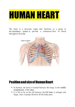
- 1. HUMAN HEART The heart is a muscular organ that functions as a pump in the circulatory system to provide a continuous flow of blood throughout the body. PositionandsizeofHumanHeart In humans, the heart is located between the lungs, in the middle compartment of the chest It is felt to be on the left because the left heart is stronger and larger, since it pumps blood to all the body parts.
- 2. mass of 250–350 grams and size of a fist. Structure Right atrium. Left atrium Left Ventricle Right Ventricle NumberofChambers In homeothermic animals, i.e. mammals, and birds, the heart is divided into four chambers: upper left and right atria; and lower left and right ventricles. In poikilothermic organisms, like Fish has two chambered heart while amphibians and reptiles have 3 chambered heart. It causes the mixing of oxygenated and deoxygenated blood.
- 3. Fish( 2- Chambered) Frog(3-Chambered) Reptile(3-Chambered) Valves
- 4. Valve between the right atrium and the right ventricle is the tricuspid valve. The mitral valve lies between the left atrium and left ventricle. It is also known as the bicuspid valve. The papillary muscles extend from the walls of the heart to valves by cartilaginous connections called chordae tendineae. These muscles prevent the valves from falling too far back when they close. Two additional semilunar valves : Pulmonary valve is located at the base of the pulmonary artery semilunar aortic valve is at the base of the aorta These valves have pocket like structure which gets filled with back flow of blood in pulmonary artery and aorta, when ventricles relax, thus sealing the valves.
- 5. BloodVessels Pulmonary Artery Pulmonary Vein Superior and inferior. Aorta Vena cava FunctioningofHeart
- 6. Cardiac cycle refers to the sequence of mechanical and electrical events that repeats with every heartbeat. It lasts approximately 0.8 seconds. Cardiac muscle tissue has auto rhythmicity, the unique ability to initiate cardiac action potential.
- 7. PhasesoftheCardiacCycle Heart rhythmically contracts( systole ) and relaxes( diastole) 1. JOINT DIASTOLE 2. ATRIAL SYSTOLE AND VENTRICULAR DIASTOLE 3. VENTRICULAR SYSTOLE 4. VENTRICULAR DIASTOLE Heart sounds—- lub dub In a normal, healthy heart, there are only two audible heart sounds: 1. S1 is the sound created by the closing of the atrioventricular valves during ventricular contraction and is normally described as a “lub,” or first heart sound. 2. The second heart sound, S2, is the sound of the closing of the semilunar valves during ventricular diastole and is described as a “dub”.