
Recommended
More Related Content
Similar to circulation.pdf for students to study an educate themselves
Similar to circulation.pdf for students to study an educate themselves (20)
Recently uploaded
Recently uploaded (20)
circulation.pdf for students to study an educate themselves
- 2. Objectives The objectives of this chapter is to enable you to: Describe the structures of the heart and the function of each Differentiate Lifestyles that affect the system Understand the various type of circulation Describe the causes, signs and symptoms, and treatments of various diseases and disorders of the cardiovascular system. Know the different devices used on the system and care given.
- 3. Terms Atrium Ventricle Tricuspid Valve Bicuspid valve Pulmonary semilunar valve Aortic semilunar valve Superior/Inferior vena cava Pulmonary artery/vein Myocardium Endocardium Pericardium Chordae tendineae Diastole Diastolic pressure Systole Hepatic portal system SA node AV node
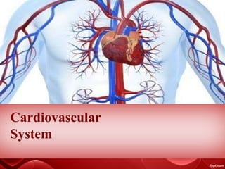
- 4. Introduction The cardiovascular system consists of the heart, blood vessels, and blood. Its primary function is to transport nutrients and oxygen-rich blood to all parts of the body and to carry deoxygenated blood back to the lungs. . At the same time, it picks up waste products from the body cells and transports them to the lungs, kidneys, and other organs for removal from the body.
- 6. The heart is a cone-shaped organ about the size of a loose fist. It is located within the mediastinum (central part of the chest) and extends from the level of the second rib to about the level of the sixth rib. It is located only slightly left of the midline of the body. The heart is bordered laterally by the lungs, posteriorly by the vertebral column, and anteriorly by the sternum. Inferiorly, the heart rests on the diaphragm.
- 7. Cardiac Membrane The heart is enclosed by a membrane called the pericardium, or pericardial sac. The pericardium has two parts. The outer part, called the fibrous pericardium, consists of a tough, fibrous material that helps protect the heart and anchor it in the chest. The inner part of the pericardium, which is called the serous pericardium, has two layers: the parietal pericardium and the visceral pericardium. The visceral pericardium is actually the outermost layer of the heart. The area between these two layers of the pericardium is known as the pericardial cavity. It contains pericardial fluid, which reduces the friction between the membranes when the heart contracts and relaxes.
- 9. Heart Wall The wall of the heart is composed of the following three layers: Epicardium. This outermost layer is the visceral pericardium. It contains fat, which helps to cushion the heart. Myocardium. This middle layer is the thickest layer of the wall and is made primarily of cardiac muscle. Endocardium. This innermost layer is thin and very smooth.
- 11. Heart Valves The heart consists of four distinct chambers: two upper chambers called “atria” and two lower chambers called “ventricles.” A wall or “septum” separates the atria and ventricles. Valves control the flow of blood within the different chambers.
- 12. The tricuspid valve is one of four valves in the heart. It’s located between the right lower heart chamber (right ventricle) and the right upper heart chamber (right atrium).The tricuspid valve opens and closes to ensure that blood flows in the correct direction. It’s also called the right atrioventricular valve
- 13. The bicuspid valve has two cusps and is located between the left atrium and the left ventricle. It prevents blood from flowing back into the left atrium when the left ventricle contracts. This valve is also known as the mitral valve and the left AV valve. Like the tricuspid valve, the bicuspid valve also has chordae tendineae (fibrous connective tissues/heart strings) attached to papillary muscles.
- 14. The pulmonary semilunar valve is located between the right ventricle and the trunk of the pulmonary arteries. It prevents blood from flowing back into the right ventricle. Because its cusps are shaped like a half moon, this valve is called a semilunar valve.
- 15. The aortic semilunar valve is between the left ventricle and the aorta. It prevents blood from flowing back into the left ventricle and is also a semilunar valve.
- 17. Factors The rate at which the heart contracts depends on many factors, such as: activity and exercise emotional factors some medical conditions a fever some medications dehydration At rest, the heart might beat around 60 times each minute. But this can increase to 100 beats per minute (bpm) or more.
- 18. Potassium ions. A low concentration of potassium ions in the blood decreases the heartrate, but a high concentration causes a dysrhythmia (abnormal heart rate). Calcium ions. A low concentration of calcium ions in the blood depresses heart actions, but a high concentration causes heart contractions called tetanic contractions, which are longer than normal heart contractions. Parasympathetic nerves. The parasympathetic nerve to the heart is the vagus nerve, and it generally keeps the heart rate relatively low. Sympathetic nerves. The sympathetic nerves increase the heart rate during times of stress. Cardiac control center. This center is located in the medulla oblongata, which is part of the brainstem. When blood pressure rises, this control center sends impulses to decrease the heart rate. When blood pressure falls, it sends impulses to increase the heart rate.
- 19. Heart Sounds During one cardiac cycle, you can hear two heart sounds. The sounds, commonly called lubb and dubb, are generated when valves in the heart snap shut. Lubb is the first heart sound and occurs when the ventricles contract and the tricuspid and bicuspid valves snap shut. Dubb is the second heart sound and occurs when the atria contract and the pulmonary and aortic semilunar valves snap shut. Physicians listen to heart sounds to diagnose certain conditions. For example, if an AV valve (tricuspid or bicuspid) is damaged, it will not close completely. This allows blood to leak back into the atria when the ventricles contract and produces an abnormal heart sound called a murmur. Murmurs may indicate serious heart conditions, although many heart murmurs are harmless.
- 20. Cardiac Conduction system The cardiac conduction system consists of a group of structures that send electrical impulses through the heart. When cardiac muscle receives an electrical impulse, it contracts. The components of the cardiac conduction system are the sinoatrial node, atrioventricular node, bundle of His, and Purkinje fibers.
- 22. Sinoatrial node (SA node). This node is located in the wall of the right atrium and generates an impulse that flows to the atrioventricular node. The SA node is also known as the natural pacemaker of the heart because it generates the heart’s rhythmic contractions.
- 23. Atrioventricular node (AV node). This node is located between the atria, just above the ventricles. After the impulse reaches the AV node, the atria contract and the impulse are sent to the bundle of His.
- 24. Bundle of His. This structure, also known as the atrioventricular, or AV, bundle, is located between the ventricles and splits into two branches, forming the left and right bundle branches, before sending the electrical impulse to the Purkinje fibers.
- 25. Purkinje fibers. These fibers are located in the walls of the ventricles. As the impulse flows through the Purkinje fibers, it causes the ventricles to contract. Physicians use a test called an electrocardiogram (ECG or EKG) to tell if the cardiac conduction system is working properly. In a normal ECG, the waves shown in Figure Below are produced. The first wave (P wave) indicates that an electrical impulse was sent through the atria, causing them to contract (depolarization). The Q, R, and S waves occur together and make up the QRS complex. This complex indicates that an electrical impulse was sent through the ventricles, causing them to contract (depolarization). Finally, the T wave indicates electrical changes that occur in the ventricles as they relax (repolarization).
- 26. Blood Vessels Blood circulation takes place in blood vessels that form a closed pathway to carry blood from the heart to cells and back again. These vessels include arteries, arterioles, veins, venules, and capillaries.
- 27. Arteries and Arterioles Arteries carry blood away from the heart and are the strongest of the blood vessels. They have a thick layer of smooth muscle that can withstand the high pressure the heart exerts on them. This pressure is necessary to carry the blood throughout the body. Small branches of arteries are called arterioles. The largest artery in the body is the aorta, which receives its blood directly from the left ventricle. The aorta branches into the coronary arteries and many other major arteries that supply blood to various parts of the body. Major arteries are illustrated in Figure Below Many arteries are paired, meaning there is a left and a right artery of the same name. Most arteries carry oxygenated blood. The exceptions to this are the pulmonary arteries, which carry deoxygenated blood from the heart to the lungs.
- 29. Capillaries Capillaries are the smallest type of blood vessel. They branch off of arterioles and have walls that are only about one cell layer thick. These thin walls make the exchange of oxygen, carbon dioxide, and nutrients possible between the blood and the body cells (see Figure Below). In fact, capillaries are the only type of blood vessels that allow substances to move into and out of the blood. Tissues that require a lot of oxygen, such as muscle and nervous tissues, have a lot of capillaries. The substances that move through the capillary walls include oxygen, carbon dioxide, nutrients, water, and metabolic wastes. These substances move through the walls through one of three processes: diffusion, filtration, or osmosis.
- 31. Veins and Venules Veins are blood vessels that carry blood toward the heart. Unlike arteries, they are not under high pressure, so they do not need thick, muscular walls. Their walls are thinner than those of arteries. Because the blood in veins is not under pressure, veins have valves that prevent backflow and keep the blood moving toward the heart (see Figure Below) Venous valve: valve opens when blood is flowing toward the heart and valve closes to prevent blood from flowing away from the heart.
- 32. Venules Venules are very small veins formed when capillaries merge together. The venules then merge to form the veins. Most veins carry deoxygenated blood. The exceptions to this are the pulmonary veins, which carry oxygenated blood from the lungs to the left ventricle of the heart. Large veins often have the same names as the arteries they run next to, but there are exceptions. For example, the veins next to the carotid arteries are the jugular veins. Large veins empty blood into the superior vena cava and the inferior vena cava (plural: venae cavae), which are the largest veins in the body. The superior vena cava generally collects blood from veins above the heart, and the inferior vena cava collects blood from veins below the heart. The major veins are summarized are illustrated in Figure Below.
- 34. The veins of the intestines carry blood from the digestive tract to the liver. The liver then processes nutrients in the blood and returns it to general circulation through the hepatic veins. The veins involved in this process are known as the hepatic portal system.
- 35. CIRCULATION Blood circulates through the body through three main circuits. The pulmonary circuit provides oxygen, the systemic circuit distributes the oxygen throughout the body, and the coronary circuit distributes the oxygen to the heart muscle.
- 36. Pulmonary Circulation The pulmonary circuit, or pulmonary circulation, is the route blood takes from the heart to the lungs and back to the heart again. The purpose of this circuit is to remove waste gases such as carbon dioxide and replenish the blood with oxygen. When blood returns to the heart from the body cells, it is low in oxygen (deoxygenated) and rich in carbon dioxide. As described earlier in this chapter, the deoxygenated blood enters the right atrium, which delivers it to the right ventricle. The right ventricle pumps the blood through the left and right pulmonary arteries to the lungs. In the lungs, blood picks up oxygen and gets rid of carbon dioxide. Blood rich in oxygen and low in carbon dioxide then returns to the heart through the four pulmonary veins. The pulmonary veins empty the oxygenated blood into the left atrium.
- 37. Systemic Circulation The systemic circuit, or systemic circulation, is the route blood takes from the heart through the body and back to the heart. The purpose of this circuit is to deliver oxygen and nutrients to the body cells. It also picks up carbon dioxide and waste products from the body cells (see Figure Above). Blood that returns from the lungs and enters the left atrium is oxygen rich (oxygenated) and has a low level of carbon dioxide. It flows from the left atrium to the left ventricle, which contracts to pump the oxygenated blood into the aorta, which branches off to various arteries to deliver the blood throughout the body.
- 38. The arteries branch into the smaller arterioles, and the arterioles branch into capillaries. In the capillaries, oxygen and nutrients picked up from the digestive system move from the blood into the body cells. Carbon dioxide and metabolic wastes move from the body cells into the blood. The blood then moves through the venules and veins and is collected into the vena cava, which delivers the blood back to the right atrium of the heart, and the whole process starts over again with pulmonary circulation.
- 39. Coronary Circulation Coronary circulation is the part of systemic circulation that supplies oxygen and nutrients to the heart and removes carbon dioxide and other wastes. The coronary arteries branch directly off the aorta immediately after the blood leaves the left atrium. Like all other arteries, the coronary arteries branch into smaller and smaller vessels, ending in capillaries, where oxygen and nutrients move into the heart cells and carbon dioxide and wastes move into the blood. The blood then travels through the venules and the cardiac veins, which merge to form a large vein called the coronary sinus. Unlike other veins, however, the coronary sinus does not empty into the vena cava. It empties directly into the right atrium.
- 40. Blockage of one or more of the coronary arteries may cause chest pain, or angina, and may lead to myocardial infarction (MI, or heart attack) if not corrected. See the Educating the Patient feature and the Pathophysiology section of this chapter for more information about these disorders.
- 41. Pressure Diastole, systole, and blood pressure Each heartbeat has two parts: Diastole: The ventricles relax and fill with blood as the atria contract, emptying all blood into the ventricles. Systole: The ventricles contract and pump blood out of the heart as the atria relax, filling with blood again.
- 42. When a person takes their blood pressure, the machine will give a high and a low number. The high number is the systolic blood pressure, and the lower number is the diastolic blood pressure. Systolic pressure: This shows how much pressure the blood creates against the artery walls during systole. Diastolic pressure: This shows how much pressure is in the arteries during diastole.
- 43. Pulse A person can feel their pulse at points where arteries pass close to the skin’s surface, such as on the wrist or neck. The pulse is the same as the heart rate. When you feel your pulse, you feel the rush of blood as the heart pumps it through the body. A healthy pulse is usually 60–100 bpm, and what is normal can vary from person to person. A very active person may have a pulse as low as 40 bpm. People with a larger body size tend to have a faster pulse, but it is not usually over 100 bpm.
- 44. Many factors affect blood pressure, including cardiac output, blood volume, vasoconstriction, vasodilation, and blood viscosity. Cardiac output is the total amount of blood the heart pumps in 1 minute. As cardiac output increases, it causes an increase in blood pressure. When cardiac output decreases, blood pressure decreases accordingly. When a person loses a large volume of blood, his blood pressure significantly decreases. If the blood pressure falls too low, the muscular blood enters the left ventricle, the wall of the ventricle is stretched. The more the wall is stretched, the harder it will contract and the more blood it will pump out. This is referred to as Starling’s law of the heart. If only a small amount of blood enters the left ventricle, it will not be stretched very much and therefore will not contract very forcefully. In this case, not much blood is pumped out of the heart.
- 45. Disease and disorder of the Cardiovascular System ANGINA Angina pectoris is chest pain that occurs when the heart does not receive enough oxygen to carry out its job of pumping blood throughout the body. It is not immediately life threatening, but if the reason for the angina is not found and corrected, the angina may become unstable (difficult to treat and less responsive to medications). This type of angina is a warning of serious or life- threatening conditions.
- 46. Causes: Angina is caused by a narrowing of the coronary arteries. Arteries may become too narrow due to coronary spasms or as a result of coronary artery disease (atherosclerosis), in which fatty deposits accumulate in the arteries
- 47. Angina tends to appear during physical activity, emotional stress, or exposure to cold temperatures, or after big meals. Symptoms of angina include: pressure, aching, or burning in the middle of the chest pressure, aching, or burning in the neck, jaw, and shoulders (usually the left shoulder) and even down the arm a sense of anxiety or uneasiness When arteries are severely narrowed, angina can also occur at rest. This is called unstable angina.
- 48. Treatment: A doctor should monitor patients with angina regularly. Most patients with angina carry sublingual nitroglycerin with them to dilate the coronary blood vessels and relieve the pain. Treatment must also address the underlying cause of the angina to prevent more serious conditions such as a heart attack or stroke. The physician may order several tests to determine the cause of angina, such as an electrocardiogram (ECG), a stress test, blood tests, chest X-rays, cardiac catheterization, or an echocardiogram.
- 49. Medication also plays an important role in treatment. Several types of medication are to ease or prevent angina. These include: nitrates beta blockers calcium-channel blockers aspirin statins ACE inhibitors ranolazine
- 50. Nursing interventions for angina pectoris Find out the intensity of anginal pain Provide fowler’s position to the patient to promote ventilation. Encourage the patient to take deep breaths, it may reduce infarct size, decrease anxiety and resolve chest pain. Provide reassurance to the client to decrease anxiety. Administer nitroglycerine sublingually as prescribed: check vital signs especially blood pressure. Check vital signs. Administer oxygen, if required. Take vital signs in every 10 to 15 minutes, till anginal pain subsides. Advice the patient to inform nursing staff if pain occurs.
- 51. CORONARY ARTERY DISEASE (CAD) Also called ATHEROSCLEROSIS, is an accumulation of fatty deposits in the arteries as a result of too much glucose in the blood. The deposits cause the arteries to become narrow, reducing the amount of blood that can flow through them. CAD affects more Americans than any other type of heart disease. The American Heart Association estimates that one in three American adults has one or more types of coronary artery disease.
- 52. Causes Coronary artery disease starts when fats, cholesterols and other substances collect on the inner walls of the heart arteries. This condition is called atherosclerosis. The buildup is called plaque. Plaque can cause the arteries to narrow, blocking blood flow. The plaque can also burst, leading to a blood clot. Besides high cholesterol, damage to the coronary arteries may be caused by: Diabetes or insulin resistance High blood pressure Not getting enough exercise (sedentary lifestyle) Smoking or tobacco use
- 53. Signs and Symptoms • There are often no signs or symptoms until a heart attack occurs. The most common symptoms include angina, shortness of breath, tightness in the chest, fatigue, and swelling in the legs of feet(edema). As with most cardiac conditions, the first line of defense against CAD is prevention. CAD may be prevented or reduced by controlling high blood pressure and high cholesterol, not smoking, eating healthy foods, engaging in regular exercise, and treating any existing conditions such as diabetes or atherosclerosis
- 54. Treatment Existing CAD is treated using lipid-lowering agents such as Mevacor® or Lipitor®, aspirin therapy, and medications to slow a rapid heart rate. The patient can help by adopting a low-fat diet, exercising moderately, and not smoking. Severe CAD may require surgery such as coronary angioplasty or coronary artery bypass grafting (CABG) to repair, widen, or detour around narrowed coronary arteries.
- 55. A MYOCARDIAL INFARCTION (MI A MYOCARDIAL INFARCTION (MI), Commonly called a heart attack, is nonreversible damage to the heart caused by a lack of oxygen. Historically, MIs have been fatal, and they often still are, but with new treatments, more and more people survive them. However, the body cannot replace the damaged cardiac cells, so a heart attack can result in permanent damage to the heart
- 56. Causes A heart attack occurs when the flow of blood to the heart is severely reduced or blocked. The blockage is usually due to a buildup of fat, cholesterol and other substances in the heart (coronary) arteries. The fatty, cholesterol-containing deposits are called plaques. The process of plaque buildup is called atherosclerosis. Sometimes, a plaque can rupture and form a clot that blocks blood flow. A lack of blood flow can damage or destroy part of the heart muscle.
- 58. While the classic symptoms of a heart attack are chest pain and shortness of breath, the symptoms can be quite varied. The most common symptoms of a heart attack include: pressure or tightness in the chest pain in the chest, back, jaw, and other areas of the upper body that lasts more than a few minutes or that goes away and comes back shortness of breath sweating nausea anxiety feeling like you’re going to faint a fast heart rate sense of impending doom
- 59. Treatment: The first treatment, if possible, is chewing an aspirin at the onset of symptoms. In an unconscious patient who has no pulse and is not breathing, cardiopulmonary resuscitation (CPR) should be administered. Other immediate treatment options include the use of a defibrillator and thrombolytic medications to destroy the blood clots that block a coronary artery. It should be noted that thrombolytic drugs are effective only if begun within 3 hours of the first symptom, so time is crucial. Surgery(angioplasty or CABG) may be necessary to replace or repair blocked coronary arteries. Long-term treatment includes anticoagulant medications such as heparin and warfarin to thin the blood, and medications such as atenolol to slow the heart rate
- 60. Nursing Interventions Administer oxygen along with medication therapy to assist with relief of symptoms. Encourage bed rest with the back rest elevated to help decrease chest discomfort and dyspnea. Encourage changing of positions frequently to help keep fluid from pooling in the bases of the lungs. Check skin temperature and peripheral pulses frequently to monitor tissue perfusion. Provide information in an honest and supportive manner. Monitor the patient closely for changes in cardiac rate and rhythm, heart sounds, blood pressure, chest pain, respiratory status, urinary output, changes in skin color, and laboratory values.
- 61. HYPERTENSION, or high blood pressure, is a consistent resting blood pressure of 140/90 mm Hg or higher. It is known as the “silent killer,” because it increases a person’s risk of heart attack, stroke, heart failure, and kidney failure, sometimes without presenting symptoms that could warn the person of medical risk.
- 62. Causes Known causes and risk factors for hypertension include narrowing of the arteries, kidney disease, endocrine disorders, pregnancy, drug use (especially cocaine and amphetamines), sleep apnea, obesity, smoking, a high-sodium diet, excessive alcohol consumption, stress, diabetes, and various medications such as oral contraceptives and cold medicines.
- 63. Signs and Symptoms Hypertension often causes no symptoms at all. When symptoms are present, they include excessive sweating, muscle cramps, fatigue, frequent urination, headaches, dizziness, and an irregular heart rate.
- 64. Treatment Hypertension cannot be cured, but it can be controlled. The first method of treatment is to treat the underlying causes, if they are known. For example, the patient may be placed on a low-sodium/low-cholesterol diet and may be encouraged to make lifestyle changes such as getting regular exercise, managing stress, and stopping smoking. The physician may also prescribe medications to slow the heart rate and/or dilate the blood vessels and diuretics to reduce blood volume. Patient compliance is the key to successful management of hypertension. Because hypertension often has no symptoms, but the prescribed medications may have noticeable side effects, patients may stop taking the medications because they feel better when they do not take it. Be sure patients understand that taking the medication(s) is crucial to their treatment and long-term health. If they experience unacceptable side effects, they should tell the physician, because many options are available, and the physician may be able to prescribe a different medication that has fewer or less noticeable side effects about Hypertension.
- 65. Nursing care Teach the patient to use a self-monitoring blood pressure cuff and to record the reading at least twice weekly in a journal for review by the physician at every office appointment. Tell the patient to take his blood pressure at the same hour each time with relatively the same type of activity preceding the measurement. Make sure that the patient understands the need to control risk factors through medication therapy, dietary modifications, exercise guidelines, stress-reduction methods, and follow-up care. Suggest stress-reduction groups, dietary changes, and an exercise program, particularly aerobic walking, to improve cardiac status and reduce obesity and serum cholesterol levels, Encourage a change in dietary habits. Explain which signs and symptoms indicate a need to contact the physician.
- 66. • DYSRHYTHMIAS are abnormal heart rhythms. The heart may beat too fast (tachycardia), too slowly (bradycardia), or irregularly. The most common type of dysrhythmia is atrial fibrillation, which is a sporadic, rapid beating of the atria that may or may not affect the ventricles. The most serious type of dysrhythmia is ventricular fibrillation, in which disorganized electrical activity in the heart causes the ventricles to quiver ineffectively instead of beating. Most sudden cardiac deaths are caused by ventricular fibrillation
- 67. Causes Most dysrhythmias result from abnormal flow of electrical impulses through the heart. Abnormal impulse conduction has many potential causes, including electric shock, certain medications, some herbal supplements, hypertension, previous heart attack, decreased blood flow to the heart, coronary artery disease, heart valve disorders, weakening of the heart muscle (cardiomyopathy), some genetic diseases, diabetes mellitus, sleep apnea, electrolyte (potassium, sodium, and calcium) imbalances, excess alcohol consumption, and drugs such as cocaine and amphetamines.
- 68. Signs and Symptoms Dysrhythmias may cause signs and symptoms including shortness of breath, dizziness or fainting, an unusually fast or slow heart rate, a fluttering feeling in the chest, and chest pain.
- 69. Treatment The first goal of treatment is to correct the underlying cause of the dysrhythmia. Other treatment options include: Vagal maneuvers to slow the heart rate. These include holding the breath, straining (bearing down as if for a bowel movement) and putting the face in cool water. Medications such as beta blockers and anti-dysrhythmics. Pacemakers. Radiofrequency catheter ablation. This procedure destroys a small amount of heart tissue to change the flow of the electrical impulses through the heart.
- 70. Maze procedure; this operation forms scars in the atria to correct the flow of electrical impulses through the heart. Implantation of an implantable cardioverter defibrillator (ICD) to regulate the heart rhythm. Surgery to correct heart defects such as narrow coronary arteries. Electric shock (defibrillation) to reset heart rhythms. Cardiopulmonary resuscitation if there is no evidence of blood flow.
- 71. AN ANEURYSM is a ballooned, weakened arterial wall. The most common locations of aneurysms are the aorta and arteries in the brain, legs, intestines, and spleen. An aortic aneurysm is a bulge in the wall of the aorta. Most aortic aneurysms occur in the abdominal aorta (abdominal aortic aneurysm), but some occur in the thoracic aorta. Most aortic aneurysms do not rupture; however, when they do, the resulting hemorrhage is a life-threatening emergency.
- 72. Causes Most causes are unknown. One identified risk for developing an aneurysm is atherosclerosis, which is usually associated with a high cholesterol diet. Smoking and obesity also increase the risk of atherosclerosis. Congenital conditions may cause an aneurysm—some individuals are born with weak aortic walls. A traumatic injury to the chest also may be a risk factor. The risk of developing an aneurysm can be reduced by not smoking, by losing excess weight, and by eating a low-fat, low-cholesterol diet. Periodic screening is an option for patients with a family history of aortic aneurysms.
- 73. Signs and Symptoms There are usually no signs or symptoms of an aneurysm, although hypertension may be present. When symptoms do exist, the most common are a pulsation in the abdomen and back pain. A sudden pain in the abdomen or back, dizziness, a fast pulse, or a loss of consciousness can be a sign that an aneurysm has ruptured. Treatment: The primary treatment is surgery to repair the aneurysm.
- 74. ENDOCARDITIS: is an inflammation of the innermost lining of the heart, including the heart valves. Causes: Bacterial infections are the most common cause of endocarditis. Patients are more susceptible to this condition if they have abnormal heart valves. Signs and Symptoms: Common signs and symptoms include weakness, fever, excessive sweating, general body aches, difficulty breathing, and blood in the urine. Treatment: The treatment for this condition is intravenous antibiotics followed by oral antibiotics for up to 6 weeks.
- 75. MYOCARDITIS: is an inflammation of the muscular layer of the heart. It is relatively uncommon but very serious because it leads to weakening of the heart wall. Causes: The most common cause of myocarditis is a viral infection, but it also may be caused by exposure to certain chemicals, allergens, and bacteria. Signs and Symptoms: Signs and symptoms include fever as well as chest pains that feel like a heart attack. Difficulty breathing, decreased urine output, fatigue, and fainting also may accompany myocarditis. Treatment: Treatment normally includes steroids to reduce inflammation, bed rest, and a low-sodium diet.
- 76. PERICARDITIS: is inflammation of the pericardium, which is the group of membranes that surround the heart. Causes: This condition is most commonly caused by complications of viral or bacterial infections. However, heart attacks and chest injuries also can lead to pericarditis. Signs and Symptoms: Symptoms include sharp, stabbing chest pains, especially during deep breaths. Fever, fatigue, and difficulty breathing while lying down are also common symptoms. Treatment: Diuretics are used to remove excess fluids around the heart. If bacteria caused the pericarditis, antibiotics are used as well. In chronic cases, surgery may be required to remove part of the membranes surrounding the heart. Because pericarditis can be very painful, treatment also generally includes painkillers.
- 77. CONGESTIVE HEART FAILURE (CHF) is a slowly developing condition in which the heart weakens over time. Eventually, the heart is no longer able to pump enough blood to meet the body’s needs.
- 78. Causes There are many risk factors for this condition, including smoking, being overweight, a diet high in cholesterol, a lack of exercise, atherosclerosis, history of MI, high blood pressure, a damaged heart valve, excessive alcohol consumption, and diabetes mellitus. Congenital heart defects (those present at birth) and drugs that weaken the heart (especially cocaine, heroin, and some antineoplastic drugs for cancer) also may contribute to the development of this disorder. Patients may reduce the risk factors for CHF by controlling high blood pressure and high cholesterol, not smoking, maintaining a healthy diet, engaging in regular exercise, and treating any existing atherosclerosis or diabetes.
- 79. Signs and Symptoms Signs and symptoms include shortness of breath; constant wheezing; prominent neck veins; fluid retention that causes swelling in the legs, feet, or abdomen; nausea; dizziness; and an irregular or rapid heartbeat. Treatment Common treatment options include medications to slow a rapid heartbeat, diuretics to decrease edema and fluid accumulation in the lungs, and medications to reduce blood pressure. In more serious cases, surgery to repair defective heart valves or other heart defects, implantation of a cardiac pacemaker, or a heart transplant may be needed.
- 80. Nursing Intervention Monitor for signs of respiratory distress Provide pulmonary hygiene as needed Administer oxygen as prescribed Keep the head of the bed elevated Monitor ABG values. Monitor for signs of altered cardiac output, including Pulmonary edema Arrhythmias, including extreme tachycardia and bradycardia
- 81. Characteristic ECG and heart sound changes Evaluate fluid status Maintain strict fluid intake and output measurements Monitor daily weights Assess for edema and severe diaphoresis Monitor electrolyte values and hematocrit level Maintain strict fluid restrictions as prescribed Administer prescribed medications which may include: Antiarrhythmias to increase cardiac performance Diuretics, to reduce venous and systemic congestion
- 82. Iron and folic acid supplements to improve nutritional status. Keep the child warm Do not allow an infant to feed for more than 45 minutes at a time Provide gavage feedings if the infant becomes fatigued before ingesting an adequate amount Promote adequate nutrition. Maintain a high-calorie, low-sodium as prescribed. Promote optimal growth and development As appropriate, refer the family to a community health nurse for follow up care after discharge.
- 83. MITRAL VALVE PROLAPSE (MVP) is a condition in which the mitral valve falls into the left atrium during systole. This prevents the valve from sealing properly. In severe cases, blood may flow back into the atrium. Although most cases are mild, MVP can become worse over time. It also increases the risk of heart valve infections and endocarditis. Causes The cause of MVP is unknown in most cases. However, it may be hereditary, and it has been linked to autonomic nervous system disorders.
- 84. Signs and Symptoms: In mild cases, symptoms may not develop. In more severe cases, palpitations, shortness of breath, and chest pain may occur. Treatment: No treatment is needed for mild cases. Medications are used to treat symptoms and to help prevent complications such as infection. In very severe cases, surgery may be required to repair the valve.
- 85. THROMBOPHLEBITIS is a condition in which a blood clot blocks or partially blocks blood flow in a vein, causing inflammation, swelling, and pain. It most commonly occurs in the leg veins. The danger of thrombophlebitis is that the blood clot may break loose, becoming an embolus that moves through the circulatory system. The embolus can cause major problems, depending on where it lodges in the body. If it blocks a blood vessel in the lungs, it becomes a pulmonary embolism (obstruction in the lungs). If it blocks a coronary artery, it may cause a myocardial infarction (heart attack). If it blocks an artery in the brain, it may cause a cerebrovascular accident (stroke).
- 86. Causes The causes and risk factors include prolonged inactivity, oral contraceptives, postmenopausal hormone replacement therapy (HRT), certain types of cancer, paralysis in the arms or legs, the presence of a venous catheter, a family history of thrombophlebitis, varicose veins, and trauma to veins. Signs and Symptoms The most common symptoms are tenderness and pain in the affected area; redness, swelling, and tenseness of the affected areas; fever; and a positive Homan’s sign (pain behind the knee that is caused by a blood clot, when the foot is force ably dorsiflexed). Treatment This disorder is most often treated by applying heat to the affected area, wearing support stockings, and elevating the legs. Anti-inflammatory medications and anticoagulants may be prescribed. In some cases, surgery may be performed to remove the clot.
- 87. VARICOSE VEINS are twisted, dilated veins usually seen in the legs. They affect women more often than men. When varicose veins occur in the rectum, they are called hemorrhoids. Causes Varicose veins may be caused by prolonged sitting or standing, damage to valves in the veins, a loss of elasticity in the veins, obesity, pregnancy, oral contraceptives, or hormone replacement therapy. Family history also seems to play a part in the development of varicose veins. In some cases, varicose veins may be prevented or at least minimized through exercise and elevation of the legs
- 88. Signs and Symptoms Signs and symptoms include discomfort in the legs, discolorations around the ankles, clusters of veins, and enlarged, dark veins seen through skin. Treatment The treatment of varicose veins includes the following: Sclerotherapy, a procedure that prevents blood from flowing through varicose veins. Laser surgery to prevent blood from flowing through affected veins. Vein stripping, which involves removing affected veins. Insertion of a catheter into the affected veins in order to destroy them. Endoscopic vein surgery to close off affected veins
- 89. Take breaks. Varicose veins appear because of prolonged standing and leg fatigue. Make sure to take breaks at work. Sit down whenever you can and rest your legs while you’re at it. Elevate your legs. Do this by lying down or by using a footstool when sitting. This will help relieve fatigue in your lower extremities, and therefore, prevent the veins from losing their elasticity. Elevate your legs at the end of the day everyday and your legs will feel so much better.
- 90. Avoid wearing tight clothes. Choose clothes that are comfortable enough for you to move around freely even while you work. Clothes that are too tight for your legs like girdles or skinny jeans can block the movement of blood up your legs. This may cause pooling of the blood, and in a short period of time, varicose veins may appear. Avoid wearing high heels. It has been found that the muscles in your calves contract less when you’re wearing high-heeled shoes. This will cause the venous blood pressure to increase, thus, straining the valves in the veins. As much as possible, choose comfortable shoes, especially for your nursing clinicals.
- 91. Avoid crossing your legs. Although it has not been scientifically proven yet, crossing your legs may cause them, especially if varicose veins run in your family. Avoid crossing your legs whenever you’re sitting, so as to keep a good circulation in your legs and prevent your legs’ venous blood pressure from increasing. Wear compression stockings. Choose the best compression stockings for you by knowing your leg measurements and the best level of tightness for you, usually around the range of 15 to 20 mmHg only. The stockings will help relieve leg fatigue and prevent varicose veins by applying pressure to your lower extremities. Eat right. The heavier you are, the more undue pressure you put on your legs, especially when you have to stand for long periods of time. Maintain healthy eating habits and make sure that you keep a daily exercise routine.
- 92. Devices use on the Circulatory System Prosthetic Cardiac Pacemaker/Artificial Pacemaker When the natural pace maker SA node does not work properly, in such patients normal heart beat can be restored and maintained with an artificial pacemaker. Temporary pace maker is used in emergency such as bradycardia— slow heart beat. Electrodes of temporary pacemaker are introduced from jugular, subclavian and femoral veins as well as from veins of the upper extremity. Permanent pacemaker is used in atrioventricular (AV) block, SA node dysfunction, etc. The artificial pacemaker consists of a pulse generator containing cell (solid state lithium cell), the lead in the form of a wire and an electrode.
- 94. Defibrillator The arrest of cardiac muscle (atrial or ventricular) with restoration of the normal rhythm is called defibrillation. which the heart muscle is contracting very rapidly but in an uncoordinated fashion. There are atrial and ventricular fibrillation. Artificial fibrillation may occur in myocardial infarction and in rheumatic heart disease.
- 95. Angioplasty (Balloon Catheterization) Angioplasty is a technique to remove the atherosclerotic plaques from the coronary arteries through ballooning. Therefore, it is better to call it coronary artery angioplasty.
- 96. Vascular Grafts (Artificial Arteries) These artificial arteries are made from porous plastic fibres of dacron or teflon. These arteries remain open for many years and function very well. However, artificial arteries are also made from rubber tubes, silver, glass, etc. but they are not good and close very soon.
- 97. Stents A special device called a stent, made of stainless steel, resembling a spring coil, can be permanently placed in an artery through a catheter (plastic tube). This is done to ensure proper blood circulation through the of coronary artery that supplies blood to the heart.
- 98. Echocardiogram An echocardiogram uses sound waves to produce images of your heart. This common test allows your doctor to see your heart beating and pumping blood. Your doctor can use the images from an echocardiogram to identify heart disease. Check for problems with the valves or chambers of your heart Check if heart problems are the cause of symptoms such as shortness of breath or chest pain Detect congenital heart defects before birth (fetal echocardiogram)
- 100. Electrocardiogram An electrocardiogram records the electrical signals in the heart. It's a common and painless test used to quickly detect heart problems and monitor the heart's health. ECG machines are standard equipment in operating rooms and ambulances. Some personal devices, such as smart watches, offer ECG monitoring An electrocardiogram is a painless, noninvasive way to help diagnose many common heart problems. A health care provider might use an electrocardiogram to determine or detect: Irregular heart rhythms (arrhythmias) If blocked or narrowed arteries in the heart (coronary artery disease) are causing chest pain or a heart attack Whether you have had a previous heart attack How well certain heart disease treatments, such as a pacemaker, are working
- 102. Cardiac catheterization is a procedure in which a thin, flexible tube (catheter) is guided through a blood vessel to the heart to diagnose or treat certain heart conditions, such as clogged arteries or irregular heartbeats. Cardiac catheterization gives doctors important information about the heart muscle, heart valves and blood vessels in the heart. Cardiac catheterization may be done during the diagnosis or treatment of: Coronary artery disease Congenital heart disease Heart failure Heart valve disease Microvascular heart disease
- 104. Coronary artery bypass graft surgery (CABG) is a procedure used to treat coronary artery disease. Coronary artery disease (CAD) is the narrowing of the coronary arteries – the blood vessels that supply oxygen and nutrients to the heart muscle. CAD is caused by a build-up of fatty material within the walls of the arteries. This build-up narrows the inside of the arteries, limiting the supply of oxygen- rich blood to the heart muscle.