
Recommended
More Related Content
What's hot
What's hot (20)
Similar to Circulatory System
Similar to Circulatory System (20)
More from Amani Riyadh
More from Amani Riyadh (20)
Recently uploaded
Recently uploaded (20)
Circulatory System
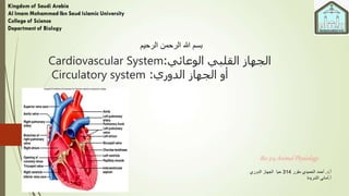
- 1. الوعائي القلبي الجهاز:Cardiovascular System الدوري الجهاز أو:Circulatory system أ.د.مقرر الحميدي أحمد314الدوري الجهاز حيا أ.الشريدة أماني Bio 314 Animal Physiology الرحيم الرحمن هللا بسم
- 2. content Introduction. Types of Blood Circulation. Heart / Heart Rate Source/ ECG. Cardiomyogulia / Heart Circulation / Blood Vessels / Blood Circulation. Blood :Blood Cells / Blood Plasma / Blood Clotting. Lymph/Lymph Functions / Spleen.
- 3. Introduction القلبي الجهازالوعائي Cardiovascular system The cardiovascular system consists of : 1. The heart. 2. The blood vessels. The main purpose or function of the circulatory system: Is to deliver nutrients, oxygen, hormones and others to cells and transport carbon dioxide and waste from cells to the respiratory and urinary system for disposal.
- 4. 1. The heart It is a muscle organ. Its the center of the vascular system with its contractions, blood distributed. Its muscles are self-constricting/ controlled by the nervous and hormonal system / in order to generate blood pressure.
- 5. 2. Blood vessels 1. Arteries: transport blood from the heart to the rest of the body. 2. Veins: transfer blood from the body's organs to the heart and capillaries. 3. Blood capillaries: which form a network of capillaries to deliver blood to and from cells in different organs of the body. Blood: It is the fluid that circulates in the blood vessels and the heart and contains all kinds of blood cells, plasma and blood proteins in blood vessels
- 6. Cont. introduction The Small blood circulation It is ( microcirculation) from the lungs to the heart. It was first described by the doctor Ibn al-Nafis in1268. Then by the scientist (Micheal Harvey) 1553. Then William Harvey 1628 he wrote about the work of the heart =(De Mout Cordis) he clear that the blood circulates in the body and that the heart is pumping to some extent, then receives it from the blood and the blood runs through the arteries of the heart and then returns to it through the veins.
- 7. blood circulation Blood circulation in animal organisms: A large number of animal organisms lack blood circulation, they are non-existent in the spongy, hydra, flatworms and cylindrical worms. Ringworms have a good circulating device (but no heart). Arthropods and molluscs they have a complete circulating system. The highest degree of circulating system are in vertebrates.
- 8. Type of blood circulation 1. Closed Circulatory System: The circulatory system in ringworms and vertebrates is closed. The blood circulates and then returns through the blood vessels through the capillaries. The valves are available that make the blood go in one direction. 2. Opened Circulatory System: The circulatory system in molluscs and arthropods is open in this type the arteries do not connect the veins by capillary bristles, but the arteries pour into the cavities between tissues called sinuses with walls consisting of a single layer of cells.
- 9. أ.د.مقرر الحميدي أحمد333حين Type of blood circulation in animals.
- 10. The Heart It is is located between the lungs and it is enclosed with a double membrane known as Pericardium, which allows it to move freely during its constriction and simplicity. The pericardial fluid fills the pericardial cavity, which helps prevent cardiac contact with the pericardial membrane.
- 11. The heart The heart is considered a dueled pump it receives in the left part of it oxidized blood from the lungs and pumps it to the arteries then to the rest of the body. while the right part of it receives blood loaded with carbon dioxide from the cells of the body and pumps it to the lungs.أ.د.مقرر الحميدي أحمد333حين
- 12. The Hear Anatomy The heart has four chambers: Two upper chambers are called the atria (one atrium): right and left. Two lower chambers called ventricles: right and left (greater in size than atria. Wall of L. ventricle is thicker than R. ventricle). A septum divides the atrium and ventricle on each side. Each septum has a valve to ensure one- way flow of blood: The right A-V valve (tricuspid) and left A-V valve (bicuspid).
- 13. The heart layer It is enclosed in an outer covering consisting of two layers called the pericardium. If take a cross section in heart, it consist of 3 layer: 1. The lining of the inner surface of the heart is called the endocardium. 2. Middle layer is myocardium 3. The external layer is called epicardium.
- 14. The heart septum The heart in all vertebrates (except amphibians and fish) is divided into four cavities (chambers) of 2 atria and 2 ventricle. The 2 atria were separated by intra atrial septum. while the 2 ventricular are separated by the inter ventricular septum. The wall of the atria is thin than the ventricles.
- 15. القلب صماماتHeart Valves The Atrioventricular valves are: 1. Tricuspid valve: located between both the atrium and the right ventricle has three plates. 2. Bicuspid valve: located between the left atrium and the left ventricle has two plates connecting the edges of the Bicuspid cords. These valves work to direct blood flow between the heart chambers and prevent the return of blood from the ventricle to the atrium. 3. There are also valves between the heart and the related blood vessels (semilunar or semicrescent vavles) such as: A. Aortic valves: located at the beginning of the aorta (Aorta) located between the left ventricle and aorta. B. Pulmonary valve or pulmonary artery valve: located between pulmonary and the right ventricle, which prevents blood from reversing direction.
- 16. Atrioventricular valves Types Tricuspid valve (Bicuspid)The semilunar or semi-crescent valve
- 17. The blood circulation in the heart The right atrium receives blood from all parts of the body except the lung through three main hollow veins: 1-The upper hollow vein (Superior vena cava) الوريدالجوف العلوي which brings blood from the upper or front parts of the body. 2- The inferior vena cava والوريداألجوفالسفلي , which brings blood from the back parts of the body. 3-The coronary sinus الجيبالتاجي , which brings blood from the vessels that nourish the heart muscles. When the right atrium is filled with blood it rushes to the right ventricle (below it), which is contracted to push blood into the lungs through the pulmonary trunk, which is branched by a cycle into two branches, the right and left pulmonary arteries.
- 18. The blood circulation in the heart: Cont… The blood then returns from the lungs through four pulmonary veins that pour into the left atrium of the heart, which, when filled, is contracted to push the blood to the left ventricle below it. When the smooth ventricle is filled with blood, it pushes blood into the body through the main aorta (Asending aorta). The main aorta is divided into coronary arteries الشرايين ,التاجية aortic arch( Aortic arch والقوساألبهري ), thoracic aorta واألبهرالصدري and abdominal aorta (Abdominal aorta واألبهرالبطني ) to extend Body organs, including the heart, are in blood except for the lungs.
- 19. أ.د.مقرر الحميدي أحمد333حين The blood circulation in the heart
- 20. The time of the heart circulation A human heart beat rate is about 75 beats/minute means that each pulse takes 60 seconds .75 beats = 0.8 seconds. During the first tenth of seconds (0.1), the two atrum are contact and the ventricles are relax, as the ventricle valves open and the semi-crescent valves close. Then, in the next 0.3 seconds, the two vents are flat and the ventricles are opened. During the remaining time of the heart cycle (0.4 seconds) known as the period of relaxation of all the heart chambers in the diastolic role, so the two vents are spun for 0.1 seconds and the vents are spun for 0.7 seconds. (see the fig).
- 21. أ.د.مقرر الحميدي أحمد333حين The time of the hear circulation
- 22. The heart beat The sound of the heart beat is from the close of the valves between the 2 atrium and the 2 ventricle The sound of the heart contraction ( Systoic sound) (Lubb) The second sound is the diastolic (Dupp) of the semilunar valves. Then a Pause between the two. So the doctor can be known the damage of the heart valves from these sounds through the hearing of doctor ear speaker..
- 24. The hear conducting system The cardiac conduction system of the heart is a series of specialized cardiac muscle cells that carries impulses (electrical signals) throughout the heart musculature, signaling the heart chambers to contract in proper sequence. The components of the conducting system are: • Sinoatrial (SA) node cells (pacemaker) • Internodal fibers • Atrioventricular (AV)node cells. • Atrioventricular bundle (bundle of His) • Right and left branches • Purkinje fibers • The impulse that signals each heartbeat begins at the 1- sinoatrial (SA) node: a crescent shaped mass of muscle cells that lies in the wall of the right atrium just below the opening of the superior vena cava.
- 25. Cont . The conducting system 1. The 1-sinoatrial node, the heart’s own pacemaker, sets the basic heart rate by generating 70-80 impulses per minute. 2. Impulses from the SA node spread in a wave along the cardiac muscle fibers of the atria signaling the atria to contract. 3. Some of these impulses travel along the intranodal pathway to the 2-atrioventricular (AV) node in the inferior part of the interatrial septum, enters the septum as 3-aterventricular bundle and divides into right and left 4-bundle branches. 4. the 4-Bundle fibers, (crura), become 5-bundles of Purkinje fibers which approach the apex of the heart, then turn superiorly into the ventricular walls
- 27. Neural regulation of heartbeat The cardiovascular (CV) center is located in the medulla oblongata The Central nerve system (CNS) controls the heart rate through innervating the S-A by branches of the sympathetic (also to myocardium) and parasympathetic divisions. The sympathetic Nervous system (SNS) impulses increase the speed of heart rate. It is activated by stress, anxiety, excitement, or exercise. The parasympathetic nervous system (PNS) impulses decrease the heart rate by the parasympathetic vagus nerve (from the brain). Almost all vessels, such as arteries, arterioles, venules and veins are innervated, except capillaries.
- 28. Neural regulation of heartbeat أ.د.مقرر الحميدي أحمد333حين
- 29. Neural regulation Sympathetic Fibers Innervate SA and AV nodes and ventricles Release noradrenaline Increase heart rate Increase contractility (constrictor nerves) Increase pressure The norepinephrine is released from sympathetic nerve terminals in the heart and in the blood vessels. Parasympathetic Nerve Innervates SA node and AV node Releases acetylcholine which may regulate sympathetic release of norepinephrine and vice versa. Slows heart rate Lowers pressure
- 30. Hormones that increase blood pressure (Vasoconstriction) When blood volume falls or blood flow decreases, the renin (an enzyme) is released by kidney. The Renin activates the secretion of angiotensinogen by the liver which then circulates in the blood and converted into angiotensin I (prohormone) under the effect of renin . Angiotensin I is activated by a lung enzyme, Angiotensin-Activating Enzyme (ACE) and becomes angiotensin II (active hormone). Angiotensin II is a powerful vasoconstrictor of blood vessel smooth muscles. It also triggers the secretion of aldosterone by the adrenal cortex. The aldosterone promotes reabsorption of both H2O and salt (mainly sodium ions) with increases the blood pressure. Angiotensin II stimulates thirst center in hypothalamus and drinking behavior. So blood volume is increased. Also angiotensin II stimulates secretion of ADH from posterior pituitary (AP). Renin-Angiotensin-Aldosterone System
- 31. Hormones that increase blood pressure (Vasoconstriction)
- 32. Hormones that increase blood pressure (Vasoconstriction) Vasopressin = Anti-diuretic hormone (ADH) It is secreted by the posterior pituitary in response to dehydration or decreased blood volume. It promotes water reabsorption by the kidney tubules H2O moves back into the blood less urine formed. It causes vasoconstriction and increases the arterial blood pressure. Norepinephrine and epinephrine Are released by sympathetic nerve terminals in the blood vessels and by the medulla of adrenal gland. It increases the rate and force of heart contractions. Circulating Epinephrine and Norepinefrine from the adrenal medullae have almost the same effects on the different organs as the effects caused by direct sympathetic stimulation, except, the effects are longer lasting.the effects are longer lasting. Other solutes: Ca2+ ions
- 33. Hormones that increase blood pressure (Vasoconstriction) Vasopressin = Anti-diuretic hormone (ADH) Norepinephrine and epinephrine
- 34. Hormones that decrease blood pressure (Vasodilation) Nitric oxide (NO) = local regulator (a gas) Nitric oxide Increased blood flow in arterioles causes the release of NO by the vascular endothelial cells (endothelium relaxing factor). It diffuses into adjacent vascular smooth muscle where it activates soluble guanylate cyclase, produces cGMP and causes vasodilatation (This causes small arteries upstream to relax). Atrial Natriuretic Peptide (ANP) It is secreted by the atria in response to the increase of atrial pressure. It causes vasodilation through the relaxation of vascular smooth muscle. Bradykinin This is a vasodilator substance which is formed in tissues during inflammation or increased tissue activity. Bradykinin is a mediator of vasodilatation in sweat glands and digestive glands when they become activated. Other solutes K+ ions, Mg2+ ions, H+ ions, CO2
- 35. Factors that affect the hear beats 1- Body temperature: where the center of body temperature regulation located in the brain's (Hypothalamus) which senses minor changes in blood temperature sends signals to the (Vasomotor center) in the spinal cord. 2- Breathing: The vascular motor center is affected by the change in the amount of oxygen and carbon dioxide in the blood. The rise of carbon dioxide increases the tension of the arteries and therefore cause the high blood pressure 3 - Psychological state: the heart rate decreases when sadness or psychological distress and increases when joy, fear and anger. It increases with motor or athletic activity. 4- Hormones: from the adrenal gland, The adrenaline and noradrenaline 5- Prescription drugs: atropine increases the heart beat rate. Muscarine, is the active substance in poisonous mushrooms, lowers the pulse of heart, (Nicotine) initially causes a decrease in the pulse rate and then followed by an increase in the heart beat rate 6- Age and sex: pulse in women faster than men, children more than adult (120 pulses/d) 7- body size: in the mouse 300-500 Pulse / in elephant 28 pulse/min, in rabbit 220 pulse/min.,In canary birds 1000 /min. in human 75/min...
- 36. الكهربائي القلبي التخطيطElectroncardiogram (ECG) Preceded by the muscular contraction of the heart during the pulse a change in the electrical voltage on both sides of the cardiac muscle fiber membranes, So the wave of muscle contraction that runs from the sinoatrial node to the top of the heart is preceded by a few ml. or a fraction of a second wave of voltage going in the same direction so that the contracting tissue is more negative than tissue that has not yet contracted and when the contracting areas are relax the it became positive and returns. On this basis, the pulse is electrically a wave of polarization during the contraction followed by a wave of repolarization during the diastolic.
- 37. Electroncardiogram ECG The electrical change accompanying the pulse moves to the surface of the body, but the amount of difference in electrical voltage on both sides of the body is minimal and can only be recorded by the electroncardiograph, which amplifies the voltage difference. The electrodes of the registered device of the voltage difference connect to the right arm, left arm and left leg and measure the difference in voltage between a couple of these electrodes and there are three ways to connect: 1. Conduct (RL) right arm (R) and left arm (L) 2. Conduction (RF) right arm (R) The left leg (F) 3. Conduct (LF) left arm (L) and left leg (F) and the device records voltage change on a graph of electrocardiogram.
- 38. Blood pressure It is the force exerted against the walls of the arteries. It is affected by heart action, blood volume, peripheral resistance, and blood viscosity. Arterial walls are able to expand and recoil because of the pressure of elastic fibers in the arterial wall. Systole = is the result of the contraction of the ventricles (normal 110-140) (pressure when heart contracts). Diastole = pressure when heart relaxes (70- 80). Arterial blood pressure rises and falls following a pattern established by the cardiac cycle. During ventricular contraction, arterial pressure is at its highest (systolic pressure). When ventricles are relaxing, arterial pressure is at its lowest (diastolic pressure).
- 39. الشرايين في الدم ضغطArterial blood pressure Blood pressure means the force that blood directs on the walls of blood vessels as a result of ventricular contraction, and pressure is measured in millimeters of mercury. From a medical point of view, pressure within the systemic arteries only. It is measured in humans usually by the brachial artery, and this is done by an arterial blood pressure meter with the number of millimeters of air capable of raising the mercury column in the glass tube of the pressure device and the rate. The appropriate pressure for a healthy human being 120/70 first (120) represents contraction pressure (Systolic pressure) and the second (70) represents (Diastolic pressure.)
- 40. Meijer the Arterial blood pressure
- 41. Hypertension Individuals with hypertension (high blood pressure) have a sustained elevation of pressure in the arteries. High Blood Pressure: >140 (systolic)/90 (diastolic) Hypertension may go undetected until complications such as heart attack, stroke, or visual problems arise. High blood pressure strains the heart, damages the arteries, and increases risk of heart attack, stroke, kidney failure, and blindness. Reduces with regular exercise, healthy diet (reduced salt, increased potassium and fiber), and moderation of alcohol intake. Importance: Blood pressure is a key factor for providing blood (thus oxygen and energy) to organs. Systolic BP must be a minimum of 70 to sustain kidney filtration and adequate blood flow to the brain.
- 42. Blood pressure Category Systolic (mm Hg) Diastolic (mm Hg) Normal Below 120 and Below 80 Prehypertension 120–139 or 80–89 Hypertension Stage 1 Stage 2 140–159 160 and above or or 90-99 100 and above
- 43. Cardiovascular diseases Cardiovascular diseases include those that affect the heart and those that affect the peripheral vascular system. The heart and blood vessels may be primarily attacked by these diseases or they may be secondarily affected as a consequence of another disease. Atherosclerosis: Arteries are narrowed by deposits of fat, cholesterol, and other substances called plaques. Once narrowed by a plaque, an artery is vulnerable to blockage by blood clots. Blockage in the coronary arteries (coronary heart disease) can lead to a heart attack. Blockage in the brain can cause a stroke.
- 45. The Blood Circuits Blood vessels that carry blood to the lung form the pulmonary circuit. الدورةالدمويةالصغرىأوالدورةالرئوية Blood vessels carrying blood to the body form the systemic circuit. الدورة الدمويةالكبرىاوالدورةالجهازية Deoxygenated Blood returning from the body enters the right atrium and passes into the right ventricle. The right ventricle pumps the blood to the lungs via the pulmonary trunk. Blood in the pulmonary circuit is oxygen poor and carbon dioxide rich Once in the lungs, the blood unloads carbon dioxide and picks up oxygen. Freshly oxygenated, the blood is carried back to the heart by the pulmonary veins pour into L. atrium. The left ventricle pumps the oxygenated blood into the aorta and from there into many distributing arteries.
- 46. Human Blood Circuits Pulmonary and systemic circuits
- 47. أ.د.مقرر الحميدي أحمد333حين Animals Blood Circuits
- 48. Cardiovascular Diseases (CVD) 1. Heart attack (myocardial infarction) Damage to, or death of, part of the heart muscle, sometimes resulting in a failure of the heart to deliver enough blood to the body; myocardial infarction. 2. Arrhythmia An irregularity in the force or rhythm of the heartbeat. 3. Sudden cardiac death A non-traumatic, unexpected death from sudden cardiac arrest, most often due to arrhythmia (in association with underlying heart disease). 4. Peripheral Vascular Disease Disease of the peripheral vessels. The lack of proper circulation (caused by blockages in arteries that feed the legs) may cause fluids to pool in the extremities. Symptoms include leg pain, cramping, numbness, tingling, coldness and loss of hair to the affected limbs. The disease process may be extensive before the person is symptomatic, as the heart and brain are more sensitive to a decreased blood flow as compared to the extremities.
- 49. Risk factors that can’t be changed: 1. Heredity Children of parents with CVD are more likely to develop it themselves 2. Being Male CVD is the leading killer of both men and women but men face a greater risk of heart attack than women, especially earlier in life. Estrogen production may offer premenopausal women some protection against CVD. 3. Age The risk of heart attack increases significantly after age 65. Over 70% of all heart attack victims are over age 65. 4. Race/Ethnicity Death rates from heart disease vary among the ethnic groups. African Americans have a much higher rate of HTN, heart disease and stroke than other groups. Asian Americans historically have had lower rates of CVD than white Americans.
- 50. Risk factors that can be changed 1. Cigarette/Tobacco Use: About 1 in 5 deaths from CVD can be contributed to smoking Women who smoke and use contraceptives have 39 times more risk of heart attack and 22 times more likely to have a stroke than women who do not smoke and take contraceptives. Smoking damages the linings of the arteries and causes platelets to be stickier. Carbon monoxide in smoke displaces oxygen in the blood, reducing the amount of oxygen available to the heart. 2. Cholesterol: Any body who has a liver produces cholesterol. It is also obtained through the foods we eat. It refers to the fatty substance that circulates the blood stream and is an important component for cell membranes, sex hormones, nerves. Cholesterol is carried in protein-lipid packages called lipoproteins (LDL and HDL). • Low-density lipoprotein (LDL) = unhealthy (“bad”) cholesterol; (containing more cholesterol than protein) excess amounts are deposited in artery walls and can clog arteries and increase the risk for CVD. • High-density lipoprotein (HDL) = healthy (“good”) cholesterol; shuttle unused cholesterol back to the liver for recycling. (less cholesterol than LDL)
- 51. Risk factors that can be changed 3. Obesity In 1998, obesity was declared a major risk factor for heart disease. Greater than 30% are above the recommended weight. 4. Triglycerides Greater than 190 mg/dl tends to increase blood thickness resulting in sluggish blood flow which makes it more difficult in delivering oxygen and nutrients to the heart.
- 52. Who to Reduce Your Heart Attack Risk? 1. Quit Smoking. 2. Know and manage your blood cholesterol level. 3. Maintain a healthy body weight. 4. Exercise regularly. 5. Healthy Eating Plan. 6. No alcohol consumption. 7. Know and manage your blood pressure. 8. Handle stress and anger effectively.
- 53. A small cycle that nourishes the heart muscles with oxidized blood where blood rushes from the left ventricle through the systemic aorta from which the right and left coronary arteries branch right and left coronary artery) that nourish the tissues of the heart muscles and then the blood returns from the tissues The heart via vein or coronary sinus to the right atrium of the heart. Coronary circulation: الدورةالتاجية الدموية
- 54. أ.د.مقرر الحميدي أحمد333حين Human Blood Pathway
- 55. Blood vessels arteries, veins and capillary Arteries and arterioles : Carry blood away from heart. Their walls are too thick for blood components to pass through. they are deep inside under the skin. Veins and Venules: Returning blood to the heart from the body. Veins have thinner walls due to the less muscle but can hold much more blood. they are not deep under the skin. Many veins in limbs have valves to prevent backflow (Varicose veins arise when pressure on valves is prolonged). They are continuous with capillaries; take in some returned fluid (rest is retained by tissues to blood via lymphatic system). Blood Capillary: They are thinner than blood vessels (only passing a red blood cell) and They connect the arteries and veins through which food and gases (CO2,O) are exchanged between the blood and cells of the body.
- 56. Blood vesicles: arteries, veins and capillary
- 57. Blood capillaries Capillaries are the smallest vessels, consisting only of a layer of endothelium through which substances are exchanged with tissue cells. • Allow exchange of gases, nutrients and wastes between blood and tissues. • In capillaries, oxygen and nutrients move out by diffusion; CO2 in (via lipid component of the membrane, channels, etc.). • Small molecules and lipid soluble molecules move by diffusion through the cell membrane. • Larger molecules, charged molecules (ions) must pass through membrane channels, exocytosis or in between 2 cells. • Water movement is controlled by the capillary hydrostatic and osmotic pressures.
- 59. Valve in vein fro blood flow
- 61. الدم وظائف Blood functions 1. Transfer oxygen from the lungs to tissue cells and transfer carbon dioxide from body cells to the lungs. 2. Transfer digested nutrients from the digestive tract to tissues 3. Transporting waste (metabolic metabolites) from the body tissue cells to be disposed of by the kidneys. 4. Transporting hormones to the target tissues that work or are affected by the hormone. 5. Blood plasma proteins act as a regulated substance to prevent sudden pH change. 6. Blood contributes to the preservation of the body's water balance 7. Blood contributes to maintaining the body's temperature 8. Blood work to defend the body against infection (because it contains antibodies) 9. Blood characterized by the characteristic of clotting to prevent loss of blood and body fluids.
- 62. الدم خواصBlood Characters Blood volume: The blood volume of each living organism varies by age and sex by different environmental and physiological conditions and varies between 6-9% of body weight. Human body weight 70 kg have 5 litter of blood. The blood circulation: Contains 50% of the blood intake, the rest in the liver is 20%, the spleen is 20%, the skin (10%). The Blood pH: (pH) ranges from (7.4-7.35 pH) The blood pH variation is small or low because it contains regulated serum solutions such as weak carbonic acid (H2CO3) which ionize in the presence of excess acid and alkali in blood in order to maintain the pH for blood as flow: NaOH + H2CO3 H2O + NaHCO3. The hemoglobin and blood plasma proteins help to save (pH).
- 63. Composition of the whole blood The blood consists of: • Plasma Water, dissolved plasma proteins, other solutes • Blood Cells: - Red blood cells (RBCs) or erythrocytes: Transport oxygen - White blood cells (WBCs) or leukocytes: Part of the immune system - Platelets or thrombocytes: Cell fragments involved in clotting
- 64. Composition of blood: Plasma and blood cells
- 66. Red blood cells (RBCs) or erythrocytes • Red blood cells (RBCs) make up 99.9% of blood’s • Small and highly specialized discs. • Thin in middle and thicker at edge • Quickly absorbs and releases oxygen they don’t contain nucleus. They contain hemoglobin protein. Hemoglobin The red pigment that gives whole blood its color • Binds and transports oxygen HbO2and CO2 • form from a Complex quaternary structure of four globular protein subunits ( 2 alfa and 2 beta globulin + heme (ferrous iron)
- 67. Red blood cells (RBCs) or erythrocytes Red blood cells are produced in the red bone marrow. )yellow marrow for the production of fat cells) such as ribs, sternum, vertebrae, skull and the end of long bones. In the fetus it is produced in the liver spleen and lymph nodes. Human erythrocytes live 124 days, 50 days in rabbit, 120 in dogs. The RBC broken by retinal cells in the spleen and liver where they consume their substance and excess hemoglobin stored in the liver and spleen in the form of a substance called Ferritin. Anemia: Anaemia of malignant anemia caused by vitamin B12 deficiency (B12). The iron deficiency necessary for the formation of hemoglobin leads to anemia Malnutrition, especially protein leads to anemia. Myeloid anemia is caused by the failure of the bone marrow to produce blood cells as a result of exposure to ionizing radiation.
- 68. White blood cells (WBCs) or leukocytes • Can migrate out of bloodstream. • Have amoeboid movement. • Attracted to chemical stimuli (+ chemotaxis). • WBC for Protecting body against germs ,antigen defense and immunity. Form in the bone marrow • The amount of WBC = 6-10milione /cm3 WBCs type: granulocytes Polymorphnuclear 1. Neutrophils =no.62% Phagocytosis 2. Basophils=0.5% produce Heparin and histamine 3. Acidophils or Eosinophils= (2.3%) phagocytes, increase in allergy, get read of antigen, A granulocytes : 1. Monocytes (5%) and 2. Lymphocytes 30%. Produce Antibodies
- 69. White blood cells (WBCs) or leukocytes
- 70. Platelets or thrombocytes Three functions of Platelets: • Release important clotting chemicals: to stop bleeding by forming a plug • Temporarily patch damaged vessel walls • Actively contract tissue after clot formation
- 71. الدم بالزماBlood Plasma Plasma is a blood fluid of a transparent color without blood cells, Compose about 55% of the total blood volume Its is composed of 90% water 10% solid materials mostly proteins and represents 6-8% of the plasma, While the rest of the solids are nutrients from digestion processes and substances consisting of metabolisms such as urea, uric acid, creatinine and Lactic acid, respiratory gases, hormones and enzymes carried by blood. Some salts, sodium ions, potassium, calcium, magnesium and carbonate, Blood plasma proteins and their functions: 3main proteins: Albumins, 55%, Globulins, 38%, fibrinogen, 7% Every 100ml contain 6-8 g of protein. Blood protein functions: 1. fiprorogen and albumin have a major role in blood clotting. 2. Glutamine in immunity 3. Proteins maintain viscosity (Viscosity of blood) 4. Osmotic pressure 5. The total blood volume. Most blood proteins are manufactured in the liver.
- 72. الدم تجلط آليةBlood Clotining processe: Blood clots are formed according to the following reactions: 1- The Pro-thrombombin is transformed into an active thromboin in the precence of thromboplastin. Ca ++ 2 - Thrombin converts dissolved fibrinogen into Fibrin as there are 15 blood clotting factors and if one of them is lost, the person becomes bleeding. The fipronogen is the number one factor I and prothrombin #2 II. Thromboplastin 1- Prothrombin active Thrombin Ca ++ -Thrombin 2-Fibrinogen Fibrin If clotting factors are isolated from blood plasma it is called serum blood The clotting process needs vitamin K (absorbed from the intestines to the liver)
- 75. الليمف وظائف: Lymph function 1. Lymph is the medium between the blood and cells of the inter-cell fluid tissue. 2. lymph transfers proteins that cannot pass through the walls of blood vessels and transfer them to the circulatory system through the thoracic lymph channel. 3. Lymph absorbs and transfers fat from the areas absorbed by the intestines to the blood. 4. Lymphatic tissues perform defensive functions in the body through T cells and B cells, which are characterized by plasma cells for their ability to release antibodies against antigens on the body.
- 76. أ.د.مقرر الحميدي أحمد333حين Lymphatic system in human
- 77. الطحالSpleen The spleen is the largest accumulation of lymph tissue in the body, 15 cm tall and 10cm x 10cm dark red, and is located in the upper left part of the abdomen behind the stomach under the diaphragm and above the left kidney, The spleen has nothing to do with the digestive system and is considered from the lymphatic system and spleen functions: 1. plays an important role in the defense Against the organisms that pass through the cells of the acrobat. 2. The lymphocytes and single-nucleus cells in the embryonic stage produce blood cells. 3. The spleen breaks down red blood cells and platelets through cellular swallowing. 4. The spleen plays an immune role through the resulting cells and antibodies of these cells. 5. The spleen is a store of blood, especially when he bleeds blood or in cases of carbon monoxide poisoning, where the spleen vacuums expand to about 350 ml of blood to compensate for the need 6. The spleen is important in the formation of the dye Bile and the storage of iron and some other metabolism.
- 78. Summary of circulatory system The circulatory system is made up of two major pieces: the heart to pump the blood around the body and the blood vessels to guide the blood to different places like the brain. Heart is a four chambered, hollow muscular organ. Coronary circulation – the circulation of blood within the heart. Pulmonary circulation – the flow of blood between the heart and lungs. Systemic circulation – the flow of blood between the heart and the cells of the body.
- 79. cont. Summary of circulatory system The force of blood on the wall of the arteries is known as blood pressure. Neural control (cardiac center) of circulation is centered in the medulla oblongata. The humoral regulation of circulation is controlled by substances secreted or absorbed into the body fluids such as angiotensin II, Renin, ADH and NO
Editor's Notes
- Final only
- Final only
- final
- final
- final
- final
- final
- final
- final
- تعداد فقط ( بالشهري)
- final
- final
- النص باللون الأخضر للاطلاع
- final
- أمراض الجهاز الدوري تعداد فقط
- final
- تعداد فقط
- تعداد فقط
- تعداد فقط
- تعداد فقط
- final
- final
- قراءة للطالبات
- final
- final
- final
- final