
Recommended
More Related Content
What's hot
What's hot (20)
Similar to Cardiovascular system
Similar to Cardiovascular system (20)
More from RobbinsHobbin
Recently uploaded
Recently uploaded (20)
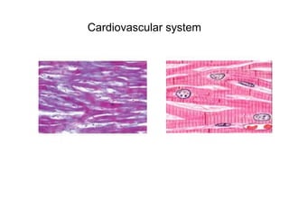
Cardiovascular system
- 2. Diagrammatic drawing of the cardiovascular and lymphatic system. The nomenclature of the main vessel types is indicated; in red are the vessels carrying oxygenated blood, in blue those carrying un-oxygenated blood and in yellow the lymphatic structures.
- 3. Schematic drawing showing the principal structural features of the larger blood vessels. On the left the major layers and associated features of a muscular artery are depicted. On the right the particular features of an elastic artery, a muscular artery, an arteriole, a venule and a vein are shown.
- 4. A. The artery is sectioned transversely, and the muscle cells of the media are cut longitudinally. Magnification 1 510. B. The artery is sectioned longitudinally, and the muscle cells are cut transversely. Magnification 1 510. C. At higher magnification, the endothelial cells can be seen, somewhat elongated in the direction of the blood flow; the dark line beneath the endothelium is the inner elastic lamina, which shows fenestration, and is straight in this preparation because it was fixed while distended. The tunica media is made of five or six arrays of muscle cells, which are transversely sectioned. The tunica adventitia displays collagen fibres and fibroblasts. Magnification 1 510.
- 5. Part of a transverse section of the aorta of a monkey, stained with haematoxylin and eosin, showing the distribution of cell nuclei. Magnification 1 100.
- 7. Part of a transverse section of a young human aorta stained with Verhoeff's stain for elastin. Note the density of the concentric fenestrated elastic lamellae. Magnification 1 100.
- 8. Transverse section of a small muscular artery, stained with Verhoeff's stain for elastin and van Gieson stain for collagen. Note the prominent inner elastic lamina, which is heavily folded because the vessel was fixed postmortem when collapsed and virtually empty. Magnification 1 200.
- 10. Transverse section of two small muscular arteries and a small vein, stained with haematoxylin and eosin. Numerous venules and capillaries are included but are indistinct at this magnification.
- 12. Transverse section of a large arteriole and venule in loose connective tissue, stained with haematoxylin and eosin.
- 14. A fenestrated capillary, surrounded by a basal lamina, a laminar process of a pericyte (left) and collagen fibrils (bottom). The endothelial cell at top contains various organelles, including two centrioles, vesicles and Golgi complexes. Magnification 1 18 000.
- 15. A fenestrated capillary in the intestinal mucosa. The fenestrations are close to the basal surface of the lining epithelium. Note the basal lamina on both the epithelial and the endothelial cell and the intervening collagen fibrils and fibroblastic process. Magnification 1 30 000.
- 16. The upper portions of the femoral and great saphenous veins laid open to show the valves. About two-thirds of the natural size.
- 17. Diagram of a microcirculatory unit based upon descriptions in Zweifach (1959, 1961, 1973) and Reynolds and Zweifach (1959). Note the terminal arteriole, thoroughfare channels, capillaries and collecting venule. The distribution of smooth muscle cells and precapillary sphincters is shown.
- 18. Diagram of an arteriovenous anastomosis. Note the thick wall of the anastomotic channel composed of layers of modified smooth muscle cells.