
Recommended
More Related Content
What's hot
What's hot (20)
Similar to Magnetic resource imaging(mri) copy
Similar to Magnetic resource imaging(mri) copy (20)
Recently uploaded
Recently uploaded (20)
Magnetic resource imaging(mri) copy
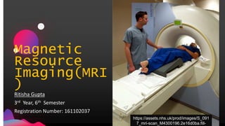
- 1. Magnetic Resource Imaging(MRI ) Ritisha Gupta 3rd Year, 6th Semester Registration Number: 161102037 https://assets.nhs.uk/prod/images/S_091 7_mri-scan_M4300196.2e16d0ba.fill-
- 2. JensMartensson • MRI shows the detailed 3D image of the organs and tissues. • Uses magnetic fields, radio waves and a computer by the polarizing the hydrogen nuclei of the water molecules present in the body. • It is also known as Nuclear Magnetic Resonance (NMR) imaging. • Based on the principles of NMR Introduction 2 https://encrypted- tbn0.gstatic.com/images?q=tbn:ANd9GcTt28t1sP4M_ufe2 NviTAlfAL-nLRYcZ4H343eBEtgr7VHkwf6U
- 3. History 1950 Spin Echoes and free induction decay detected by Erwin Hahn 1971 NMR Raymond Damadian reported that tumors and normal tissue can be distinguished in vivo by Nuclear Magnetic Resonance ("NMR") 1972 Magnetic Resonance Phenomena Felix Bloch and Edward Purcell independently discovered the phenomenon and were awarded Nobel Prize. 1973 NMR Gradients Lauterbur publishes method for generating NMR gradients. 2003 Nobel Prize Paul Lauterbur and Sir Peter Mansfield was awarded Nobel Prize "for discoveries concerning magnetic resonance imaging." 03
- 4. 4 200 3 Paul Lauterbur and Sir Peter Mansfield • Lauterbur was awarded Nobel Prize for using the concept of spatial localization using magnetic field gradients which allowed the rapid acquisition of 2D images. • Sir Peter Mansfield was also awarded Nobel Prize for developing techniques for efficient gradient utilization and fast imaging
- 5. JensMartensson Concept and Technique Water Water present in an adult is 60%. Isotopes To be added.......... Magnetic Field Scanner produces strong magnetic fields to polarize Hydrogen Ions Radio- Frequency Scanner produces weak RF fields to rotate the nuclei. Radio Signal Collection and MRI Generation Radio Signals(with Radio Antenna) and Magnetic fields are collected from different angles to produce 3D image. page 5 1 . 2 . 3 . 4 . 5 .
- 6. Source: https://www.cs.sfu.ca/~stella/papers/blairthesis/main/node11.html Principle of MRI page 6
- 13. JensMartensson On the Basis Of Functions & Applications 13
- 14. JensMartensson Functional MRI- Mostly used in the brain surgeries and diseases related to brain. Interventional MRI- Used in biopsies of suspected lesions, to remove tissues non-invasively. Cardiac MRI- Used in pericardial diseases, ventricular disorders. Magnetic Resonance Angiography- Used for abdomen, pelvis, arms, etc. Magnetic Resonance Venography- Checks the flow of blood in veins Diffusion MRI- With the help of Diffused Tensor Imaging (DTI), a brain map can be created showing the direction of nerve fibres using white matter. 14
- 19. JensMartensson • An MRI can be used to detect the method of disease throughout the body, defining the brain anatomy, problems associated with the vertebrae or inter-vertebral discs of the spine, etc. • MRI can be done of various organs like chest, heart, spine, brain etc. • But the patients having any metallic materials within the body like pacemaker of heart, metallic chips, surgical chips, prosthetic devices, metallic bone plates etc cannot undergo this technique. • The patients with any history of claustrophobia are also not allowed to undergo the MRI. CONCLUSION:
- 20. JensMartensson • https://www.thoughtco.com/magnetic-resonance-imaging-mri-1992133 • https://www.medicinenet.com/mri_scan/article.htm#how_do_i_prepare_for_an_mri_how_is_i t_performed • https://www.itnonline.com/article/recent-advances-mri-technology • http://www.hypres.com/wp-content/uploads/2015/07/HYPRES_The-Future-of-MRI_2015.pdf • https://cfmi.georgetown.edu/downloads/training/2/MRI-basics.pdf https://www.onlinelibrary.wiley.com/doi/full/10.1002/jmrs.226#references-section • https://www.cis.rit.edu/htbooks/mri/inside.htm • https://www.researchgate.net/publication/259922846_Introduction_to_the_Basics_of_Magne tic_Resonance_Imaging • https://www.cs.sfu.ca/~stella/papers/blairthesis/main/node11.html#SECTION0032000000000 0000000 References: 20