
Recommended
More Related Content
Similar to MRI Imaging Guide: Basics, Components, Uses & Risks
Similar to MRI Imaging Guide: Basics, Components, Uses & Risks (20)
Recently uploaded
Recently uploaded (20)
MRI Imaging Guide: Basics, Components, Uses & Risks
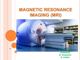
- 1. MAGNETIC RESONANCE IMAGING (MRI) Presented By K.Neelima D. Yasaswini S. Likitha
- 2. CONTENTS: INTRODUCTION ABOUT MRI BASIC PRINCIPLE COMPONENETS OF MRI WORKING MODEL OF MRI IMAGE WEIGHT MRI VS CT SCAN USES RISKS OF MRI SCAN
- 3. INTRODUCTION: Prof Peter Mansfield was awarded Noble prize in 2003 for his discovery in MRI with Prof Paul C Lautenberg of USA. MRI is a type of scan that uses strong magnetic fields and radio waves to produce detailed images of inside of the body. MRI perhaps the best application of superconductivity which directly affected the humanity across the globe. Prof Peter Mansfield Paul C Lautenberg
- 4. ABOUT MRI An MRI scanner is a large tube that contains powerful magnets. You lie inside the tube during scan. Magnetic resonance imaging is used to produce three dimensional detailed anatomical images. It is based on sophisticated technology It is often used for disease detection.
- 5. BASIC PRINCIPLE: Human body is entirely comprised of cells. Which contain water principally made of Hydrogen ions. The Magnet embedded with in the MRI Scanner can act on these positively charged hydrogen ions. When the magnet is switched off the protons will gradually return to their original state in a process known as precession.
- 6. COMPONENTS OF MRI Main magnet formed by super conducting coils. Gradient coil RF coils Computer system
- 7. WORKING OF MRI MRI’s employ powerful magnets which produce strong magnetic field that forces protons in the body. Radio frequency current is pulsed through the patient. Contrast agents may be given to the patients. (GADOLINIUM)
- 8. IMAGE WEIGHT: The Magnetic fields produced by the scanners can be manipulated to produce two distinct types of images. T1 weighted and T2 weighted. The resulting images will show different tissue types in different densities.
- 9. MRI VS CT SCAN CT SCAN MRI Uses X- rays for imaging Uses large external field, RF pulse and 3 different gradient fields. Exposure to ionizing radiation MRI machines do not emit ionizing radiation. Resolution problem. Good resolution & 3-D reconstruction. Injection of contrast medium. (dye) can cause kidney. Gadolinium contrast is relatively nontoxic. Problems or result in allergic or injection – site reactions in some people More cost Less cost than MRI. Quick process and easily available Lengthy process and non availability.
- 10. USES: MRI scanning is excellent for visualizing soft tissue. They do not cost any radiation exposure to the patient. It is used for disease detection, diagnosis and treatment monitoring. MRI can differentiate between white matter and grey matter.
- 11. RISKS: It does employ a strong magnetic field. The magnetic field extends beyond the machine and exerts powerful forces on objects of iron, some steels and other magnetic objects. The people with claustrophobia may find it difficult to tolerate long scan times inside the machine.
- 12. THANK YOU