The Making of an MRI
•
3 likes•161 views

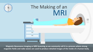
Magnetic Resonance Imaging or MRI Scanning as we commonly call it is a process where strong magnetic fields and radio waves are used to produce detailed images of the inside of a human body.Contact Us: Open MRI of Orlando 668 N. Orlando Avenue, Suite 1005 Maitland, FL 32751., Tel No: (407) 740-8848, Fax: 407-740-0324, Email: NewPatient@OpenMRIofOrlando.com

Recommended
More Related Content
What's hot
What's hot (20)
Similar to The Making of an MRI
Similar to The Making of an MRI (20)
Recently uploaded
Recently uploaded (20)
The Making of an MRI
- 1. The Making of an MRI Magnetic Resonance Imaging or MRI Scanning as we commonly call it is a process where strong magnetic fields and radio waves are used to produce detailed images of the inside of a human body.
- 2. How MRI Scans Help MRI Scans provide clear images of the abdomen, blood vessels, brain, chest, pelvis, tissues, joints and the spinal cord. With the help of these images, doctors can accurately diagnose a patient's medical condition. Doctors usually recommend getting an MRI Scan to detect conditions like joint or muscle disorders, cancer, etc.
- 3. MRI Scans are Commonly Generated for Head & Neck Region – 6% Upper & Lower Extremities – 20% Chest & Cardiac System – 3% Abdomen & Pelvis – 8% Spine – 26% Brain – 25%
- 4. Components of an MRI Scanner Although there are different makes and models of MRI Scanners available these days, the basic structure remains the same. The Magnet is the most important component of the MRI Scanners. The most commonly used magnet is the Superconducting Magnet, which is extremely powerful. The 3 Gradient Magnets present inside these machines, with significantly lower strength, are used to create a variable field that allows different body parts to be scanned. The Patient Table slides the patients in and out of the MRI Scanner. The area of the body that is to be scanned determines the patient's position.
- 5. A set of Coils helps transmit radio frequency waves into the patients' bodies. There are different coils present for different parts of the body. An extremely powerful Computer System is used to gather the data generated during the MRI scanning process and create images from the collected data.
- 6. The Scanning Process 1 2 3 The patient made to lie down and is positioned on the movable patient table. The coil containing devices are placed around or close to the area that is to be scanned. If injectable contrast is to be used for the scans, an IV line is inserted into the patient's vein.
- 7. 4 5 6 The patient is then slid inside the MRI Scanner, into the active magnetic field. The hydrogen atoms present in the patient's body align themselves in the direction of the magnetic field. Radio frequency waves transmitted by the coils cause protons from some hydrogen cells to spin at a particular frequency. 7 The computer receives signals from these protons in the form of mathematical data, which is then converted into images.
- 8. 8 9 The patient made to lie down and is positioned on the movable patient table. The coil containing devices are placed around or close to the area that is to be scanned. Sources of Reference: http://www.openmrioforlando.com/features.php http://www.magnetic-resonance.org/ch/21-01.html http://science.howstuffworks.com/mri1.htm http://www.radiologyinfo.org/en/info.cfm?pg=bodymr