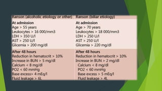
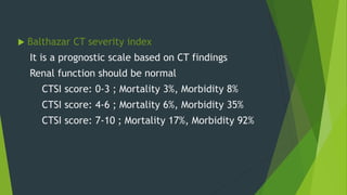
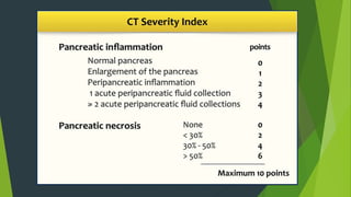
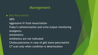
Acute Pancreatitis
By
Orangzeb Khatri
(4th year MBBS,Dow Medical College Karachi. )
Early medical experts found it extremely difficult to
understand the true function of the organ and they
actually considered pancreas to be nothing more
than a shock absorber.
Anatomy overview:
Pancreas is a retroperitoneal organ, lying on posterior
abdominal wall in the C-shaped curvature of duodenum.
Lies at the level of L1 and L2.
Posterior to stomach separated from it by lesser sac.
Divided into 4 parts: head, neck, body and tail.
Around 80g
Blood supply:
Splenic artery
Inferior pancreatic artery
Superior pancreaticoduodenal
artery
Inferior pancreaticoduodenal
artery
Pancreatica magna artery
Caudal pancreatic artery
Venous Drainage:
Splenic vein
SMV
Portal vein
Lymphatic Drainage:
Celiac nodes
Superior mesenteric nodes
Pancreaticosplenic nodes
Nerve Supply:
Parasympathetic by vagus
Sympathetic by splanchnic plexus
Ductal System:
Main pancreatic duct
(duct of Wirsung)
it joins CBD and empties into major
duodenal papilla (ampulla of Vater) in
2nd part of duodenum guarded by
sphincter of Oddi
Accessory pancreatic duct
(duct of Santorini)
It opens into minor duodenal papilla
superior to opening of main pancreatic
duct
Pancreas is both exocrine and
endocrine gland
Exocrine portion consists of acinar
cells and ductular cells, secretes
digestive juices i.e. trypsinogen,
lipase, amylase nuclease, elastase,
bicarbonate etc.
Endocrine portion consists of islets of
Langerhans concentrated most in tail,
secretes insulin, glucagon,
somatostatin and PP
Acute Pancreatitis
Acute inflammation of pancreas
Premature activation of pancreatic enzymes leading
to autodigestion
Can occur at any age but peak incidence is in young
man and older females
Types:
 Mild AP
Interstitial inflammation and edema without persistent organ
failure or local complications
Mortality rate of 1%
 Severe AP
Pancreatic necrosis accompanied by SIRS, multiple-organ
failure and local complications
Mortality rate of 20-50%
Mortality within 1st week due to multiple-organ failure
Mortality after 1st week is due to septic complication
Etiology
GET SMASHED
 Gall stone (MCC in Pakistan)
 Ethanol (MCC in western world)
 Trauma
 Steroids
 Mumps (coxsakie B and CMV)
 Autoimmune
 Scorpion venom
 Hyperlipidaemia
 Hypercalcemia
 Heredity
 ERCP induced
 Drugs (thiazide, furosemide,
estrogen, azathioprine,
sulphonamide etc. )
Diagnosis
It requires two of the following three features:
1. Clinical evidence
2. Biochemical Evidence
3. Radiologic Evidence
Clinical Presentation
 Abdominal pain
Site: Epigastric
Onset: Sudden
Character: Stabbing
Radiation: Back
Associated symptom: Nausea and Vomiting
Timing: Constant
Exacerbating/relieving factor: Movement/Leaning
Severity: Very severe
Physical Examination
 General Physical Examination
Tachycardia
Tachypnea
Hypotension
Fever
Pale
Sweating
Jaundice
Abdominal Examination
 Inspection
Grey-Turner’s sign
Cullen’s sign
Pandiaraja’s sign (ecchymosis of right axilla)
Mild distension
 Palpation
Guarding and Tenderness
 Auscultation
Absent gut sounds
One may find pleural effusion in basal part of chest
on percussion
Biochemical Test
 Serum amylase
3-5 times above normal limit (<100 U/L)
peaks in 1-2 hrs, returns to normal in 3-5 days
persistent elevation for greater than 10 days
suggests local complication i.e. pseudocyst and
abscess
 Serum lipase
3-5 times above upper normal limit (0-50 IU/L)
more sensitive and specific than amylase
also helpful in delayed presentation
Radiology
 Abdominal X-ray
Sentinel-loop sign
colon cut-off sign
 U/S
Does not establish the diagnosis of AP but it must
be done within 24hrs to rule out biliary tract
pathology
Sentinel-loop sign
Colon cut-off sign
 CT scan
Best single imaging investigation
not done in all pts, its indications are:
If the diagnosis is uncertain
Progressive clinical deterioration
Signs of sepsis
Signs of any organ failure
Any local complication
 typical findings
focal or diffuse parenchymal enlargement
changes in density because of edema
indistinct pancreatic margins owing to
inflammation
surrounding retroperitoneal fat stranding
 liquefactive necrosis of pancreatic parenchyma
lack of parenchymal enhancement
often multifocal
 infected necrosis
difficult to distinguish from aseptic liquefactive
necrosis
presence of gas is helpful
FNA helpful
 abscess formation
circumscribed fluid collection
little or no necrotic tissues (thus distinguishing
it from infected necrosis)
 hemorrhage
high-attenuation fluid in the retroperitoneum
or peripancreatic tissues
Renal-halo sign
Renal-halo sign
Prognosis
 Ranson’s criteria
It is the most frequently utilized predictor of
mortality
Its limitation is that it can not predict severity on
admission (requires 48h)
Ranson’s score 3 or greater suggests severe
Pancreatitis
 Balthazar CT severity index
It is a prognostic scale based on CT findings
Renal function should be normal
CTSI score: 0-3 ; Mortality 3%, Morbidity 8%
CTSI score: 4-6 ; Mortality 6%, Morbidity 35%
CTSI score: 7-10 ; Mortality 17%, Morbidity 92%
Manegement
 Mild Pancreatitis
NPO
Aggressive IV fluid resuscitation
Foley’s catheterization and urine output monitoring
Analgesics
Antiemetics
Antibiotics are not indicated
Cholecystectomy in case of gall stone pancreatitis
CT scan only when condition is deterioration
 Severe Pancreatitis
Admission to HDU
NPO
Aggressive IV fluid resuscitation
Foley’s catheterization and urine output monitoring
Analgesics
Antiemetics
Supplementation SaO2 >95%
Monitoring of vital signs, CVP, urine output and blood gases
LFTs, RFTs, serum Ca++, blood glucose, coagulation profile
CT scan
ERCP within 72h of severe gall stone pancreatitis or sings of cholangitis
Supportive therapy for organ failure
Antibiotics: Metronidazole + Imipenem not for >14 days
Nutritional support
Elective cholecystectomy in case of gall stone pancreatitis
It can have severe complications and
high mortality despite treatment.
It is believed that “Alexander the
great” died of pancreatitis
Complications
 Systemic (common in 1st week)
Shock
Arrhythmia
ARDS
Pleural Effusion
Renal Failure
DIC
Paralytic ileus
Encephalopathy
Metabolic
Hypocalcemia
Hyperglycemia
Hyperlipedemia
 Local (common after 1st week)
Acute fluid collection
Pancreatic necrosis
Pancreatic abscess
Pancreatic pseudocyst
Pancreatic ascites
Pleural effusion
portal vein thrombosis
Pseudo-aneurysm
Pancreatic Pseudocyst
Collection of Pancreatic enzymes rich fluid walled off by
granulation tissue
Psudo because not lined by epithelium
Most common cystic lesion (75%)
Requires 4 or more weeks to develop
Presents with persistent abdominal pain and persistent
elevated amylase level
A mass may be palpated on abdominal exam
Diagnosed by US and CT
Differentiated from cystic neoplasm by aspiration
CEA level is high in Cystic Neoplasm
Amylase level is high in pseudocyst but not diagnostic
Cytology reveals inflammatory cells in pseudocyst
Management:
Indications
Symptomatic
Complication
>6 cm and thick walled
Lasting for >12 weeks
To differentiate from tumor
Lasting for >12 weeks
To differentiate from tumor
Percutaneous Transgastric cystgastrostomy
Endoscopic transgastric cystgastrostomy
Surgical approach
Cystogastrostomy
Cystodudenostomy
Roux-en-Y Cystojejunostomy
Resection of tail of pancreas

Acute pancreatitis

  • 1.
    Acute Pancreatitis By Orangzeb Khatri (4thyear MBBS,Dow Medical College Karachi. )
  • 2.
    Early medical expertsfound it extremely difficult to understand the true function of the organ and they actually considered pancreas to be nothing more than a shock absorber.
  • 3.
    Anatomy overview: Pancreas isa retroperitoneal organ, lying on posterior abdominal wall in the C-shaped curvature of duodenum. Lies at the level of L1 and L2. Posterior to stomach separated from it by lesser sac. Divided into 4 parts: head, neck, body and tail. Around 80g
  • 5.
    Blood supply: Splenic artery Inferiorpancreatic artery Superior pancreaticoduodenal artery Inferior pancreaticoduodenal artery Pancreatica magna artery Caudal pancreatic artery
  • 6.
    Venous Drainage: Splenic vein SMV Portalvein Lymphatic Drainage: Celiac nodes Superior mesenteric nodes Pancreaticosplenic nodes Nerve Supply: Parasympathetic by vagus Sympathetic by splanchnic plexus
  • 7.
    Ductal System: Main pancreaticduct (duct of Wirsung) it joins CBD and empties into major duodenal papilla (ampulla of Vater) in 2nd part of duodenum guarded by sphincter of Oddi Accessory pancreatic duct (duct of Santorini) It opens into minor duodenal papilla superior to opening of main pancreatic duct
  • 8.
    Pancreas is bothexocrine and endocrine gland Exocrine portion consists of acinar cells and ductular cells, secretes digestive juices i.e. trypsinogen, lipase, amylase nuclease, elastase, bicarbonate etc. Endocrine portion consists of islets of Langerhans concentrated most in tail, secretes insulin, glucagon, somatostatin and PP
  • 9.
    Acute Pancreatitis Acute inflammationof pancreas Premature activation of pancreatic enzymes leading to autodigestion Can occur at any age but peak incidence is in young man and older females
  • 10.
    Types:  Mild AP Interstitialinflammation and edema without persistent organ failure or local complications Mortality rate of 1%  Severe AP Pancreatic necrosis accompanied by SIRS, multiple-organ failure and local complications Mortality rate of 20-50% Mortality within 1st week due to multiple-organ failure Mortality after 1st week is due to septic complication
  • 11.
    Etiology GET SMASHED  Gallstone (MCC in Pakistan)  Ethanol (MCC in western world)  Trauma  Steroids  Mumps (coxsakie B and CMV)  Autoimmune  Scorpion venom  Hyperlipidaemia  Hypercalcemia  Heredity  ERCP induced  Drugs (thiazide, furosemide, estrogen, azathioprine, sulphonamide etc. )
  • 12.
    Diagnosis It requires twoof the following three features: 1. Clinical evidence 2. Biochemical Evidence 3. Radiologic Evidence
  • 13.
    Clinical Presentation  Abdominalpain Site: Epigastric Onset: Sudden Character: Stabbing Radiation: Back Associated symptom: Nausea and Vomiting Timing: Constant Exacerbating/relieving factor: Movement/Leaning Severity: Very severe
  • 14.
    Physical Examination  GeneralPhysical Examination Tachycardia Tachypnea Hypotension Fever Pale Sweating Jaundice
  • 15.
    Abdominal Examination  Inspection Grey-Turner’ssign Cullen’s sign Pandiaraja’s sign (ecchymosis of right axilla) Mild distension  Palpation Guarding and Tenderness  Auscultation Absent gut sounds
  • 16.
    One may findpleural effusion in basal part of chest on percussion
  • 17.
    Biochemical Test  Serumamylase 3-5 times above normal limit (<100 U/L) peaks in 1-2 hrs, returns to normal in 3-5 days persistent elevation for greater than 10 days suggests local complication i.e. pseudocyst and abscess  Serum lipase 3-5 times above upper normal limit (0-50 IU/L) more sensitive and specific than amylase also helpful in delayed presentation
  • 18.
    Radiology  Abdominal X-ray Sentinel-loopsign colon cut-off sign  U/S Does not establish the diagnosis of AP but it must be done within 24hrs to rule out biliary tract pathology
  • 19.
  • 20.
  • 21.
     CT scan Bestsingle imaging investigation not done in all pts, its indications are: If the diagnosis is uncertain Progressive clinical deterioration Signs of sepsis Signs of any organ failure Any local complication
  • 22.
     typical findings focalor diffuse parenchymal enlargement changes in density because of edema indistinct pancreatic margins owing to inflammation surrounding retroperitoneal fat stranding  liquefactive necrosis of pancreatic parenchyma lack of parenchymal enhancement often multifocal
  • 23.
     infected necrosis difficultto distinguish from aseptic liquefactive necrosis presence of gas is helpful FNA helpful  abscess formation circumscribed fluid collection little or no necrotic tissues (thus distinguishing it from infected necrosis)  hemorrhage high-attenuation fluid in the retroperitoneum or peripancreatic tissues Renal-halo sign
  • 24.
  • 25.
    Prognosis  Ranson’s criteria Itis the most frequently utilized predictor of mortality Its limitation is that it can not predict severity on admission (requires 48h) Ranson’s score 3 or greater suggests severe Pancreatitis
  • 27.
     Balthazar CTseverity index It is a prognostic scale based on CT findings Renal function should be normal CTSI score: 0-3 ; Mortality 3%, Morbidity 8% CTSI score: 4-6 ; Mortality 6%, Morbidity 35% CTSI score: 7-10 ; Mortality 17%, Morbidity 92%
  • 29.
    Manegement  Mild Pancreatitis NPO AggressiveIV fluid resuscitation Foley’s catheterization and urine output monitoring Analgesics Antiemetics Antibiotics are not indicated Cholecystectomy in case of gall stone pancreatitis CT scan only when condition is deterioration
  • 30.
     Severe Pancreatitis Admissionto HDU NPO Aggressive IV fluid resuscitation Foley’s catheterization and urine output monitoring Analgesics Antiemetics
  • 31.
    Supplementation SaO2 >95% Monitoringof vital signs, CVP, urine output and blood gases LFTs, RFTs, serum Ca++, blood glucose, coagulation profile CT scan ERCP within 72h of severe gall stone pancreatitis or sings of cholangitis Supportive therapy for organ failure Antibiotics: Metronidazole + Imipenem not for >14 days Nutritional support Elective cholecystectomy in case of gall stone pancreatitis
  • 32.
    It can havesevere complications and high mortality despite treatment. It is believed that “Alexander the great” died of pancreatitis
  • 33.
    Complications  Systemic (commonin 1st week) Shock Arrhythmia ARDS Pleural Effusion Renal Failure DIC Paralytic ileus Encephalopathy Metabolic Hypocalcemia Hyperglycemia Hyperlipedemia
  • 34.
     Local (commonafter 1st week) Acute fluid collection Pancreatic necrosis Pancreatic abscess Pancreatic pseudocyst Pancreatic ascites Pleural effusion portal vein thrombosis Pseudo-aneurysm
  • 35.
    Pancreatic Pseudocyst Collection ofPancreatic enzymes rich fluid walled off by granulation tissue Psudo because not lined by epithelium Most common cystic lesion (75%) Requires 4 or more weeks to develop Presents with persistent abdominal pain and persistent elevated amylase level A mass may be palpated on abdominal exam
  • 36.
    Diagnosed by USand CT Differentiated from cystic neoplasm by aspiration CEA level is high in Cystic Neoplasm Amylase level is high in pseudocyst but not diagnostic Cytology reveals inflammatory cells in pseudocyst Management: Indications Symptomatic Complication >6 cm and thick walled Lasting for >12 weeks To differentiate from tumor
  • 37.
    Lasting for >12weeks To differentiate from tumor Percutaneous Transgastric cystgastrostomy Endoscopic transgastric cystgastrostomy Surgical approach Cystogastrostomy Cystodudenostomy Roux-en-Y Cystojejunostomy Resection of tail of pancreas