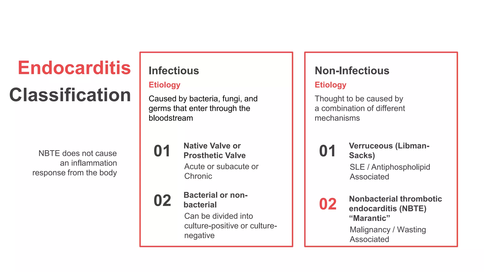
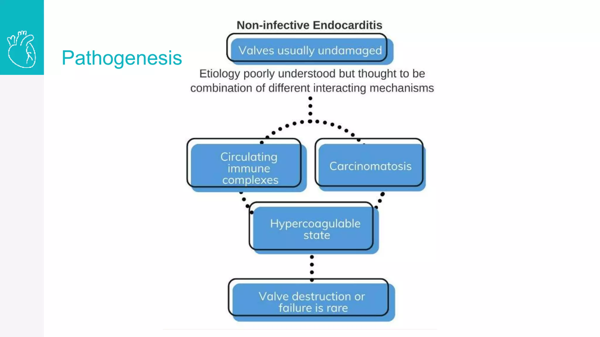
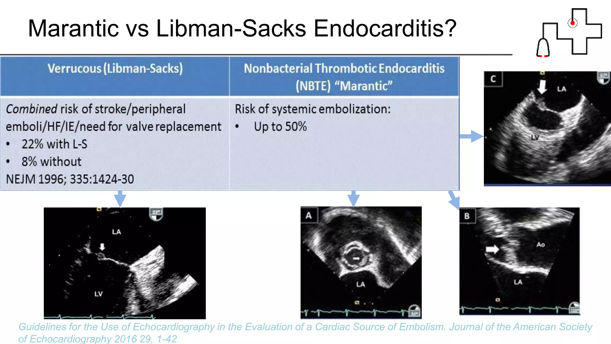
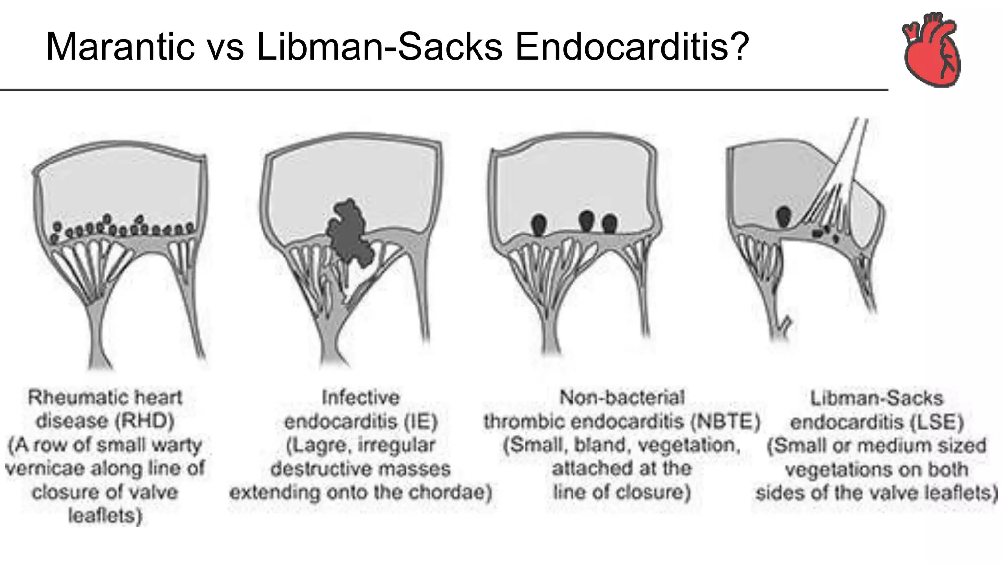
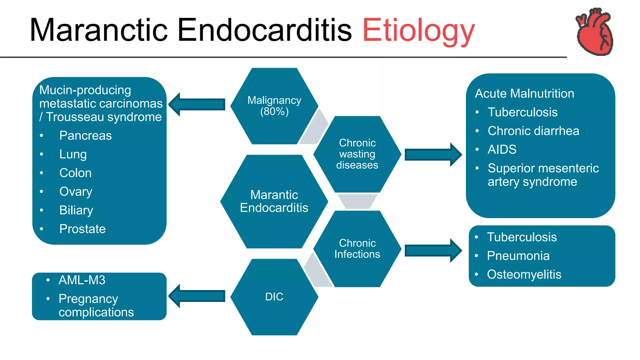
3. Endocardium
• Inner layer of the
cardiac wall and
covers the inner
surface of the
cardiac chambers
• The superficial
surface of the
cardiac valves
4. Endocardium
Histology
• Endothelium
endothelial cells (squamous epithelial cells)
• Subendothelial connective tissue layer
collagen fibrils and fibroblasts
more prominent in the atria, in particular, the left atrium
• Elastic tissue layer
elastic fibers, collagen fibers and smooth muscle cells
more prominent in the left atrium and the left ventricular septum
• Subendocardial tissue layer
connects endocardium to the myocardium
collagen and elastic fibers, small blood vessels and Purkinje fibers
6. Endocarditis
▹ Primarily of 1 or more heart
valves
▹ The mural endocardium
▹ A septal defect
Inflammation of the endocardial
surfaces of the heart
7. Endocarditis
Classification
NBTE does not cause
an inflammation
response from the body
Infectious
Etiology
Caused by bacteria, fungi, and
germs that enter through the
bloodstream
Non-Infectious
Etiology
Thought to be caused by
a combination of different
mechanisms
01
Acute or subacute or
Chronic
Native Valve or
Prosthetic Valve
02
Can be divided into
culture-positive or culture-
negative
Bacterial or non-
bacterial
01
SLE / Antiphospholipid
Associated
Verruceous (Libman-
Sacks)
02
Malignancy / Wasting
Associated
Nonbacterial thrombotic
endocarditis (NBTE)
“Marantic”
8. • Commonly affects undamaged valves (in contrast with infective
endocarditis)
• A separate entity to culture-negative endocarditis which is due to
infectious etiologies that are not readily identified
• Pathogenesis is unknown, but thought to be endothelial injury in the
setting of a hypercoagulable state
Deposition of sterile platelet thrombi on heart valves
(mostly aortic and mitral)
Marantic
Endocarditis
Pathogenesis
A form of noninfectious
endocarditis. a spectrum of
noninfectious lesions of the
heart valves that is most
commonly seen in advanced
malignancy
Nonbacterial thrombotic
endocarditis (NBTE)
9. Marantic Endocarditis Pathogenesis
Presentation
Cancer is spread
widely throughout
the body
Carcinomatosis
Wasting,
disseminated
intravascular
coagulation,
Hypercoagulable
State
But can occur in
patients with
valvular
pathology
Valve usually
undamaged
Valve Dysfunction
is rare
Systemic
Embolism
10. Marantic Endocarditis Characteristics
• A rare condition most often found postmortem with rates from 0.9
to 1.6 percent
• Up to 2.7% to 10% in adenocarcinomas and mucin-secreting
carcinomas, most commonly affects patients between the fourth
and eighth decades of life
• No gender predilection
• Libman-Sacks endocarditis could span up to 43% under TEE in
patients with SLE
16. How can we differentiate NBTE from IE?
• No pathognomonic
signs/symptoms or echo features
that are specific to NBTE
• Diagnosis can only be
differentiated with demonstration of
platelet thrombi on histologic exam
• High clinical suspicion is required
for diagnosis!
17. Marantic vs Libman-Sacks Endocarditis?
Guidelines for the Use of Echocardiography in the Evaluation of a Cardiac Source of Embolism. Journal of
the American Society of Echocardiography 2016 29, 1-42
18. Marantic vs Libman-Sacks Endocarditis?
Guidelines for the Use of Echocardiography in the Evaluation of a Cardiac Source of Embolism. Journal of the American Society
of Echocardiography 2016 29, 1-42
22. Presentation
Imaging
Signs and symptoms
Echocardiography
1 2
3 4
• Vegetations are frequently left-sided with
• two-thirds of cases involving the mitral valve, a quarter
the aortic
TEE is more sensitive than TTE
• Three sets of blood cultures and serological testing to
rule out Infective Endocarditis
• Workup for disseminated intravascular coagulation (DIC)
should be performed in every case of suspected NBTE
No laboratory tests that confirm the diagnosis of NBTE
• CT of the brain or MRI may be performed in those
with suspected cerebral embolization
Presents with strokes not radiologically specific to NBTE
• Major clinical manifestations of NBTE result from
systemic emboli in up to 50%
• Cardiac murmurs infrequently in less than half of
patients
• Fever is uncommon, may be an underlying
manifestation of malignancy
Often asymptomatic
Laboratory
23. Diagnosis
Gold Standard
• Made pathologically by
demonstration of thrombi
on autopsy or specimens
• Routine acquisition of
valvular tissue is not
practical
Clinical Diagnosis
• A constellation of clinical,
echocardiographic, and
absence of microbiologic
findings
• Demonstration of valvular
vegetations on
echocardiography in the
absence of systemic
infection in patients who
are at high risk
24. Diagnostic Challenges
Diagnosis of
NBTE is
difficult to
establish
antemortem
• Paucity of
symptoms
• Late
presentation
Fever due to
the
underlying
malignancy
can lower
the clinical
suspicion for
NBTE
Small
remnants on
affected
valves
(≤3 mm)
following
embolization
not readily
identified by
echocardiogr
aphy
False
negatives
25. The Mitral valve revealing thickened leaflets
3D TEE PLAX
Transoesophageal echocardiography short axis
showing the thickening in motion
3D TEE PSAX Mitral
26. Transoesophageal echocardiography showing large
oscillating structures in long axis
3D TEE PLAX
The aortic valve revealing severely thickened cusps
3D TEE PSAX Aortic
28. Anticoagulation
NBTE patients are routinely anticoagulated
• Rather than warfarin or a direct
thrombin or factor Xa inhibitor
• Anticoagulation should be
continued indefinitely
Therapeutic dose subcutaneous
LMWH or intravenous UFH should
be used
• Provided there is no
contraindication (e.g., active central
nervous system bleeding)
• Due to high rates of recurrent and
extensive embolization in this
population
With or without evidence of
systemic emboli
01 02
29. Surgery
May be considered in select cases
• Heart failure
• Acute valve rupture
• Prevention of recurrent
embolization
Indications for surgery same as IE
• Benefits should be weighed against
the risks in the context of the life
expectancy
• Vegetation excision or valve
replacement
Where the risk benefit is favorable
01 02
30. Treatment for underlying disease
Disease treatment often ineffective in the absence of effective systemic therapy
• Antibiotics do not need to be
continued unless infective
endocarditis is diagnosed
Many patients may be placed on
empiric antibiotics during
investigation
• It is unknown whether NBTE
improves with cancer therapy
• Anticoagulation should continue
regardless of the response
Metastatic nature of most
malignancies at presentation
01 02
31. Followup & Prognosis
Optimal followup has not been defined and prognosis of NBTE has not been formally
evaluated — should be individualized
Due to the strong association between
NBTE and advanced malignancy
Prognosis is grim despite
anticoagulation
NBTE complications
• Infective endocarditis
• Embolization despite
anticoagulation
Complications of therapy
• Bleeding
• Thrombocytopenia
Patients should be followed
clinically for complications
01 02