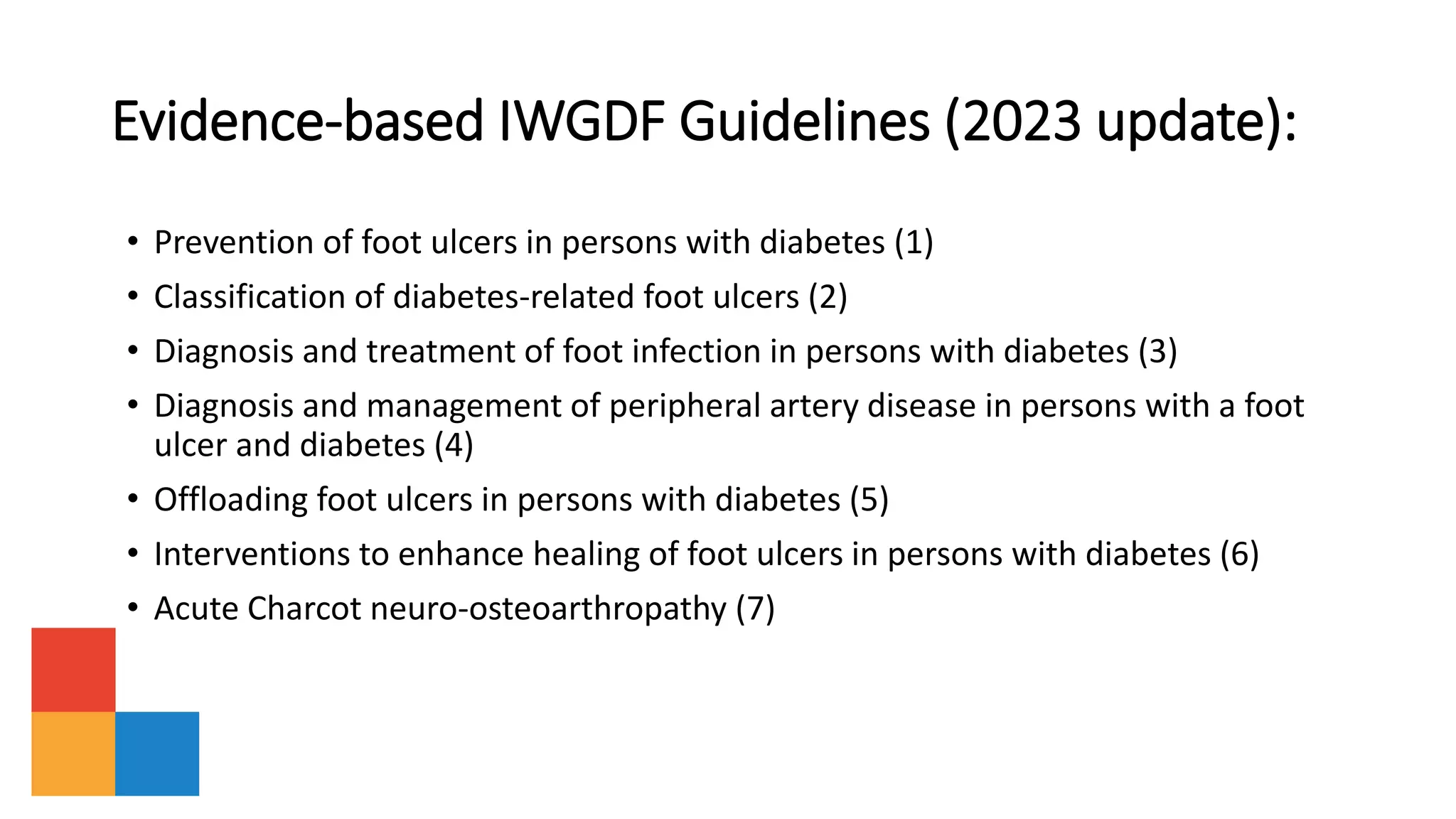
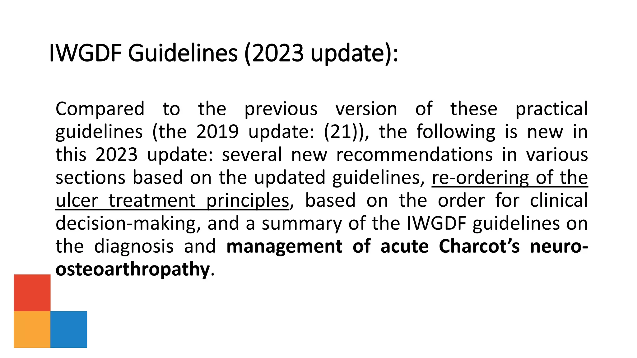
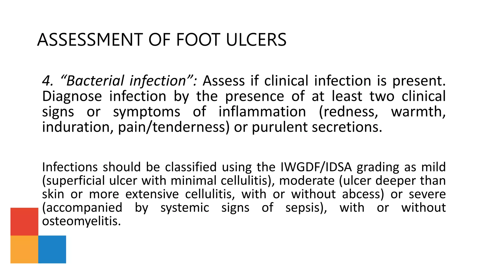
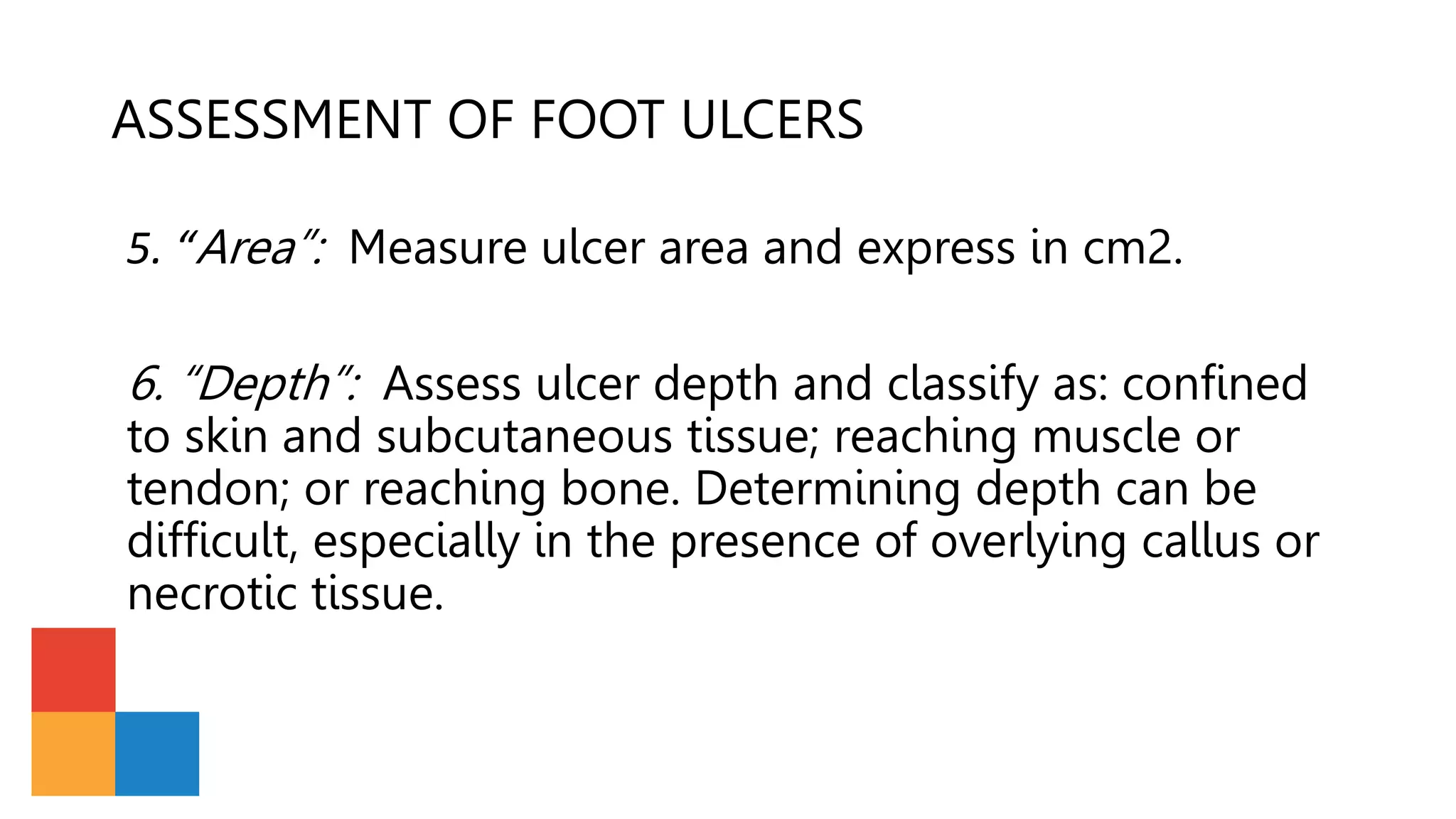
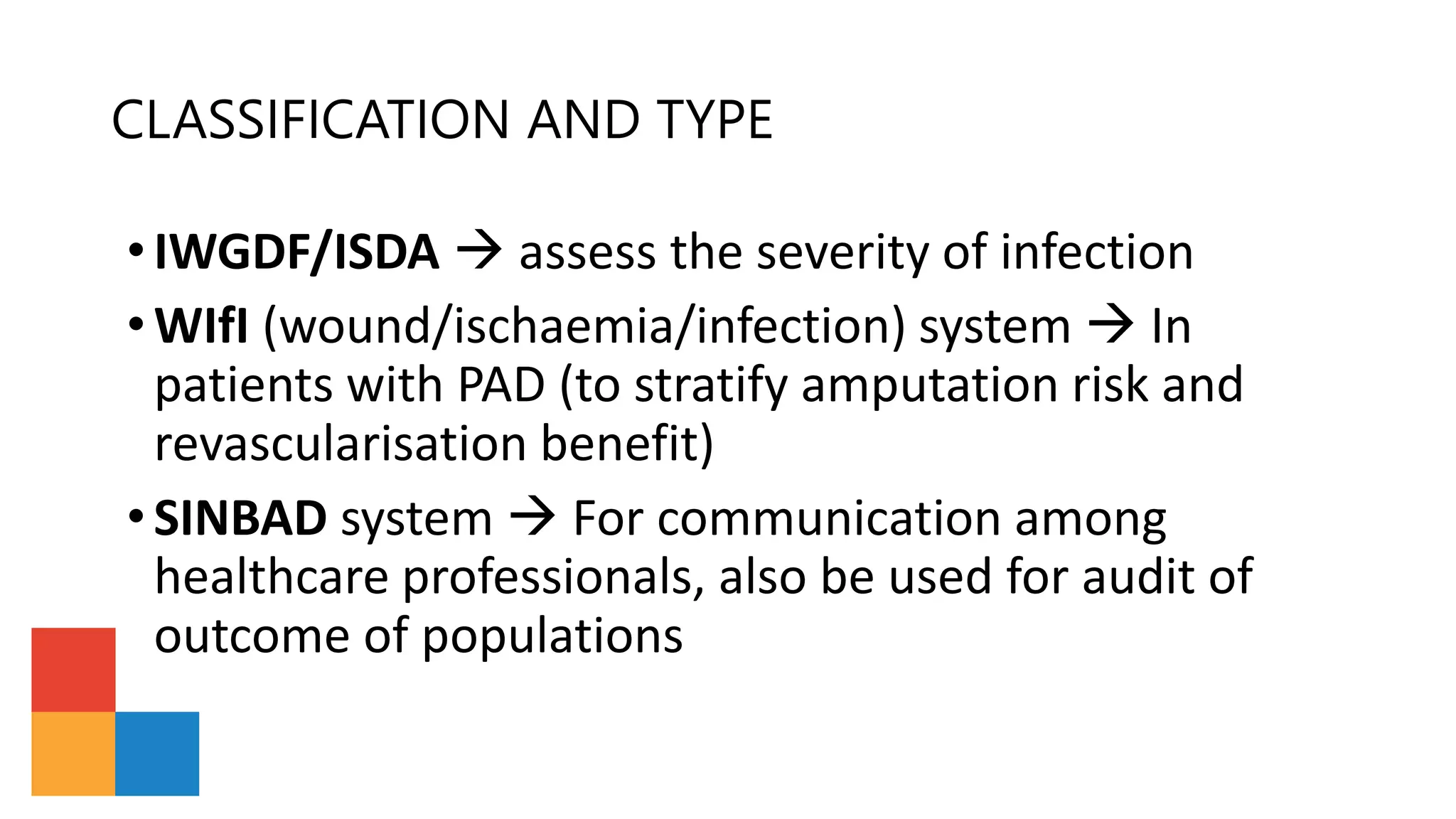
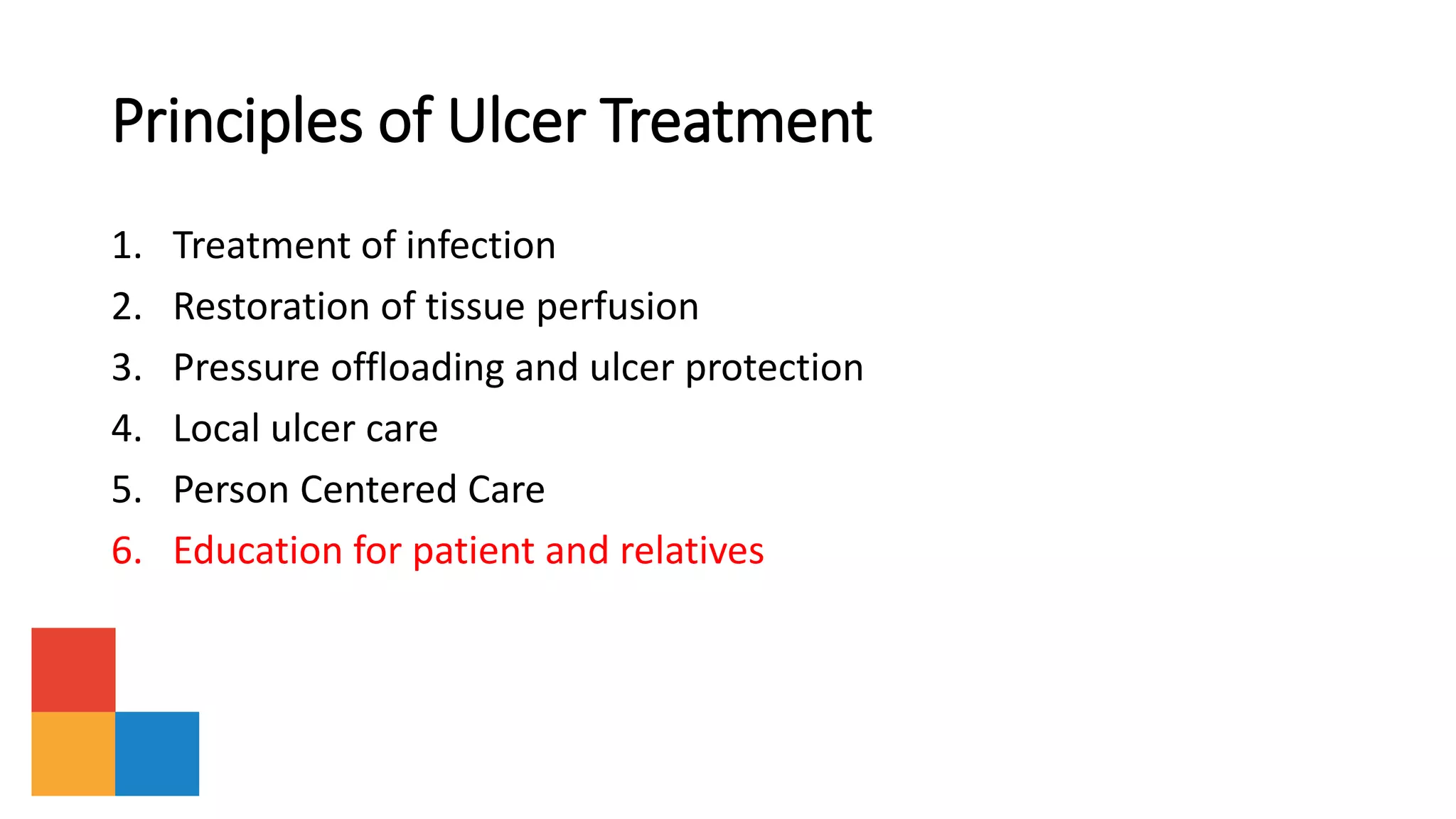
SlideShare a Scribd company logo
1 of 33
2 0 1 9 & 2 0 2 3
Introduction
IWGDF Practical Guidelines describe the basic
principles of prevention and management of
diabetic foot disease.
Evidence-based IWGDF Guidelines (2023 update):
• Prevention of foot ulcers in persons with diabetes (1)
• Classification of diabetes-related foot ulcers (2)
• Diagnosis and treatment of foot infection in persons with diabetes (3)
• Diagnosis and management of peripheral artery disease in persons with a foot
ulcer and diabetes (4)
• Offloading foot ulcers in persons with diabetes (5)
• Interventions to enhance healing of foot ulcers in persons with diabetes (6)
• Acute Charcot neuro-osteoarthropathy (7)
IWGDF Guidelines (2023 update):
Compared to the previous version of these practical
guidelines (the 2019 update: (21)), the following is new in
this 2023 update: several new recommendations in various
sections based on the updated guidelines, re-ordering of the
ulcer treatment principles, based on the order for clinical
decision-making, and a summary of the IWGDF guidelines on
the diagnosis and management of acute Charcot’s neuro-
osteoarthropathy.
Diabetic Foot Disease
• Diabetic Foot Disease is among the most serious
complications of diabetes mellitus
• It is a source of major suffering and financial cost for the
patient, also a considerable burden on the patients family,
healthcare professionals and facilities and society in general
• Strategies includes: prevention, patient and staff
education, multidisciplinary treatment, and close
monitoring
Patophysiology
• The pathway to ulceration are similar in most patients.
• These ulcers frequently results from a person with diabetes
simultaneously having two or more risk factors, with
diabetic peripheral neuropathy and peripheral artery
disease (PAD)
Neuropathy
•The neuropathy  leads to insensitive and
sometimes deformed foot.
•In neuropathy, minor trauma can precipitate
ulceration of the foot (e.g: ill-fitting shoes or an acute
mechanical or thermal injury)
Loss of protective sensation, foot deformities, and limited joint
mobility can result in abnormal biomechanical loading of the foot. This
produces high mechanical stress in some areas, the response to which
is usually thickened skin (callus). The callus then leads to a further
increase in the loading of the foot, often with subcutaneous
haemorrhage and eventually skin ulceration.
Peripheral Artery Disease (PAD)
• PAD generally caused by atherosclerosis, is present in up to 50% of
patients with a diabetic foot ulcer.
• A small percentage of foot ulcers in patients with severe PAD are
purely ischaemic; these are usually painful and may follow minor
trauma
• The majority of foot ulcers, however, are either purely neuropathic
or neuro-ischaemic, i.e., caused by combined neuropathy and
ischaemia
FOOT ULCER PREVENTION
There are five key elements to prevent foot ulcers:
1.Identify the person with an at-risk foot
2.Regularly inspect and examine the feet of a person at-risk for foot
ulceration
3.Provide structured education for patients, their family and
healthcare professionals
4.Encourage routine wearing of appropriate footwear
5.Treat risk factors for ulceration
Areas of the foot at highest risk for ulceration
Footwear should be sufficiently wide
to accommodate the foot without
excessive pressure on the skin
The proper way to cut toe nails
ASSESSMENT & TREATMENT
OF FOOT ULCERS
ASSESSMENT OF FOOT ULCERS
Classification of the foot ulcer: the foot ulcer should be classified
following the assessment of the six items of the SINBAD system.
1. “SITE”: Describe where the ulcer is located on the foot. This
includes description of forefoot, midfoot or hindfoot, but it is
also suggested to differentiate between plantar, interdigital,
medial, lateral or dorsal.
ASSESSMENT OF FOOT ULCERS
2. “ISCHEMIA”: Assess if pedal blood flow is intact (at least one
palpable pulse), or if there is clinical evidence of reduced blood flow.
Further, examine the arterial pedal wave forms (with a Doppler
instrument), measure the ankle and toe pressures, and calculate the
ankle-brachial index (ABI) and toe-brachial index (TBI).
ASSESSMENT OF FOOT ULCERS
3. “Neuropathy”: Assess if protective sensation is intact or
lost  DOING A SENSORY FOOT EXAMINATION
ASSESSMENT OF FOOT ULCERS
4. “Bacterial infection”: Assess if clinical infection is present.
Diagnose infection by the presence of at least two clinical
signs or symptoms of inflammation (redness, warmth,
induration, pain/tenderness) or purulent secretions.
Infections should be classified using the IWGDF/IDSA grading as mild
(superficial ulcer with minimal cellulitis), moderate (ulcer deeper than
skin or more extensive cellulitis, with or without abcess) or severe
(accompanied by systemic signs of sepsis), with or without
osteomyelitis.
ASSESSMENT OF FOOT ULCERS
5. “Area”: Measure ulcer area and express in cm2.
6. “Depth”: Assess ulcer depth and classify as: confined
to skin and subcutaneous tissue; reaching muscle or
tendon; or reaching bone. Determining depth can be
difficult, especially in the presence of overlying callus or
necrotic tissue.
CLASSIFICATION AND TYPE
•IWGDF/ISDA  assess the severity of infection
•WIfI (wound/ischaemia/infection) system  In
patients with PAD (to stratify amputation risk and
revascularisation benefit)
•SINBAD system  For communication among
healthcare professionals, also be used for audit of
outcome of populations
Principles of Ulcer Treatment
1. Treatment of infection
2. Restoration of tissue perfusion
3. Pressure offloading and ulcer protection
4. Local ulcer care
5. Person Centered Care
6. Education for patient and relatives
1. Treatment of Infection
• Superficial ulcer with limited soft tissue (mild) infection:
• Cleanse, debride all necrotic tissue and surrounding callus
• Start empiric oral antibiotic therapy targeted at Staphylococcus aureus and
streptococci (unless there are reasons to consider other, or additional, likely
pathogens)
• Deep or extensive (potentially limb-threatening) infection (moderate
or severe infection):
• Urgently evaluate for need for surgical intervention to remove necrotic tissue,
including infected
• bone, release compartment pressure or drain abscesses
• Assess for PAD; if present consider urgent treatment, including
revascularization
• Initiate empiric, parenteral, broad-spectrum antibiotic therapy, aimed at
common gram-positive and gram-negative bacteria, including obligate
anaerobes
• Adjust (constrain and target, if possible) the antibiotic regimen based on both
the clinical response to empirical therapy and culture and sensitivity results
2. Restoration of Tissue Perfusion
• Revascularization if:
• An ankle pressure <50mm Hg or an ABI <0.5  consider urgent vascular imaging
• A toe pressure is <30mmHg or TcpO2 is <25 mmHg
• When an ulcer fails to show signs of healing within 6 weeks, despite optimal management
• The aim of revascularization:
• to restore direct flow to at least one of the foot arteries, preferably the artery that supplies
the anatomical region of the wound
• Select a revascularization technique based on both individual factors (the patients
factor and the local operator expertise
• After a revascularization procedure  effectiveness should be
evaluated with an objective measurement of perfusion
• Pharmacological treatments to improve perfusion have not been
proven to be beneficial
• Emphasise efforts to reduce cardiovascular risk (cessation of smoking,
control of hypertension and dyslipidaemia, use of anti-platelet drugs)
• Non-removable knee-high offloading device
• a total contact cast (TCC)
• Removable knee-high offloading device
• Ankle-high offloading device
• If biomechanical relief are not available  using felted foam, but only
in combination with appropriate footwear
3. Pressure offloading and ulcer protection
4. Local Ulcer Care
• Regular inspection of the ulcer by a trained health care provider is
essential
• Debride the ulcer and remove surrounding callus (preferably with
sharp surgical instruments), and repeat as needed
• Select dressings to control excess exudation and maintain moist
environment
• Do not soak the feet, as this may induce skin maceration.
• Consider negative pressure to help heal post-operative wounds
Consider one of the following adjunctive treatments in non-infected
ulcers that fail to heal after 4-6 weeks despite optimal clinical care:
• A sucrose octasulfate impregnated dressing in neuro-ischemic ulcers (without
severe ischemia)
• A multi-layered patch of autologous leucocytes, platelets and fibrin in ulcers
with or without moderate ischemia
• Placental membrane allografts in ulcers with or without moderate ischemia
• Systemic oxygen therapy as an adjunctive treatment in ischaemic ulcers that
do not heal despite revascularisation
• The following treatments are not well-supported for routine ulcer
management:
• Biologically active products (collagen, growth factors, bio- engineered tissue)
in neuropathic ulcers
• Silver, or other antimicrobial agent, containing dressings or topical
applications
5. Person Centered Care
• Optimise glycaemic control, if necessary, with insulin.
• Treat oedema or malnutrition, if present.
• Treat cardiovascular risk factors.
• Treat depression or other psycho-social difficulties.
6. Education for Patients and Relatives
• Instruct patients (and relatives or carers) on appropriate foot ulcer
self-care and how to recognize and report signs and symptoms of new
or worsening infection (e.g., onset of fever, changes in local wound
conditions, worsening hyperglycaemia)
• During a period of enforced bed rest, instruct on how to prevent an
ulcer on the contra- lateral foot
ACTIVE CHARCOT NEURO-OSTEOARTHROPATHY (CNO)
• In any person with diabetes mellitus and with a red, hot, swollen foot, the diagnosis of active
CNO should be considered. As described in our Charcot guidelines, CNO is a sterile inflammatory
process in persons with neuropathy that results in injury to bones, joints and soft tissues
• The diagnosis is based on the aforementioned clinical findings of inflammation after
exclusion of other causes and abnormalities on imaging. If these abnormalities are not
seen on plain X-ray, an MRI should be performed; if an MRI is not possible, perform a
CT-scan and/or a radionucleotide scan. When such advanced imaging is not possible the
person should be treated as having a probable active CNO.
• If not treated adequately, it can result in progressive fracturing and dislocations resulting
in a deformed foot
• The diagnosis is based on the aforementioned clinical findings of inflammation after
exclusion of other causes and abnormalities on imaging. If these abnormalities are not
seen on plain X-ray, an MRI should be performed; if an MRI is not possible, perform a
CT-scan and/or a radionucleotide scan
ACTIVE CHARCOT NEURO-OSTEOARTHROPATHY (CNO)
• In order to promote remission of the disease and to prevent (progressive)
deformity, the affected extremity should be offloaded and immobilized.
• Treatment should start once the diagnosis is considered and continue
until clinical remission with consolidation of fractures is achieved. As long
as there are clinical signs of inflammation the offloading should be
continued.
• Currently there is no medical therapy that can shorten the duration of
disease or prevent deformities, such interventions are therefore not
recommended. Vitamin D and calcium should be supplemented
according to local guidelines for persons with an elevated risk of
inadequate vitamin D levels.
• The knee-high cast can be stopped when there are no clinical signs of
inflammation with radiographic consolidation of fractures (if present) on
plain X-ray.
THANK YOU

More Related Content

What's hot

Diabetic Foot
Diabetic  FootDiabetic  Foot
Diabetic Footdrkmliau
 
The Diabetic Foot: What You Need to Know
The Diabetic Foot: What You Need to KnowThe Diabetic Foot: What You Need to Know
The Diabetic Foot: What You Need to KnowOmar Haqqani
 
Glasgow Coma Scale Presentation
Glasgow Coma Scale PresentationGlasgow Coma Scale Presentation
Glasgow Coma Scale PresentationHayden G
 
Comprehensive Diabetic Foot Examination
Comprehensive Diabetic Foot ExaminationComprehensive Diabetic Foot Examination
Comprehensive Diabetic Foot ExaminationDonald Pelto
 
Negative pressure wound therapy
Negative pressure wound therapy Negative pressure wound therapy
Negative pressure wound therapy Drkabiru2012
 
Acute and sub-acute Osteomyelitis
Acute and sub-acute OsteomyelitisAcute and sub-acute Osteomyelitis
Acute and sub-acute OsteomyelitisAIIMS Bhopal
 
Medical Management in OsteoarthritisOsteoarthritis
 Medical Management in OsteoarthritisOsteoarthritis Medical Management in OsteoarthritisOsteoarthritis
Medical Management in OsteoarthritisOsteoarthritisUtai Sukviwatsirikul
 
History taking in Medicine
History taking in MedicineHistory taking in Medicine
History taking in Medicinedrnooruddin
 
Major Trauma management in emergency room Tribhuvan university teaching hospi...
Major Trauma management in emergency room Tribhuvan university teaching hospi...Major Trauma management in emergency room Tribhuvan university teaching hospi...
Major Trauma management in emergency room Tribhuvan university teaching hospi...sameerdm
 
The diabetic foot; state of the art
The diabetic foot; state of the artThe diabetic foot; state of the art
The diabetic foot; state of the artMEEQAT HOSPITAL
 
Polymyalgia rheumatica and giant cell arteiritis
Polymyalgia rheumatica and giant cell arteiritisPolymyalgia rheumatica and giant cell arteiritis
Polymyalgia rheumatica and giant cell arteiritisdattasrisaila
 
Principles of arthrotomy & arthrocentesis
Principles of arthrotomy & arthrocentesisPrinciples of arthrotomy & arthrocentesis
Principles of arthrotomy & arthrocentesisAsi-oqua Bassey
 
Diabetic foot by Dr. Basil Tumaini and Dr. May Shoo
Diabetic foot by Dr. Basil Tumaini and Dr. May ShooDiabetic foot by Dr. Basil Tumaini and Dr. May Shoo
Diabetic foot by Dr. Basil Tumaini and Dr. May ShooBasil Tumaini
 
Peripheral Vascular Injuries
Peripheral Vascular InjuriesPeripheral Vascular Injuries
Peripheral Vascular InjuriesLateef Khan
 
Discussion on psoriatic arthritis
Discussion on psoriatic arthritisDiscussion on psoriatic arthritis
Discussion on psoriatic arthritissakib_lostvalley
 

What's hot (20)

Diabetic Foot
Diabetic  FootDiabetic  Foot
Diabetic Foot
 
The Diabetic Foot: What You Need to Know
The Diabetic Foot: What You Need to KnowThe Diabetic Foot: What You Need to Know
The Diabetic Foot: What You Need to Know
 
Glasgow Coma Scale Presentation
Glasgow Coma Scale PresentationGlasgow Coma Scale Presentation
Glasgow Coma Scale Presentation
 
Comprehensive Diabetic Foot Examination
Comprehensive Diabetic Foot ExaminationComprehensive Diabetic Foot Examination
Comprehensive Diabetic Foot Examination
 
Negative pressure wound therapy
Negative pressure wound therapy Negative pressure wound therapy
Negative pressure wound therapy
 
Acute and sub-acute Osteomyelitis
Acute and sub-acute OsteomyelitisAcute and sub-acute Osteomyelitis
Acute and sub-acute Osteomyelitis
 
Medical Management in OsteoarthritisOsteoarthritis
 Medical Management in OsteoarthritisOsteoarthritis Medical Management in OsteoarthritisOsteoarthritis
Medical Management in OsteoarthritisOsteoarthritis
 
History taking in Medicine
History taking in MedicineHistory taking in Medicine
History taking in Medicine
 
Major Trauma management in emergency room Tribhuvan university teaching hospi...
Major Trauma management in emergency room Tribhuvan university teaching hospi...Major Trauma management in emergency room Tribhuvan university teaching hospi...
Major Trauma management in emergency room Tribhuvan university teaching hospi...
 
Offloading in diabetic foot
Offloading in diabetic footOffloading in diabetic foot
Offloading in diabetic foot
 
The diabetic foot; state of the art
The diabetic foot; state of the artThe diabetic foot; state of the art
The diabetic foot; state of the art
 
Metabolic response to trauma
Metabolic response to traumaMetabolic response to trauma
Metabolic response to trauma
 
Polymyalgia rheumatica and giant cell arteiritis
Polymyalgia rheumatica and giant cell arteiritisPolymyalgia rheumatica and giant cell arteiritis
Polymyalgia rheumatica and giant cell arteiritis
 
Soft tissue healing
Soft tissue healingSoft tissue healing
Soft tissue healing
 
Principles of arthrotomy & arthrocentesis
Principles of arthrotomy & arthrocentesisPrinciples of arthrotomy & arthrocentesis
Principles of arthrotomy & arthrocentesis
 
CHRONIC OSTEOMYELITIS
CHRONIC  OSTEOMYELITISCHRONIC  OSTEOMYELITIS
CHRONIC OSTEOMYELITIS
 
Diabetic foot by Dr. Basil Tumaini and Dr. May Shoo
Diabetic foot by Dr. Basil Tumaini and Dr. May ShooDiabetic foot by Dr. Basil Tumaini and Dr. May Shoo
Diabetic foot by Dr. Basil Tumaini and Dr. May Shoo
 
Diabetic neuropathy
Diabetic neuropathyDiabetic neuropathy
Diabetic neuropathy
 
Peripheral Vascular Injuries
Peripheral Vascular InjuriesPeripheral Vascular Injuries
Peripheral Vascular Injuries
 
Discussion on psoriatic arthritis
Discussion on psoriatic arthritisDiscussion on psoriatic arthritis
Discussion on psoriatic arthritis
 

Similar to IWGDF PRACTICAL GUIDELINES 2019 + 2023 update.pptx

Diabetic foot
Diabetic foot Diabetic foot
Diabetic foot FaizNaeem1
 
Management of diabetic foot
Management of diabetic footManagement of diabetic foot
Management of diabetic footRahulkumar5300
 
Diabetic foot disease ‫‬
Diabetic foot disease ‫‬Diabetic foot disease ‫‬
Diabetic foot disease ‫‬ismail naameh
 
Kinzhnik — OGANIZING MEDICAL CARE FOR DFS PATIENT
Kinzhnik — OGANIZING MEDICAL CARE FOR DFS PATIENTKinzhnik — OGANIZING MEDICAL CARE FOR DFS PATIENT
Kinzhnik — OGANIZING MEDICAL CARE FOR DFS PATIENTPavel Fedotov
 
Michael Knizhnik — OGANIZING MEDICAL CARE FOR DFS PATIENT
Michael Knizhnik — OGANIZING MEDICAL CARE FOR DFS PATIENTMichael Knizhnik — OGANIZING MEDICAL CARE FOR DFS PATIENT
Michael Knizhnik — OGANIZING MEDICAL CARE FOR DFS PATIENTPavel Fedotov
 
Nursing care of dsf sr abeer
Nursing care of dsf sr abeerNursing care of dsf sr abeer
Nursing care of dsf sr abeerMEEQAT HOSPITAL
 
DIABETIC FOOT ULCER
DIABETIC FOOT ULCERDIABETIC FOOT ULCER
DIABETIC FOOT ULCERHaziq Mars
 
trophic ulcer.pptx
trophic ulcer.pptxtrophic ulcer.pptx
trophic ulcer.pptxTEJARAM19
 
Osteomyelitis and its management
Osteomyelitis and its managementOsteomyelitis and its management
Osteomyelitis and its managementShweta Sharma
 
Diabetic Foot .pptx
Diabetic Foot .pptxDiabetic Foot .pptx
Diabetic Foot .pptxWafa sheikh
 
DIABETIC FOOT ULCER AND SOFT TISSUE INFECTIONS-converted.pdf
DIABETIC FOOT ULCER AND SOFT TISSUE INFECTIONS-converted.pdfDIABETIC FOOT ULCER AND SOFT TISSUE INFECTIONS-converted.pdf
DIABETIC FOOT ULCER AND SOFT TISSUE INFECTIONS-converted.pdfRidzwanSafai1
 
nursing care of patient musculoskeletal.ppt
nursing care of patient musculoskeletal.pptnursing care of patient musculoskeletal.ppt
nursing care of patient musculoskeletal.pptahmed ibrahem
 

Similar to IWGDF PRACTICAL GUIDELINES 2019 + 2023 update.pptx (20)

Diabetic foot
Diabetic foot Diabetic foot
Diabetic foot
 
Management of diabetic foot
Management of diabetic footManagement of diabetic foot
Management of diabetic foot
 
Diabetic foot disease ‫‬
Diabetic foot disease ‫‬Diabetic foot disease ‫‬
Diabetic foot disease ‫‬
 
Diabetic foot
Diabetic footDiabetic foot
Diabetic foot
 
Kinzhnik — OGANIZING MEDICAL CARE FOR DFS PATIENT
Kinzhnik — OGANIZING MEDICAL CARE FOR DFS PATIENTKinzhnik — OGANIZING MEDICAL CARE FOR DFS PATIENT
Kinzhnik — OGANIZING MEDICAL CARE FOR DFS PATIENT
 
Michael Knizhnik — OGANIZING MEDICAL CARE FOR DFS PATIENT
Michael Knizhnik — OGANIZING MEDICAL CARE FOR DFS PATIENTMichael Knizhnik — OGANIZING MEDICAL CARE FOR DFS PATIENT
Michael Knizhnik — OGANIZING MEDICAL CARE FOR DFS PATIENT
 
Diabetic foot ulcer / surgical wounds
Diabetic foot ulcer / surgical woundsDiabetic foot ulcer / surgical wounds
Diabetic foot ulcer / surgical wounds
 
Nursing care of DSF
Nursing care of DSF Nursing care of DSF
Nursing care of DSF
 
Nursing care of dsf sr abeer
Nursing care of dsf sr abeerNursing care of dsf sr abeer
Nursing care of dsf sr abeer
 
Diabetic foot
Diabetic foot Diabetic foot
Diabetic foot
 
Diabetic foot
Diabetic footDiabetic foot
Diabetic foot
 
Diabetic foot
Diabetic footDiabetic foot
Diabetic foot
 
DIABETIC FOOT ULCER
DIABETIC FOOT ULCERDIABETIC FOOT ULCER
DIABETIC FOOT ULCER
 
trophic ulcer.pptx
trophic ulcer.pptxtrophic ulcer.pptx
trophic ulcer.pptx
 
Osteomyelitis and its management
Osteomyelitis and its managementOsteomyelitis and its management
Osteomyelitis and its management
 
Diabeticfoot
DiabeticfootDiabeticfoot
Diabeticfoot
 
Diabetic foot
Diabetic footDiabetic foot
Diabetic foot
 
Diabetic Foot .pptx
Diabetic Foot .pptxDiabetic Foot .pptx
Diabetic Foot .pptx
 
DIABETIC FOOT ULCER AND SOFT TISSUE INFECTIONS-converted.pdf
DIABETIC FOOT ULCER AND SOFT TISSUE INFECTIONS-converted.pdfDIABETIC FOOT ULCER AND SOFT TISSUE INFECTIONS-converted.pdf
DIABETIC FOOT ULCER AND SOFT TISSUE INFECTIONS-converted.pdf
 
nursing care of patient musculoskeletal.ppt
nursing care of patient musculoskeletal.pptnursing care of patient musculoskeletal.ppt
nursing care of patient musculoskeletal.ppt
 

More from Mau Maulana

MATERI STS AQIDAH KLS 4 Semst II aga islam
MATERI STS AQIDAH KLS 4 Semst II aga islamMATERI STS AQIDAH KLS 4 Semst II aga islam
MATERI STS AQIDAH KLS 4 Semst II aga islamMau Maulana
 
Trauma Score.pptx
Trauma Score.pptxTrauma Score.pptx
Trauma Score.pptxMau Maulana
 
PEMERIKSAAN_SAdari tes1pptx.pptx
PEMERIKSAAN_SAdari tes1pptx.pptxPEMERIKSAAN_SAdari tes1pptx.pptx
PEMERIKSAAN_SAdari tes1pptx.pptxMau Maulana
 
GASTER perforasi 1.pptx
GASTER perforasi 1.pptxGASTER perforasi 1.pptx
GASTER perforasi 1.pptxMau Maulana
 
Perforasi Gaster coba1.pptx
Perforasi Gaster coba1.pptxPerforasi Gaster coba1.pptx
Perforasi Gaster coba1.pptxMau Maulana
 
abdominal-trauma.ppt
abdominal-trauma.pptabdominal-trauma.ppt
abdominal-trauma.pptMau Maulana
 
3 Screening and Nutritional Assessment.pptx.pptx
3 Screening and Nutritional Assessment.pptx.pptx3 Screening and Nutritional Assessment.pptx.pptx
3 Screening and Nutritional Assessment.pptx.pptxMau Maulana
 
keseimbangancairanelektrolitaw-140717024451-phpapp01.pdf
keseimbangancairanelektrolitaw-140717024451-phpapp01.pdfkeseimbangancairanelektrolitaw-140717024451-phpapp01.pdf
keseimbangancairanelektrolitaw-140717024451-phpapp01.pdfMau Maulana
 
Lanjutan TPN.pdf
Lanjutan TPN.pdfLanjutan TPN.pdf
Lanjutan TPN.pdfMau Maulana
 

More from Mau Maulana (16)

MATERI STS AQIDAH KLS 4 Semst II aga islam
MATERI STS AQIDAH KLS 4 Semst II aga islamMATERI STS AQIDAH KLS 4 Semst II aga islam
MATERI STS AQIDAH KLS 4 Semst II aga islam
 
pancreatic.pdf
pancreatic.pdfpancreatic.pdf
pancreatic.pdf
 
hcc.pdf
hcc.pdfhcc.pdf
hcc.pdf
 
Trauma Score.pptx
Trauma Score.pptxTrauma Score.pptx
Trauma Score.pptx
 
btc.pdf
btc.pdfbtc.pdf
btc.pdf
 
PEMERIKSAAN_SAdari tes1pptx.pptx
PEMERIKSAAN_SAdari tes1pptx.pptxPEMERIKSAAN_SAdari tes1pptx.pptx
PEMERIKSAAN_SAdari tes1pptx.pptx
 
GASTER perforasi 1.pptx
GASTER perforasi 1.pptxGASTER perforasi 1.pptx
GASTER perforasi 1.pptx
 
Perforasi Gaster coba1.pptx
Perforasi Gaster coba1.pptxPerforasi Gaster coba1.pptx
Perforasi Gaster coba1.pptx
 
ppt niu.pptx
ppt niu.pptxppt niu.pptx
ppt niu.pptx
 
Antidotum.pptx
Antidotum.pptxAntidotum.pptx
Antidotum.pptx
 
knee joint.pptx
knee joint.pptxknee joint.pptx
knee joint.pptx
 
abdominal-trauma.ppt
abdominal-trauma.pptabdominal-trauma.ppt
abdominal-trauma.ppt
 
Snake bite.pptx
Snake bite.pptxSnake bite.pptx
Snake bite.pptx
 
3 Screening and Nutritional Assessment.pptx.pptx
3 Screening and Nutritional Assessment.pptx.pptx3 Screening and Nutritional Assessment.pptx.pptx
3 Screening and Nutritional Assessment.pptx.pptx
 
keseimbangancairanelektrolitaw-140717024451-phpapp01.pdf
keseimbangancairanelektrolitaw-140717024451-phpapp01.pdfkeseimbangancairanelektrolitaw-140717024451-phpapp01.pdf
keseimbangancairanelektrolitaw-140717024451-phpapp01.pdf
 
Lanjutan TPN.pdf
Lanjutan TPN.pdfLanjutan TPN.pdf
Lanjutan TPN.pdf
 

Recently uploaded

Historical philosophical, theoretical, and legal foundations of special and i...
Historical philosophical, theoretical, and legal foundations of special and i...Historical philosophical, theoretical, and legal foundations of special and i...
Historical philosophical, theoretical, and legal foundations of special and i...jaredbarbolino94
 
Types of Journalistic Writing Grade 8.pptx
Types of Journalistic Writing Grade 8.pptxTypes of Journalistic Writing Grade 8.pptx
Types of Journalistic Writing Grade 8.pptxEyham Joco
 
Alper Gobel In Media Res Media Component
Alper Gobel In Media Res Media ComponentAlper Gobel In Media Res Media Component
Alper Gobel In Media Res Media ComponentInMediaRes1
 
Proudly South Africa powerpoint Thorisha.pptx
Proudly South Africa powerpoint Thorisha.pptxProudly South Africa powerpoint Thorisha.pptx
Proudly South Africa powerpoint Thorisha.pptxthorishapillay1
 
POINT- BIOCHEMISTRY SEM 2 ENZYMES UNIT 5.pptx
POINT- BIOCHEMISTRY SEM 2 ENZYMES UNIT 5.pptxPOINT- BIOCHEMISTRY SEM 2 ENZYMES UNIT 5.pptx
POINT- BIOCHEMISTRY SEM 2 ENZYMES UNIT 5.pptxSayali Powar
 
ECONOMIC CONTEXT - PAPER 1 Q3: NEWSPAPERS.pptx
ECONOMIC CONTEXT - PAPER 1 Q3: NEWSPAPERS.pptxECONOMIC CONTEXT - PAPER 1 Q3: NEWSPAPERS.pptx
ECONOMIC CONTEXT - PAPER 1 Q3: NEWSPAPERS.pptxiammrhaywood
 
How to Make a Pirate ship Primary Education.pptx
How to Make a Pirate ship Primary Education.pptxHow to Make a Pirate ship Primary Education.pptx
How to Make a Pirate ship Primary Education.pptxmanuelaromero2013
 
Pharmacognosy Flower 3. Compositae 2023.pdf
Pharmacognosy Flower 3. Compositae 2023.pdfPharmacognosy Flower 3. Compositae 2023.pdf
Pharmacognosy Flower 3. Compositae 2023.pdfMahmoud M. Sallam
 
Enzyme, Pharmaceutical Aids, Miscellaneous Last Part of Chapter no 5th.pdf
Enzyme, Pharmaceutical Aids, Miscellaneous Last Part of Chapter no 5th.pdfEnzyme, Pharmaceutical Aids, Miscellaneous Last Part of Chapter no 5th.pdf
Enzyme, Pharmaceutical Aids, Miscellaneous Last Part of Chapter no 5th.pdfSumit Tiwari
 
Interactive Powerpoint_How to Master effective communication
Interactive Powerpoint_How to Master effective communicationInteractive Powerpoint_How to Master effective communication
Interactive Powerpoint_How to Master effective communicationnomboosow
 
KSHARA STURA .pptx---KSHARA KARMA THERAPY (CAUSTIC THERAPY)————IMP.OF KSHARA ...
KSHARA STURA .pptx---KSHARA KARMA THERAPY (CAUSTIC THERAPY)————IMP.OF KSHARA ...KSHARA STURA .pptx---KSHARA KARMA THERAPY (CAUSTIC THERAPY)————IMP.OF KSHARA ...
KSHARA STURA .pptx---KSHARA KARMA THERAPY (CAUSTIC THERAPY)————IMP.OF KSHARA ...M56BOOKSTORE PRODUCT/SERVICE
 
Biting mechanism of poisonous snakes.pdf
Biting mechanism of poisonous snakes.pdfBiting mechanism of poisonous snakes.pdf
Biting mechanism of poisonous snakes.pdfadityarao40181
 
Employee wellbeing at the workplace.pptx
Employee wellbeing at the workplace.pptxEmployee wellbeing at the workplace.pptx
Employee wellbeing at the workplace.pptxNirmalaLoungPoorunde1
 
भारत-रोम व्यापार.pptx, Indo-Roman Trade,
भारत-रोम व्यापार.pptx, Indo-Roman Trade,भारत-रोम व्यापार.pptx, Indo-Roman Trade,
भारत-रोम व्यापार.pptx, Indo-Roman Trade,Virag Sontakke
 
Final demo Grade 9 for demo Plan dessert.pptx
Final demo Grade 9 for demo Plan dessert.pptxFinal demo Grade 9 for demo Plan dessert.pptx
Final demo Grade 9 for demo Plan dessert.pptxAvyJaneVismanos
 
Introduction to ArtificiaI Intelligence in Higher Education
Introduction to ArtificiaI Intelligence in Higher EducationIntroduction to ArtificiaI Intelligence in Higher Education
Introduction to ArtificiaI Intelligence in Higher Educationpboyjonauth
 

Recently uploaded (20)

Historical philosophical, theoretical, and legal foundations of special and i...
Historical philosophical, theoretical, and legal foundations of special and i...Historical philosophical, theoretical, and legal foundations of special and i...
Historical philosophical, theoretical, and legal foundations of special and i...
 
Types of Journalistic Writing Grade 8.pptx
Types of Journalistic Writing Grade 8.pptxTypes of Journalistic Writing Grade 8.pptx
Types of Journalistic Writing Grade 8.pptx
 
Alper Gobel In Media Res Media Component
Alper Gobel In Media Res Media ComponentAlper Gobel In Media Res Media Component
Alper Gobel In Media Res Media Component
 
Proudly South Africa powerpoint Thorisha.pptx
Proudly South Africa powerpoint Thorisha.pptxProudly South Africa powerpoint Thorisha.pptx
Proudly South Africa powerpoint Thorisha.pptx
 
ESSENTIAL of (CS/IT/IS) class 06 (database)
ESSENTIAL of (CS/IT/IS) class 06 (database)ESSENTIAL of (CS/IT/IS) class 06 (database)
ESSENTIAL of (CS/IT/IS) class 06 (database)
 
POINT- BIOCHEMISTRY SEM 2 ENZYMES UNIT 5.pptx
POINT- BIOCHEMISTRY SEM 2 ENZYMES UNIT 5.pptxPOINT- BIOCHEMISTRY SEM 2 ENZYMES UNIT 5.pptx
POINT- BIOCHEMISTRY SEM 2 ENZYMES UNIT 5.pptx
 
ECONOMIC CONTEXT - PAPER 1 Q3: NEWSPAPERS.pptx
ECONOMIC CONTEXT - PAPER 1 Q3: NEWSPAPERS.pptxECONOMIC CONTEXT - PAPER 1 Q3: NEWSPAPERS.pptx
ECONOMIC CONTEXT - PAPER 1 Q3: NEWSPAPERS.pptx
 
Model Call Girl in Tilak Nagar Delhi reach out to us at 🔝9953056974🔝
Model Call Girl in Tilak Nagar Delhi reach out to us at 🔝9953056974🔝Model Call Girl in Tilak Nagar Delhi reach out to us at 🔝9953056974🔝
Model Call Girl in Tilak Nagar Delhi reach out to us at 🔝9953056974🔝
 
How to Make a Pirate ship Primary Education.pptx
How to Make a Pirate ship Primary Education.pptxHow to Make a Pirate ship Primary Education.pptx
How to Make a Pirate ship Primary Education.pptx
 
Pharmacognosy Flower 3. Compositae 2023.pdf
Pharmacognosy Flower 3. Compositae 2023.pdfPharmacognosy Flower 3. Compositae 2023.pdf
Pharmacognosy Flower 3. Compositae 2023.pdf
 
TataKelola dan KamSiber Kecerdasan Buatan v022.pdf
TataKelola dan KamSiber Kecerdasan Buatan v022.pdfTataKelola dan KamSiber Kecerdasan Buatan v022.pdf
TataKelola dan KamSiber Kecerdasan Buatan v022.pdf
 
Enzyme, Pharmaceutical Aids, Miscellaneous Last Part of Chapter no 5th.pdf
Enzyme, Pharmaceutical Aids, Miscellaneous Last Part of Chapter no 5th.pdfEnzyme, Pharmaceutical Aids, Miscellaneous Last Part of Chapter no 5th.pdf
Enzyme, Pharmaceutical Aids, Miscellaneous Last Part of Chapter no 5th.pdf
 
Interactive Powerpoint_How to Master effective communication
Interactive Powerpoint_How to Master effective communicationInteractive Powerpoint_How to Master effective communication
Interactive Powerpoint_How to Master effective communication
 
KSHARA STURA .pptx---KSHARA KARMA THERAPY (CAUSTIC THERAPY)————IMP.OF KSHARA ...
KSHARA STURA .pptx---KSHARA KARMA THERAPY (CAUSTIC THERAPY)————IMP.OF KSHARA ...KSHARA STURA .pptx---KSHARA KARMA THERAPY (CAUSTIC THERAPY)————IMP.OF KSHARA ...
KSHARA STURA .pptx---KSHARA KARMA THERAPY (CAUSTIC THERAPY)————IMP.OF KSHARA ...
 
Biting mechanism of poisonous snakes.pdf
Biting mechanism of poisonous snakes.pdfBiting mechanism of poisonous snakes.pdf
Biting mechanism of poisonous snakes.pdf
 
Employee wellbeing at the workplace.pptx
Employee wellbeing at the workplace.pptxEmployee wellbeing at the workplace.pptx
Employee wellbeing at the workplace.pptx
 
भारत-रोम व्यापार.pptx, Indo-Roman Trade,
भारत-रोम व्यापार.pptx, Indo-Roman Trade,भारत-रोम व्यापार.pptx, Indo-Roman Trade,
भारत-रोम व्यापार.pptx, Indo-Roman Trade,
 
Final demo Grade 9 for demo Plan dessert.pptx
Final demo Grade 9 for demo Plan dessert.pptxFinal demo Grade 9 for demo Plan dessert.pptx
Final demo Grade 9 for demo Plan dessert.pptx
 
Introduction to ArtificiaI Intelligence in Higher Education
Introduction to ArtificiaI Intelligence in Higher EducationIntroduction to ArtificiaI Intelligence in Higher Education
Introduction to ArtificiaI Intelligence in Higher Education
 
OS-operating systems- ch04 (Threads) ...
OS-operating systems- ch04 (Threads) ...OS-operating systems- ch04 (Threads) ...
OS-operating systems- ch04 (Threads) ...
 

IWGDF PRACTICAL GUIDELINES 2019 + 2023 update.pptx

  • 1. 2 0 1 9 & 2 0 2 3
  • 2. Introduction IWGDF Practical Guidelines describe the basic principles of prevention and management of diabetic foot disease.
  • 3. Evidence-based IWGDF Guidelines (2023 update): • Prevention of foot ulcers in persons with diabetes (1) • Classification of diabetes-related foot ulcers (2) • Diagnosis and treatment of foot infection in persons with diabetes (3) • Diagnosis and management of peripheral artery disease in persons with a foot ulcer and diabetes (4) • Offloading foot ulcers in persons with diabetes (5) • Interventions to enhance healing of foot ulcers in persons with diabetes (6) • Acute Charcot neuro-osteoarthropathy (7)
  • 4. IWGDF Guidelines (2023 update): Compared to the previous version of these practical guidelines (the 2019 update: (21)), the following is new in this 2023 update: several new recommendations in various sections based on the updated guidelines, re-ordering of the ulcer treatment principles, based on the order for clinical decision-making, and a summary of the IWGDF guidelines on the diagnosis and management of acute Charcot’s neuro- osteoarthropathy.
  • 5. Diabetic Foot Disease • Diabetic Foot Disease is among the most serious complications of diabetes mellitus • It is a source of major suffering and financial cost for the patient, also a considerable burden on the patients family, healthcare professionals and facilities and society in general • Strategies includes: prevention, patient and staff education, multidisciplinary treatment, and close monitoring
  • 6. Patophysiology • The pathway to ulceration are similar in most patients. • These ulcers frequently results from a person with diabetes simultaneously having two or more risk factors, with diabetic peripheral neuropathy and peripheral artery disease (PAD)
  • 7. Neuropathy •The neuropathy  leads to insensitive and sometimes deformed foot. •In neuropathy, minor trauma can precipitate ulceration of the foot (e.g: ill-fitting shoes or an acute mechanical or thermal injury)
  • 8. Loss of protective sensation, foot deformities, and limited joint mobility can result in abnormal biomechanical loading of the foot. This produces high mechanical stress in some areas, the response to which is usually thickened skin (callus). The callus then leads to a further increase in the loading of the foot, often with subcutaneous haemorrhage and eventually skin ulceration.
  • 9. Peripheral Artery Disease (PAD) • PAD generally caused by atherosclerosis, is present in up to 50% of patients with a diabetic foot ulcer. • A small percentage of foot ulcers in patients with severe PAD are purely ischaemic; these are usually painful and may follow minor trauma • The majority of foot ulcers, however, are either purely neuropathic or neuro-ischaemic, i.e., caused by combined neuropathy and ischaemia
  • 10. FOOT ULCER PREVENTION There are five key elements to prevent foot ulcers: 1.Identify the person with an at-risk foot 2.Regularly inspect and examine the feet of a person at-risk for foot ulceration 3.Provide structured education for patients, their family and healthcare professionals 4.Encourage routine wearing of appropriate footwear 5.Treat risk factors for ulceration
  • 11. Areas of the foot at highest risk for ulceration
  • 12. Footwear should be sufficiently wide to accommodate the foot without excessive pressure on the skin The proper way to cut toe nails
  • 14. ASSESSMENT OF FOOT ULCERS Classification of the foot ulcer: the foot ulcer should be classified following the assessment of the six items of the SINBAD system. 1. “SITE”: Describe where the ulcer is located on the foot. This includes description of forefoot, midfoot or hindfoot, but it is also suggested to differentiate between plantar, interdigital, medial, lateral or dorsal.
  • 15. ASSESSMENT OF FOOT ULCERS 2. “ISCHEMIA”: Assess if pedal blood flow is intact (at least one palpable pulse), or if there is clinical evidence of reduced blood flow. Further, examine the arterial pedal wave forms (with a Doppler instrument), measure the ankle and toe pressures, and calculate the ankle-brachial index (ABI) and toe-brachial index (TBI).
  • 16. ASSESSMENT OF FOOT ULCERS 3. “Neuropathy”: Assess if protective sensation is intact or lost  DOING A SENSORY FOOT EXAMINATION
  • 17. ASSESSMENT OF FOOT ULCERS 4. “Bacterial infection”: Assess if clinical infection is present. Diagnose infection by the presence of at least two clinical signs or symptoms of inflammation (redness, warmth, induration, pain/tenderness) or purulent secretions. Infections should be classified using the IWGDF/IDSA grading as mild (superficial ulcer with minimal cellulitis), moderate (ulcer deeper than skin or more extensive cellulitis, with or without abcess) or severe (accompanied by systemic signs of sepsis), with or without osteomyelitis.
  • 18. ASSESSMENT OF FOOT ULCERS 5. “Area”: Measure ulcer area and express in cm2. 6. “Depth”: Assess ulcer depth and classify as: confined to skin and subcutaneous tissue; reaching muscle or tendon; or reaching bone. Determining depth can be difficult, especially in the presence of overlying callus or necrotic tissue.
  • 19. CLASSIFICATION AND TYPE •IWGDF/ISDA  assess the severity of infection •WIfI (wound/ischaemia/infection) system  In patients with PAD (to stratify amputation risk and revascularisation benefit) •SINBAD system  For communication among healthcare professionals, also be used for audit of outcome of populations
  • 20. Principles of Ulcer Treatment 1. Treatment of infection 2. Restoration of tissue perfusion 3. Pressure offloading and ulcer protection 4. Local ulcer care 5. Person Centered Care 6. Education for patient and relatives
  • 21. 1. Treatment of Infection • Superficial ulcer with limited soft tissue (mild) infection: • Cleanse, debride all necrotic tissue and surrounding callus • Start empiric oral antibiotic therapy targeted at Staphylococcus aureus and streptococci (unless there are reasons to consider other, or additional, likely pathogens)
  • 22. • Deep or extensive (potentially limb-threatening) infection (moderate or severe infection): • Urgently evaluate for need for surgical intervention to remove necrotic tissue, including infected • bone, release compartment pressure or drain abscesses • Assess for PAD; if present consider urgent treatment, including revascularization • Initiate empiric, parenteral, broad-spectrum antibiotic therapy, aimed at common gram-positive and gram-negative bacteria, including obligate anaerobes • Adjust (constrain and target, if possible) the antibiotic regimen based on both the clinical response to empirical therapy and culture and sensitivity results
  • 23. 2. Restoration of Tissue Perfusion • Revascularization if: • An ankle pressure <50mm Hg or an ABI <0.5  consider urgent vascular imaging • A toe pressure is <30mmHg or TcpO2 is <25 mmHg • When an ulcer fails to show signs of healing within 6 weeks, despite optimal management • The aim of revascularization: • to restore direct flow to at least one of the foot arteries, preferably the artery that supplies the anatomical region of the wound • Select a revascularization technique based on both individual factors (the patients factor and the local operator expertise
  • 24. • After a revascularization procedure  effectiveness should be evaluated with an objective measurement of perfusion • Pharmacological treatments to improve perfusion have not been proven to be beneficial • Emphasise efforts to reduce cardiovascular risk (cessation of smoking, control of hypertension and dyslipidaemia, use of anti-platelet drugs)
  • 25. • Non-removable knee-high offloading device • a total contact cast (TCC) • Removable knee-high offloading device • Ankle-high offloading device • If biomechanical relief are not available  using felted foam, but only in combination with appropriate footwear 3. Pressure offloading and ulcer protection
  • 26. 4. Local Ulcer Care • Regular inspection of the ulcer by a trained health care provider is essential • Debride the ulcer and remove surrounding callus (preferably with sharp surgical instruments), and repeat as needed • Select dressings to control excess exudation and maintain moist environment • Do not soak the feet, as this may induce skin maceration. • Consider negative pressure to help heal post-operative wounds
  • 27. Consider one of the following adjunctive treatments in non-infected ulcers that fail to heal after 4-6 weeks despite optimal clinical care: • A sucrose octasulfate impregnated dressing in neuro-ischemic ulcers (without severe ischemia) • A multi-layered patch of autologous leucocytes, platelets and fibrin in ulcers with or without moderate ischemia • Placental membrane allografts in ulcers with or without moderate ischemia • Systemic oxygen therapy as an adjunctive treatment in ischaemic ulcers that do not heal despite revascularisation
  • 28. • The following treatments are not well-supported for routine ulcer management: • Biologically active products (collagen, growth factors, bio- engineered tissue) in neuropathic ulcers • Silver, or other antimicrobial agent, containing dressings or topical applications
  • 29. 5. Person Centered Care • Optimise glycaemic control, if necessary, with insulin. • Treat oedema or malnutrition, if present. • Treat cardiovascular risk factors. • Treat depression or other psycho-social difficulties.
  • 30. 6. Education for Patients and Relatives • Instruct patients (and relatives or carers) on appropriate foot ulcer self-care and how to recognize and report signs and symptoms of new or worsening infection (e.g., onset of fever, changes in local wound conditions, worsening hyperglycaemia) • During a period of enforced bed rest, instruct on how to prevent an ulcer on the contra- lateral foot
  • 31. ACTIVE CHARCOT NEURO-OSTEOARTHROPATHY (CNO) • In any person with diabetes mellitus and with a red, hot, swollen foot, the diagnosis of active CNO should be considered. As described in our Charcot guidelines, CNO is a sterile inflammatory process in persons with neuropathy that results in injury to bones, joints and soft tissues • The diagnosis is based on the aforementioned clinical findings of inflammation after exclusion of other causes and abnormalities on imaging. If these abnormalities are not seen on plain X-ray, an MRI should be performed; if an MRI is not possible, perform a CT-scan and/or a radionucleotide scan. When such advanced imaging is not possible the person should be treated as having a probable active CNO. • If not treated adequately, it can result in progressive fracturing and dislocations resulting in a deformed foot • The diagnosis is based on the aforementioned clinical findings of inflammation after exclusion of other causes and abnormalities on imaging. If these abnormalities are not seen on plain X-ray, an MRI should be performed; if an MRI is not possible, perform a CT-scan and/or a radionucleotide scan
  • 32. ACTIVE CHARCOT NEURO-OSTEOARTHROPATHY (CNO) • In order to promote remission of the disease and to prevent (progressive) deformity, the affected extremity should be offloaded and immobilized. • Treatment should start once the diagnosis is considered and continue until clinical remission with consolidation of fractures is achieved. As long as there are clinical signs of inflammation the offloading should be continued. • Currently there is no medical therapy that can shorten the duration of disease or prevent deformities, such interventions are therefore not recommended. Vitamin D and calcium should be supplemented according to local guidelines for persons with an elevated risk of inadequate vitamin D levels. • The knee-high cast can be stopped when there are no clinical signs of inflammation with radiographic consolidation of fractures (if present) on plain X-ray.