
Recommended
Recommended
More Related Content
What's hot
What's hot (19)
Similar to Respiratory system
Similar to Respiratory system (20)
More from Komal Kp
More from Komal Kp (14)
Recently uploaded
Recently uploaded (20)
Respiratory system
- 1. Human Respiratory system By, K. P. Komal, Asst. Prof. GSC, CTA. Page 1 Lecture Notes On Respiratory system By, K. P. KOMAL ASSISTANT PROFESSOR DEPARTMENT OF BIOCHEMISTRY GOVERNMENT SCIENCE COLLEGE, CHITRADURGA. 577501 KARNATAKA STATE.
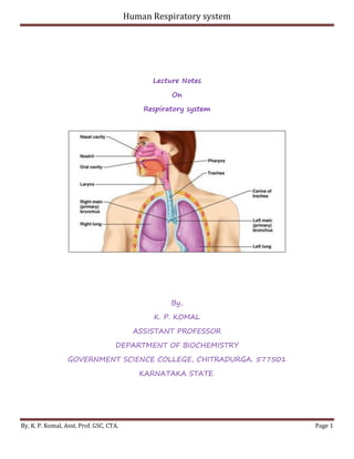
- 2. Human Respiratory system By, K. P. Komal, Asst. Prof. GSC, CTA. Page 2 Human Respiratory System The human respiratory system consists of a complex set of organs and tissues that capture oxygen from the environment and transport the oxygen into the lungs. The organs and tissues that comprise the human respiratory system include the nose, pharynx, trachea, and lungs. Nose The respiratory system of humans begins with the nose, where air is conditioned by warming and moistening. Bone partitions separate the nasal cavity into chambers, where air swirls about in currents. Hairs and hair like cilia trap dust particles and purify the air. Pharynx The nasal chambers open into a cavity at the rear of the mouth called the pharynx (throat). From the pharynx, two tubes called Eustachian tubes open to the middle ear to equalize air pressure there. The pharynx also contains tonsils and adenoids, which are pockets of lymphatic tissue used to trap and filter microorganisms. Trachea After passing through the pharynx, air passes into the windpipe, or trachea. The trachea has a framework of smooth muscle with about 16 to 20 open rings of cartilage shaped like a C. These rings give rigidity to the trachea and ensure that it remains open. The opening to the trachea is a slit like structure called the glottis. A thin flap of tissue called the epiglottis folds over the opening during swallowing and prevents food from entering the trachea. At the upper end of the trachea, several folds of cartilage form the larynx, or voice box. In the larynx, flaplike pairs of tissues called vocal cords vibrate when a person exhales and produce sounds.
- 3. Human Respiratory system By, K. P. Komal, Asst. Prof. GSC, CTA. Page 3 At its lower end, the trachea branches into two large bronchi (singular, bronchus). These tubes also have smooth muscle and cartilage rings. The bronchi branch into smaller bronchioles, forming a bronchial “tree.” The bronchioles terminate in the alveoli. Lungs Human lungs are composed of approximately 300 million alveoli. Red blood cells pass through the capillaries in single file, and oxygen from each alveolus enters the red blood cells and binds to the hemoglobin. In addition, carbon dioxide contained in the plasma and red blood cells leaves the capillaries and enters the alveoli when a breath is taken. Most carbon dioxide reaches the alveoli as bicarbonate ions, and about 25 percent of it is bound loosely to hemoglobin. When a person inhales, the rib muscles and diaphragm contract, thereby increasing the volume of the chest cavity. This increase leads to reduced air pressure in the chest cavity, and air rushes into the alveoli, forcing them to expand and fill. The lungs passively obtain air from the environment by this process. During exhalation, the rib muscles and diaphragm relax, the chest cavity volume diminishes, and the internal air pressure increases. The compressed air forces the alveoli to close, and air flows out. The nerve activity that controls breathing arises from impulses transported by nerve fibers passing into the chest cavity and terminating at the rib muscles and diaphragm. These impulses are regulated by the amount of carbon dioxide in the blood: A high carbon dioxide concentration leads to an increased number of nerve impulses and a more rapid breathing rate. Function of the Respiratory System The function of the respiratory system is to deliver air to the lungs. Oxygen in the air diffuses out of the lungs and into the blood, while carbon dioxide diffuses in the opposite direction, out of the blood and into the lungs. Respiration includes the following processes:
- 4. Human Respiratory system By, K. P. Komal, Asst. Prof. GSC, CTA. Page 4 External respiration is the process of gas exchange between the atmosphere and the body tissues. In order to accomplish this task, the following events occur: 1. Pulmonary ventilation is the process of breathing—inspiration (inhaling air) and expiration (exhaling air). 2. Gas transport, carried out by the cardiovascular system, is the process of distributing the oxygen throughout the body and collecting CO 2 and returning it to the lungs. Internal respiration is the process of gas exchange between the blood, the interstitial fluids (fluids surrounding the cells), and the cells. Inside the cell, cellular respiration generates energy (ATP), using O 2 and glucose and producing waste CO 2.
- 5. Human Respiratory system By, K. P. Komal, Asst. Prof. GSC, CTA. Page 5 Oxygen Transport in the Blood Even though oxygen is transported via the blood, it is not very soluble in liquids. A small amount of oxygen does dissolve in the blood and is transported in the bloodstream, but it is only about 1.5% of the total amount.
- 6. Human Respiratory system By, K. P. Komal, Asst. Prof. GSC, CTA. Page 6 The majority of oxygen molecules are carried from the lungs to the body’s tissues by a specialized transport system, which relies on the erythrocyte—the red blood cell. Erythrocytes contain a metalloprotein, hemoglobin, which serves to bind oxygen molecules to the erythrocyte (Figure 1). Heme is the portion of hemoglobin that contains iron, and it is heme that binds oxygen. One hemoglobin molecule contains iron-containing Heme molecules, and because of this, each hemoglobin molecule is capable of carrying up to four molecules of oxygen. As oxygen diffuses across the respiratory membrane from the alveolus to the capillary, it also diffuses into the red blood cell and is bound by hemoglobin. The following reversible chemical reaction describes the production of the final product, oxyhemoglobin (Hb–O2), which is formed when oxygen binds to hemoglobin. Oxyhemoglobin is a bright red-colored molecule that contributes to the bright red color of oxygenated blood. Hb + O2 ↔ Hb − O2 In this formula, Hb represents reduced hemoglobin, that is, hemoglobin that does not have oxygen bound to it. There are multiple factors involved in how readily heme binds to and dissociates from oxygen, which will be discussed in the subsequent sections. Figure 1. Erythrocyte and Hemoglobin. Hemoglobin consists of four subunits, each of which contains one molecule of iron.
- 7. Human Respiratory system By, K. P. Komal, Asst. Prof. GSC, CTA. Page 7 Function of Hemoglobin Hemoglobin is composed of subunits, a protein structure that is referred to as a quaternary structure. Each of the four subunits that make up hemoglobin is arranged in a ring-like fashion, with an iron atom covalently bound to the heme in the center of each subunit. Binding of the first oxygen molecule causes a conformational change in hemoglobin that allows the second molecule of oxygen to bind more readily. As each molecule of oxygen is bound, it further facilitates the binding of the next molecule, until all four heme sites are occupied by oxygen. The opposite occurs as well: After the first oxygen molecule dissociates and is “dropped off” at the tissues, the next oxygen molecule dissociates more readily. When all four heme sites are occupied, the hemoglobin is said to be saturated. When one to three heme sites are occupied, the hemoglobin is said to be partially saturated. Therefore, when considering the blood as a whole, the percent of the available heme units that are bound to oxygen at a given time is called hemoglobin saturation. Hemoglobin saturation of 100 percent means that every heme unit in all of the erythrocytes of the body is bound to oxygen. In a healthy individual with normal hemoglobin levels, hemoglobin saturation generally ranges from 95 percent to 99 percent. Oxygen Dissociation from Hemoglobin Partial pressure is an important aspect of the binding of oxygen to and disassociation from heme. An oxygen–hemoglobin dissociation curve is a graph that describes the relationship of partial pressure to the binding of oxygen to heme and its subsequent dissociation from heme (Figure 2).
- 8. Human Respiratory system By, K. P. Komal, Asst. Prof. GSC, CTA. Page 8 Gases travel from an area of higher partial pressure to an area of lower partial pressure. In addition, the affinity of an oxygen molecule for heme increases as more oxygen molecules are bound. Therefore, in the oxygen–hemoglobin saturation curve, as the partial pressure of oxygen increases, a proportionately greater number of oxygen molecules are bound by heme. Not surprisingly, the oxygen–hemoglobin saturation/dissociation curve also shows that the lower the partial pressure of oxygen, the fewer oxygen molecules are bound to heme. As a result, the partial pressure of oxygen plays a major role in determining the degree of binding of oxygen to heme at the site of the respiratory membrane, as well as the degree of dissociation of oxygen from heme at the site of body tissues. The mechanisms behind the oxygen–hemoglobin saturation/dissociation curve also serve as automatic control mechanisms that regulate how much oxygen is delivered to different tissues throughout the body. This is important because some tissues have a higher metabolic rate than others. Highly active tissues, such as muscle, rapidly use oxygen to produce ATP, lowering the partial pressure of oxygen in the tissue to about 20 mm Hg. The partial pressure of oxygen inside capillaries is about 100 mm Hg, so the difference between the two becomes quite high, about 80 mm Hg. As a result, a greater number of oxygen molecules dissociate from hemoglobin and enter the tissues. The reverse is true of tissues, such as adipose (body fat), which have lower metabolic rates. Because less oxygen is used by these cells, the partial pressure of oxygen within such tissues remains relatively high, resulting in fewer oxygen molecules dissociating from hemoglobin and entering the tissue interstitial fluid. Although venous blood is said to be deoxygenated, some oxygen is still bound to hemoglobin in its red blood cells. This
- 9. Human Respiratory system By, K. P. Komal, Asst. Prof. GSC, CTA. Page 9 provides an oxygen reserve that can be used when tissues suddenly demand more oxygen. Figure 2. Oxygen-Hemoglobin Dissociation and Effects of pH and Temperature. These three graphs show (a) the relationship between the partial pressure of oxygen and hemoglobin saturation, (b) the effect of pH on the oxygen–hemoglobin dissociation curve, and (c) the effect of temperature on the oxygen– hemoglobin dissociation curve.
- 10. Human Respiratory system By, K. P. Komal, Asst. Prof. GSC, CTA. Page 10 Factors other than partial pressure also affect the oxygen–hemoglobin saturation/dissociation curve. For example, a higher temperature promotes hemoglobin and oxygen to dissociate faster, whereas a lower temperature inhibits dissociation. However, the human body tightly regulates temperature, so this factor may not affect gas exchange throughout the body. The exception to this is in highly active tissues, which may release a larger amount of energy than is given off as heat. As a result, oxygen readily dissociates from hemoglobin, which is a mechanism that helps to provide active tissues with more oxygen. Certain hormones, such as androgens, epinephrine, thyroid hormones, and growth hormone, can affect the oxygen–hemoglobin saturation/disassociation curve by stimulating the production of a compound called 2,3-bisphosphoglycerate (BPG) by erythrocytes. BPG is a byproduct of glycolysis. Because erythrocytes do not contain mitochondria, glycolysis is the sole method by which these cells produce ATP. BPG promotes the disassociation of oxygen from hemoglobin. Therefore, the greater the concentration of BPG, the more readily oxygen dissociates from hemoglobin, despite its partial pressure. The pH of the blood is another factor that influences the oxygen–hemoglobin saturation/dissociation curve. The Bohr effect is a phenomenon that arises from the relationship between pH and oxygen’s affinity for hemoglobin: A lower, more acidic pH promotes oxygen dissociation from hemoglobin. In contrast, a higher, or more basic, pH inhibits oxygen dissociation from hemoglobin. The greater the amount of carbon dioxide in the blood, the more molecules that must be converted, which in turn generates hydrogen ions and thus lowers blood pH. Furthermore, blood pH may become more acidic when certain byproducts of cell metabolism, such as lactic acid, carbonic acid, and carbon dioxide, are released into the bloodstream.
- 11. Human Respiratory system By, K. P. Komal, Asst. Prof. GSC, CTA. Page 11 Carbon Dioxide Transport in the Blood Carbon dioxide is transported by three major mechanisms. The first mechanism of carbon dioxide transport is by blood plasma, as some carbon dioxide molecules dissolve in the blood. The second mechanism is transport in the form of bicarbonate (HCO3 –), which also dissolves in plasma. The third mechanism of carbon dioxide transport is similar to the transport of oxygen by erythrocytes Dissolved Carbon Dioxide Although carbon dioxide is not considered to be highly soluble in blood, a small fraction—about 7 to 10 percent—of the carbon dioxide that diffuses into the blood from the tissues dissolves in plasma. The dissolved carbon dioxide then travels in the bloodstream and when the blood reaches the pulmonary capillaries, the dissolved carbon dioxide diffuses across the respiratory membrane into the alveoli, where it is then exhaled during pulmonary ventilation. Bicarbonate Buffer A large fraction—about 70 percent—of the carbon dioxide molecules that diffuse into the blood is transported to the lungs as bicarbonate. Most bicarbonate is produced in erythrocytes after carbon dioxide diffuses into the capillaries, and subsequently into red blood cells. Carbonic anhydrase (CA) causes carbon dioxide and water to form carbonic acid (H2CO3), which dissociates into two ions: bicarbonate (HCO3 –) and hydrogen (H+). The following formula depicts this reaction: CO2 + H2O CA ↔ H2CO3↔H+ + HCO3 − Bicarbonate tends to build up in the erythrocytes, so that there is a greater concentration of bicarbonate in the erythrocytes than in the surrounding blood plasma. As a result, some of the bicarbonate will leave the erythrocytes and move down its concentration gradient into the plasma in exchange for chloride (Cl–) ions. This phenomenon is referred to as the chloride shift and occurs because by exchanging one
- 12. Human Respiratory system By, K. P. Komal, Asst. Prof. GSC, CTA. Page 12 negative ion for another negative ion, neither the electrical charge of the erythrocytes nor that of the blood is altered. At the pulmonary capillaries, the chemical reaction that produced bicarbonate (shown above) is reversed, and carbon dioxide and water are the products. Much of the bicarbonate in the plasma re-enters the erythrocytes in exchange for chloride ions. Hydrogen ions and bicarbonate ions join to form carbonic acid, which is converted into carbon dioxide and water by carbonic anhydrase. Carbon dioxide diffuses out of the erythrocytes and into the plasma, where it can further diffuse across the respiratory membrane into the alveoli to be exhaled during pulmonary ventilation. Carbaminohemoglobin About 20 percent of carbon dioxide is bound by hemoglobin and is transported to the lungs. Carbon dioxide does not bind to iron as oxygen does; instead, carbon dioxide binds amino acid moieties on the globin portions of hemoglobin to form carbaminohemoglobin, which forms when hemoglobin and carbon dioxide bind. When hemoglobin is not transporting oxygen, it tends to have a bluish-purple tone to it, creating the darker maroon color typical of deoxygenated blood. The following formula depicts this reversible reaction: CO2 + Hb ↔ HbCO2 Similar to the transport of oxygen by heme, the binding and dissociation of carbon dioxide to and from hemoglobin is dependent on the partial pressure of carbon dioxide. Because carbon dioxide is released from the lungs, blood that leaves the lungs and reaches body tissues has a lower partial pressure of carbon dioxide than is found in the tissues. As a result, carbon dioxide leaves the tissues because of its higher partial pressure, enters the blood, and then moves into red blood cells, binding to hemoglobin. In contrast, in the pulmonary capillaries, the partial pressure of carbon dioxide is high compared to within the alveoli. As a result, carbon dioxide dissociates readily from hemoglobin and diffuses across the respiratory membrane into the air.
- 13. Human Respiratory system By, K. P. Komal, Asst. Prof. GSC, CTA. Page 13 In addition to the partial pressure of carbon dioxide, the oxygen saturation of hemoglobin and the partial pressure of oxygen in the blood also influence the affinity of hemoglobin for carbon dioxide. The Haldane effect is a phenomenon that arises from the relationship between the partial pressure of oxygen and the affinity of hemoglobin for carbon dioxide. Hemoglobin that is saturated with oxygen does not readily bind carbon dioxide. However, when oxygen is not bound to heme and the partial pressure of oxygen is low, hemoglobin readily binds to carbon dioxide. Regulation of H+ by the Lungs Acid–base imbalances in the blood’s pH can be altered by changes in breathing to expel more CO2 and raise pH back to normal. Acid–base imbalance occurs when a significant insult causes the blood pH to shift out of its normal range (7.35 to 7.45). An excess of acid in the blood is called acidemia and an excess of base is called alkalemia. The process that causes the imbalance is classified based on the etiology of the disturbance (respiratory or metabolic) and the direction of change in pH ( acidosis or alkalosis). There are four basic processes and one or a combination may occur at any given time. 1. Metabolic acidosis 2. Respiratory acidosis 3. Metabolic alkalosis 4. Respiratory alkalosis Blood carries oxygen, carbon dioxide, and hydrogen ions (H+) between tissues and the lungs. The majority of CO2 transported in the blood is dissolved in plasma (60% is dissolved bicarbonate). A smaller fraction is transported in the red blood cells that combine with the globin portion of hemoglobin as carbaminohemoglobin. This is the chemical portion of the red
- 14. Human Respiratory system By, K. P. Komal, Asst. Prof. GSC, CTA. Page 14 blood cell that aids in the transport of oxygen and nutrients around the body, but, this time, it is carbon dioxide that is transported back to the lung. Acid–base imbalances that overcome the buffer system can be compensated in the short term by changing the rate of ventilation. This alters the concentration of carbon dioxide in the blood, shifting the above reaction according to Le Chatelier’s principle, which in turn alters the pH. The basic reaction governed by this principle is as follows: H2O+CO2⇋H2CO3⇋H++CO−3 When the blood pH drops too low (acidemia), the body compensates by increasing breathing to expel more CO2; this shifts the above reaction to the left such that less hydrogen ions are free; thus, the pH will rise back to normal. For alkalemia, the opposite occurs.