SlideShare a Scribd company logo
1 of 30
Download to read offline
Digestive system notes 2017
1 By, K. P. Komal, Asst. Professor of Biochemistry, chitradurga.
Lecture Notes
On
Digestive system
By,
K. P. KOMAL
ASSISTANT PROFESSOR
DEPARTMENT OF BIOCHEMISTRY
GOVERNMENT SCIENCE COLLEGE, CHITRADURGA. 577501
KARNATAKA STATE.
Digestive system notes 2017
2 By, K. P. Komal, Asst. Professor of Biochemistry, chitradurga.
Human Digestive System
In the human digestive system, large organic masses are broken down into
smaller particles that the body can use as fuel. This is a complex process. The
breakdown of the nutrients requires the coordination of several enzymes secreted from
specialized cells within the mouth, stomach, intestines, and liver. The major organs or
structures that coordinate digestion within the human body include the mouth,
esophagus, stomach, small and large intestines, and liver.
Mouth
In the human body, the mouth (oral cavity) is a specialized organ for receiving
food and breaking up large organic masses. In the mouth, food is changed mechanically
by biting and chewing. Humans have four kinds of teeth: incisors are chisel-shaped
teeth in the front of the mouth for biting; canines are pointed teeth for tearing; and
premolars and molars are flattened, ridged teeth for grinding, pounding, and crushing
food.
In the mouth, food is moistened by saliva, a sticky fluid that binds food particles
together into a soft mass. Three pairs of salivary glands—parotid, submaxillary, and
sublingual—secrete saliva into the mouth. The saliva contains an enzyme
called amylase, which digests starch molecules into smaller molecules of the disaccharide
maltose.
During chewing, the tongue moves food about and manipulates it into a mass
called a bolus. The bolus is pushed back into the pharynx (throat) and is forced through
the opening to the esophagus.
Esophagus
The esophagus is a thick-walled muscular tube located behind the windpipe that
extends through the neck and chest to the stomach. The bolus of food moves through
the esophagus by peristalsis: a rhythmic series of muscular contractions that propels the
bolus along. The contractions are assisted by the pull of gravity.
Digestive system notes 2017
3 By, K. P. Komal, Asst. Professor of Biochemistry, chitradurga.
Stomach
The esophagus joins the stomach at a point just below the diaphragm. A valvelike
ring of muscle called the cardiac sphinctersurrounds the opening to the stomach. The
sphincter relaxes as the bolus passes through and then quickly closes.
The stomach is an expandable pouch located high in the abdominal cavity. Layers
of stomach muscle contract and churn the bolus of food with gastric juices to form a
soupy liquid called chyme.
The stomach stores food and prepares it for further digestion. In addition, the
stomach plays a role in protein digestion. Gastric glands called chief cells secrete
pepsinogen. Pepsinogen is converted to the enzyme pepsin in the presence of
hydrochloric acid. Hydrochloric acid is secreted by parietal cells in the stomach lining.
The pepsin then digests large proteins into smaller proteins called peptides. To protect
the stomach lining from the acid, a third type of cell secretes mucus that lines the
stomach cavity. An overabundance of acid due to mucus failure may lead to an ulcer.
Small intestine
The soupy mixture called chyme spurts from the stomach through a sphincter
into the small intestine. An adult’s small intestine is about 23 feet long and is divided
into three sections: the first 10 to 12 inches form the duodenum; the next 10 feet
form the jejunum;and the final 12 feet form the ileum. The inner surface of the small
intestine contains numerous fingerlike projections called villi (the singular is villus). Each
villus has projections of cells called microvillito increase the surface area.
Most chemical digestion takes place in the duodenum. In this region, enzymes
digest nutrients into simpler forms that can be absorbed. Intestinal enzymes are
supplemented by enzymes from the pancreas, a large, glandular organ near the
stomach. In addition, bile enters the small intestine from the gallbladder to assist in fat
digestion.
Digestive system notes 2017
4 By, K. P. Komal, Asst. Professor of Biochemistry, chitradurga.
The enzymes functioning in carbohydrate digestion include amylase (for starch),
maltase (for maltose), sucrase (for sucrose), and lactase (for lactose). For fats, the
principal enzyme is lipase. Before lipase can act, the large globules of fat must be broken
into smaller droplets by bile. Bile is a mixture of salts, pigments, and cholesterol that is
produced by the liver and stored in the gallbladder, a saclike structure underneath the
liver.
Protein digestion is accomplished by several enzymes, including two pancreatic
enzymes: trypsin and chymotrypsin. Peptides are broken into smaller peptides, and
peptidases reduce the enzymes to amino acids. Nucleases digest nucleic acids into
nucleotides in the small intestine also.
Most absorption in the small intestine occurs in the jejunum. The products of
digestion enter cells of the villi, move across the cells, and enter blood vessels
called capillaries. Diffusion accounts for the movement of many nutrients, but
facilitated diffusion is responsible for the movement of glucose and amino acids. The
products of fat digestion pass as small droplets of fat into lacteals, which are branches
of the lymphatic system.
Absorption is completed in the final part of the small intestine,
theileum. Substances that have not been digested or absorbed then pass into the large
intestine.
Large intestine
The small intestine joins the large intestine in the lower-right abdomen of the
body. The two organs meet at a blind sac called thececum and a small fingerlike organ
called the appendix.Evolutionary biologists believe the cecum and appendix are vestiges
of larger organs that may have been functional in human ancestors.
The large intestine is also known as the colon. It is divided into ascending,
transverse, and descending portions, each about one foot in length. The colon’s chief
functions are to absorb water and to store, process, and eliminate the residue following
Digestive system notes 2017
5 By, K. P. Komal, Asst. Professor of Biochemistry, chitradurga.
digestion and absorption. The intestinal matter remaining after water has been
reclaimed is known as feces. Feces consist of nondigested food (such as cellulose), billions
of mostly harmless bacteria, bile pigments, and other materials. The feces are stored in
the rectum and passed out through the anus to complete the digestion process.
Liver
The liver has an important function in processing the products of human
digestion. For example, cells of the liver remove excess glucose from the bloodstream
and convert the glucose to a polymer called glycogen for storage.
The liver also functions in amino acid metabolism. In a process called
deamination, it converts some amino acids to compounds that can be used in energy
metabolism. In doing so, the liver removes the amino groups from amino acids and uses
the amino groups to produce urea. Urea is removed from the body in the
urine. Fats are processed into two-carbon units that can enter the Kreb’s cycle for
energy metabolism. The liver also stores vitamins and minerals, forms many blood
proteins, synthesizes cholesterol, and produces bile for fat digestion.
The metabolism of carbohydrates is the process of getting the carbohydrates in
the foods we eat into the right format to provide fuel to our body's cells. This process
involves digestion, absorption and transportation. Most commonly, carbohydrate
metabolism results in the production of glucose molecules which are the most efficient
source of energy (ATP) for our muscles and our brains. Energy or fuel from our food is
used for cell growth, repair and normal cell functioning.
Carbohydrate
Digestion
Carbohydrates are most commonly consumed as polysaccharides (e.g. starch, fibre
or cellulose) or disaccharides (e.g. lactose, sucrose, galactose) and therefore need to be
broken down into their simpler monosaccharide forms which the body can utilise.
Digestive system notes 2017
6 By, K. P. Komal, Asst. Professor of Biochemistry, chitradurga.
The digestion process of polysaccharides such as starch will begin in the mouth
where it is hydrolysed by salivary amylase. The amount of starch hydrolysed in this
environment is often quite small as most food does not stay in the mouth long. Once
the food bolus reaches the stomach the salivary enzymes are denatured. As a result,
digestion predominantly occurs in the small intestine with pancreatic amylase
hydrolysing the starch to dextrin and maltose.
Enzymes classed as glucosidases on the brush border of the small intestine break
down the dextrin and maltase, lactase and sucrase convert the other disaccharides into
their two monosaccharide units.
Absorption & transport
The monosaccharide units, glucose, galactose and fructose are transported through
the wall of the small intestine into the portal vein which then takes them straight to
the liver. The mode of transport varies between the monosaccharides. Both glucose and
fructose are absorbed relatively quickly, depending on what other nutrients are eaten at
the same time. For example a meal or food containing protein and fat causes the sugars
to be absorbed slower than when consumed on their own.
 Glucose, at low concentrations is transported through the mucosal lining into the
epithelial cells of the intestine by active transport, via a sodium dependant
transporter. At higher concentrations, a second facilitative transporter becomes
involved. From the epithelial cells glucose is moved into the surrounding capillaries
by facilitated diffusion.
 Galactose is transported in the same way as glucose, utilising the same
transporters. As galactose is not found as a monosaccharide in nature, absorbed
galactose primarily comes from the breakdown of lactose.
 Fructose moves entirely via facilitated diffusion. The process utilises a different
transporter to glucose when entering the enterocytes, however both fructose and
glucose utilise the same transporter to exit the enterocyte into the capillaries. The
Digestive system notes 2017
7 By, K. P. Komal, Asst. Professor of Biochemistry, chitradurga.
absorption of fructose is much slower than that of glucose and is quantitatively
limited. Consumption of large amounts of fructose has been shown to produce a
level of fructose malabsorption in almost all cases. Co-ingestion of glucose with
fructose has been shown to facilitate fructose absorption.
Once in the liver galactose and fructose are removed from the blood and
converted into other metabolites. When eaten in moderate quantities, most fructose is
taken up by the liver and converted to glucose, glycogen and lactate. A fraction may
also be oxidised or converted into fatty acids and uric acid. Only a small amount of
fructose reaches the bloodstream, so blood fructose concentrations are always low.
Galactose is primarily converted into glucose and stored as glycogen.
On the other hand most of the glucose derived from food is transported via the
blood stream to the peripheral tissues where, under normal circumstances, the
hormone insulin enables it to be taken up by the cells and used as an energy source via
the glycolysis pathway. As glucose is the most important fuel source for the body and in
particular the brain the body attempts to keep a basal circulating blood glucose of
around 4-5mmol/L. This homeostasis mechanism is predominantly controlled by the
actions of glycogen and insulin.
Storage
Surplus glucose is initially stored as glycogen in the liver or muscles. The liver can
store approximately 100g of glycogen which is used to maintain basal blood glucose
levels between meals, whilst the muscles typically store 400-500g often used during
movement. Once these reserves are saturated, excess glucose is converted to fat for
longer term storage.
Digestive system notes 2017
8 By, K. P. Komal, Asst. Professor of Biochemistry, chitradurga.
Proteins
Digestion and Absorption:
The major constituents of the food are carbohydrates, proteins and lipids. They
are digested and absorbed in the stomach and intestine. Some of the digested/degraded
components of the food stuffs may either be reutilized or may be excreted out. Chewing
of food, movements of the stomach and intestine facilitate the grinding of the food
materials and bring them in contact with gastric secretions.
Proteolytic enzymes are absent in the salivary secretions, hence there is no
digestion of proteins in the mouth. Proteolysis takes place in the gastro-intestinal tract
(i.e. stomach and intestine). When the proteins enter the stomach they stimulate the
secretion of the hormone called gastrin which in turn stimulates the secretion of HCl by
parietal cells of the stomach and pepsinogen from the chief cells.
Gastric juice is acidic i.e. the pH is 1.5—2.5. Acidic pH of the stomach has an
antiseptic action that kills the bacteria and other microorganisms. At this pH the
dietary proteins also get denatured. In presence of HCl, pepsinogen is converted to
Digestive system notes 2017
9 By, K. P. Komal, Asst. Professor of Biochemistry, chitradurga.
pepsin by autocatalysis resulting in removal of some of the amino acids from the amino
terminal end. Pepsin is an endopeptidase for tyr, phe, trp.
In the stomach the proteins are converted as follows:
Protein → Metaprotein → Proteone → Peptone → Peptide
As the food passes from the stomach to small intestine the low pH of the food
triggers the secretion of the hormone ‘secretin’ into the blood. It stimulates the
pancreas to secrete HCO3 into the small intestine in order to neutralize HCl. The
secretion of HCO3 into the intestine abruptly raises the pH from 2.5 to 7.0. The entry
of amino acids into the duodenum releases the hormone ‘cholecystokinin’ which in turn
triggers the release of pancreatic juice (that contains many pancreatic enzymes like
trypsinogen, chymotrypsinogen, procarboxypeptidases) by the exocrine cells of the
pancreas (ecbolic and hydrolatic). Most of these enzymes are produced as zymogens
(inactive enzymes) by the pancreas in order to protect the exocrine cells from being
digested.
Subsequent to the entry of trypsinogen into the small intestine it is activated to
trypsin by enterokinase secreted by the intestinal cells. Trypsin is formed from
trypsinogen by the removal of hexapeptide from the N-terminal end.
The newly formed trypsin activates the remaining trypsinogen, Trypsin is an
endopeptidase, specific for (acts on) the peptide bonds contributed by the basic amino
acids like arginine, histidine and lysine. Chymotrypsin is secreted in an inactive from
called chymotrypsinogen which is activated by trypsin. Chymotrypsin is an
endopeptidase specific to aromatic amino acids i.e. phenylalanine, tyrosine, tryptophan.
Digestive system notes 2017
10 By, K. P. Komal, Asst. Professor of Biochemistry, chitradurga.
Carboxypeptidase secreted as procarboxypeptidase is activated again by trypsin. It
is an exopeptidase that cleaves the amino acids from the carboxy terminal end. Amino
peptidase secreted as pro-aminopeptidase is once again activated by trypsin. It is an
exopeptidase that cleaves the amino acids from the free amino terminal end. Dipeptides
acts only on dipeptides and hydrolyses it into 2 amino acids.
Even after the action of all these enzymes most of the proteins remain
undigested. Protein like collagen, fibrin etc., escape digestion and are excreted out.
Absorption of the Digested Proteins:
There are four distinct carrier systems in the intestinal epithelium for the absorption of
the digested proteins. These are:
(1) Carrier system for neutral amino acids
(2) Carrier system for basic amino acids
(3) Carrier system for acidic amino acids
(4) Carrier system for glycine and imino acid (proline)
Digestive system notes 2017
11 By, K. P. Komal, Asst. Professor of Biochemistry, chitradurga.
The digested amino acids are carried across the mucosal cell membrane from the
intestinal lumen to the cytoplasm of the cell by one of the above carrier systems specific
to that particular amino acid. Absorption of amino acids is an up-hill process (i.e.
against gradient as compared to the Na+ absorption which is downhill i.e. along the
gradient).
There are four systems that operate to absorb amino acids from the mucosal cells into
the blood. They are:
(1) A — system (alanine system)
(2) L — system (leucine system)
(3) Ly – system (lysine system)
(4) Ala-ser-cyst – system
Amino acids are taken up by the blood capillaries of the mucosa and are
transported in the plasma to the liver. Some amounts of amino acids are also absorbed
through the lymph. Glucagon stimulates the absorption of amino acids through ‘A’
system mediated by cAMP. Insulin stimulates the trans cellular transport of amino acids
to minimize the loss in the urine. The proximal tubule cells reabsorb and return them to
the blood stream. It is done by glutathione, a tripeptide. Three ATPs are utilized for the
absorption of each amino acid.
Absorbed amino acids do not stimulate antibody production whereas an intact
protein absorbed becomes antigenic. Intestinal membranes allow the transport of
proteins across them ex.—In a neonate the intestinal mucosa is permeable to y-globulin
(immunoglobulin) of colostrum.
The immune system in a neonate is not well developed thus absorption of intact
y-globulin into the blood does not elicit any immune response instead it results in the
defence of the neonate against infections. In adults, some proteins may be absorbed
intact through the intestinal mucosa leading to the formation of antibodies and
Digestive system notes 2017
12 By, K. P. Komal, Asst. Professor of Biochemistry, chitradurga.
anaphylactic reactions or other such immunological phenomena after the absorption of
those proteins. Thus in such cases allergies to food proteins occur.
Digestion of Fats/ lipids:
Stomach:
Lipase present in the stomach is unable to hydrolyze fats owing to the high
acidity of the gastric contents. Therefore, the major part of the ingested fat is digested
in the small intestine.
Small Intestine:
The ingested fat reaching the duodenum is mixed with the bile and pancreatic
juice which contains lipase. The bile salts emulsify the fat before the action of lipase. The
emulsification is also brought about by monoglycerides, phospholipid and lysolecithin.
The secreted inactive pancreatic lipase is activated by bile and Ca. The surface
area of the emulsified fat becomes increased for which the rate of reaction of lipase is
increased. Pancreatic lipase hydrolyzes 1- and 3-positions of the triglycerides leaving a
mixture of 2- monoglycerides, 1, 2- and 2, 3-diglycerides as well as the soaps of the
free fatty acids.
The pancreatic juice also contains phospholipase and cholesterol-esterase which
hydrolyze phospholipid and esterified cholesterol. Intestinal juice also contains a lipase
whose action is not of much importance as most of the fat is hydrolyzed by the
pancreatic lipase.
Absorption of Fats:
Several theories have been proposed for the mechanism of absorption of fats after
digestion.
The important theories are:
A. Lipolytic hypothesis.
Digestive system notes 2017
13 By, K. P. Komal, Asst. Professor of Biochemistry, chitradurga.
B. Partition theory.
C. More recent theory.
A. Lipolytic Hypothesis:
1. According to this theory, fat is completely hydrolyzed to fatty acids and
glycerol which are absorbed.
2. The fatty acids combining with bile salts form a miscible complex which is ab-
sorbed into the intestinal mucosa.
3. The fatty acids are then separated from bile acids and converted into
triglycerides by combining with glycerol.
4. The triglycerides are passed to the lacteals. They then enter the lymphatic’s
and reach the systemic circulation via thoracic duct.
B. Partition Theory:
1. According to this theory, 30 per cent of the triglycerides are hydrolyzed to
fatty acids and glycerol while 70 per cent remain un-hydrolyzed.
2. The un-hydrolyzed triglycerides are emulsified by monoglycerides and
diglycerides in combination with bile salts to form minute particles known as
“micelles” of size about 0.1 to 0.5µ.
3. The resulting mixture is absorbed into the intestines, passed on to the lacteals
and then to the lymphatic’s. The mixture then reaches the systemic circulation via
thoracic duct.
4. The free fatty acids are absorbed as bile salt-fatty acid complex into the
intestinal mucosa. The fatty acids are absorbed into the portal blood to reach the
liver.
Digestive system notes 2017
14 By, K. P. Komal, Asst. Professor of Biochemistry, chitradurga.
C. Recent Theory:
1. The removal of the ester group of 2- mono-acylglycerol requires isomerization
to a primary ester linkage. This is a slow process. As a result, monoacylglycerols
are the major end products of fat digestion and less than one-fourth of the
ingested fat is completely broken down to glycerol and fatty acids.
2. Within the intestinal wall, 2-monoacylglycerols are converted to
triacylglycerol’s and l-monoacylglycerols are further hydrolyzed to form free
glycerol and fatty acids.
3. The fatty acids are then activated by thiokinase in presence of ATP and
coenzyme A for the resynthesize of triacylglycerol’s.
4. The free glycerol in the intestinal lumen is about 22 per cent of total amount
of triacylglycerol originally present. This passes directly to the portal vein.
Digestive system notes 2017
15 By, K. P. Komal, Asst. Professor of Biochemistry, chitradurga.
5. The glycerol within the intestinal wall is activated by glycerokinase in presence
of ATP to form glycerol-3-phosphate for the synthesis of triacylglycerol followed
by the combination with acyl-CoA present in the intestinal wall.
6. All long chain fatty acids present in the intestinal wall are reincorporated into
triacylglycerol’s which are transported to the lymphatic vessels of the abdominal
region (the so-called lacteals) for distribution to the rest of the body.
7. The great majority of absorbed fat appears in the form of chylomicrons which
appear first at the lymphatic vessels of the abdominal region and later in the
systemic blood. The chylomicrons contain triacylglycerol, free and esterified
cholesterol, phospholipid and 0.5 per cent protein.
All of the factors relating to digestion and absorption of fat are mentioned in Fig. 16.8.
Digestive system notes 2017
16 By, K. P. Komal, Asst. Professor of Biochemistry, chitradurga.
Absorption of Phospholipids:
Phospholipids are split by phospholipases and their acyl chains are incorporated
into chylomicrons, choline, the hydrophilic component, may be transported directly to
the liver via the hepatic portal vein.
Absorption of Cholesterol:
It is absorbed into the lymphatic’s and recovered mainly as cholesteryl esters.
Digestive system notes 2017
17 By, K. P. Komal, Asst. Professor of Biochemistry, chitradurga.
Composition and Functions of Various Digestive Juice:
There are five digestive juices; saliva, gastric juice, pancreatic juice, succus
entericus (intestinal juice) and bile.
The necessity for so many digestive juices is that –
(a) One juice does not contain all the enzymes necessary for digesting all the
different types of foodstuffs. For instance, saliva contains only carbohydrate-splitting
enzymes; whereas gastric juice contains both fat- and protein -splitting enzymes but
none acting on carbohydrates, and
(b) The second reason is that, one particular digestive juice cannot digest a
particular type of food up to completion.
It will digest only up to a certain stage and then, the products will be handed
over to the next digestive juice for further digestion. In this way digestion is completed.
For instance, gastric juice digests protein up to the stage of peptone, pancreatic juice
carries the digestion of peptone further up to lower peptide. The latter is digested
completely up to amino acids by succus entericus.
One more interesting fact about the digestive juices is that, their reactions are not
all same. Saliva is slightly acid, gastric juice is strongly acid, but pancreatic juice is
strongly alkaline. This, more or less, alternate acid and alkaline reaction, prevents any
serious alteration of blood reaction. This is a special device for maintaining blood
reaction constant.
1. Saliva:
Characteristics:
 Total Amount – 1,200 -1,500 ml in 24 hours. A large proportion of this 24-
hour volume is secreted at meal time, when secretory rate is highest.
 Consistency – Slightly cloudy, due to the presence of cells and mucin.
Digestive system notes 2017
18 By, K. P. Komal, Asst. Professor of Biochemistry, chitradurga.
 Reaction – Usually slightly acid (pH 6.02 – 7.05). On standing or boiling it loses
CO2 and becomes alkaline. This alkaline reaction causes precipitation of salivary
constituents, as tartar on the teeth or calculus in salivary duct.
 Specific Gravity – 1.002 – 1.012.
 Freezing Point – 0.07 – 0.34°C.
Composition:
1. Water – 99.5%.
2. Solids – 0.5%.
i. Cellular Constituents:
Yeast cells, bacteria, protozoa, polymorphonuclear leucocytes, desquamated epithelial
cells, etc.
ii. Inorganic Salts:
About 0.2% consists of NaCI, KCI, acid and alkaline sodium phosphate, CaCO3, calcium
phosphate, potassium thiocyanate. Smoker’s saliva is rich in thiocyanate. In case of
poisoning with metals like lead, mercury, etc., they are secreted in saliva.
iii. Orgainc – 0.3%.
a. Enzymes P tyalin (salivary amylase) lipase, carbonic anhydrase, phosphatase
and a bacteriolytic enzyme, lysozyme.
b. Mucin.
c. Urea, amino acids, cholesterol and vitamins (in small amounts).
d. The soluble specific blood group substances also form part of the organic
constituents of saliva and they have the same characteristics as the agglutinogen
on the erythrocyte. In human beings, A, B, O and Lea substances have been
demonstrated. Their concentration in saliva is from 10 to 20 mgm per litre.
3. Gases:
Saliva contains about 1 ml oxygen, 2.5 ml nitrogen and 50 ml of CO2 per 100
ml. Bicarbonates, phosphates and the proteins act as buffers. Chlorides activate amylase.
Digestive system notes 2017
19 By, K. P. Komal, Asst. Professor of Biochemistry, chitradurga.
The thiocyanate (KCNS) is a product of excretion. It is formed in the body from the
cyanogen radicle (-CN) derived from proteins. Its formation is a process of detoxication
of the poisonous cyanides and hence is an example of protective synthesis. With ferric
chloride it gives a rich brown colour.
An enzyme kallikrein is present in saliva which acts upon plasma protein to
produce a substance known as kallidin or bradykinin. This produces vasodilatation of
salivary gland during secretion and is also responsible for the sudden fall and
consequent rise in blood pressure observed after injection of saliva in animals.
Functions:
1. Mechanical Functions:
a. It keeps the mouth moist and helps speech. Decrease in salivary secretion as
occurs after nervousness, causes impairment of speech.
b. It helps in the process of mastication of the foodstuff and in preparing it into a
bolus, suitable for deglutition. Here, saliva also acts as a lubricant.
c. Constant flow of saliva washes down the food debris and thereby does not allow
the bacteria to grow. In acute fevers, where the salivary secretion is inhibited, the
food debris is not properly washed away and the bacteria multiply. These collect
as the sordes at the root of the teeth and upon the tongue.
2. Digestive Functions:
Saliva contains two enzymes:
a. Ptyalin:
Splits starch up to maltose in the following manner [Fig. 9.24]
Digestive system notes 2017
20 By, K. P. Komal, Asst. Professor of Biochemistry, chitradurga.
b. Maltase:
Maltase (in traces) converts maltose into glucose.
3. Excretory Functions:
Saliva excretes urea, heavy metals (Hg, Pb, Bi, As, etc.), thiocyanates, certain
drugs like iodide, etc. Alkaloids, such as morphine, antibiotics, such as penicillin,
streptomycin, etc. are also excreted in the saliva. The excretion of ethyl alcohol by the
salivary gland has prompted the recommendation that such a test should be used for
medico-legal purpose. [It also excretes certain virulent micro-organisms, such as the
virus of hydrophoboea, acute anterior poliomyelitis, mumps, etc.]
4. Helps in the Sensation of Taste:
Taste is a chemical sensation. Unless the substances are in solution, the taste buds
cannot be stimulated. Saliva acts as a solvent and is thus essential for taste.
5. Helps Water Balance:
Saliva keeps the mouth moist. When moisture is reduced in the mouth, certain
nerve endings at the back of the tongue are stimulated and the sensation of thirst
arises. When body water is lost (sweating, diarrhoea, etc.) – saliva is reduced and thirst
is felt. The subject feels the necessity of drinking water and thus water balance is
restored.
6. Helps Heat Loss:
This is mainly found in animals (dog, sheep, etc.). When they become hot or
excited more saliva is secreted causing greater heat loss.
7. Buffering Action:
Mainly bicarbonate and to a lesser extent phosphate and mucin present in saliva
act as buffers. There is an increase in bicarbonate concentration during food intake.
8. Bacteriolytic Action:
Digestive system notes 2017
21 By, K. P. Komal, Asst. Professor of Biochemistry, chitradurga.
Cell membrane of different varieties of bacteria contains polysaccharides,
lysozyme, the enzyme present in the saliva is polysaccharides, and thus it dissolves the
cell wall of many bacteria and finally kills them.
2. Gastric Juice:
Composition:
The average composition of human gastric juice is as follows:
1. Water – 99.45%.
2. Total Solids – 0.55%.
a. Inorganic – 0.15% (NaCI, KCI, CaCI,, calcium phosphate, Mag. Phosphate,
bicarbonate, etc.).
b. Organic – 0.40%.
i. Mucin.
ii. Intrinsic factor,
iii. Enzymes.
a. Pepsin.
i. Other proteolytic enzymes of the gastric juice are; cathepsin, gastricin,
parapepsin I and II.
ii. Gastric rennin.
iii. Gastric lipase.
iv. Other gastric enzymes are present in minute amounts and are; lysozyme,
gelatinase, urease, carbonic anhydrase.
Characteristics:
i. Total Quantity – About 500 -1,000 ml per meal (1,200 ml -1,500 ml per day).
ii. Reaction – Strongly acid.
iii. Free HCI – -0.4 -0.5%
iv. Total Acidity – – 0.45 – 0.6%. It includes free HCI, as well as HCI combined with
proteins. It also includes other acids, such as lactic acid. As ordinarily examined, the
Digestive system notes 2017
22 By, K. P. Komal, Asst. Professor of Biochemistry, chitradurga.
gastric contents show a lower acidity (0.15% to 0.25% HCI), because the HCI is partly
neutralised by mucin and other substances.
v. pH – 0.9-1.5
vi. Specific Gravity – 1.002 -1.004.
vii. Freezing Point – 0.59°C.
Functions:
i. Pepsin – The enzyme pepsin, with HCI, digests proteins up to the stage of
peptone.
ii. Rennin – Rennin coagulates caseinogen of milk.
iii. Gastric Lipase – Gastric lipase digests fat to some degree.
iv. HCI Acts as an Antiseptic – HCI acts as an antiseptic and causes some
hydrolysis of all the foodstuffs.
v. Excretion – Toxins, heavy metals, certain alkaloids, etc., are excreted through
gastric juice.
3. Pancreatic Juice:
Characteristics:
i. Total Quantity – About 500 ml per meal. About 1,500 ml in 24 hours.
ii. Reaction – Alkaline.
iii. Specific Gravity – 1.010 to 1.030
iv. pH – 8.0 – 8.3 (in dog).
Constituents:
i. Inorganic Constituents:
The distinguishing chemical characteristic is its high bicarbonate content. The
principal bases are sodium and potassium. Small amounts of calcium, magnesium and
zinc are also present.
ii. Organic Constituents:
Enzymes:
Digestive system notes 2017
23 By, K. P. Komal, Asst. Professor of Biochemistry, chitradurga.
The enzymes of pancreatic juice are trypsinogen, chymotrypsinogen, procar-
boxypeptidase, nucleotidases (ribonuclease and deoxyribonuclease), elastase, collagenase,
pancreatic lipase, lecithinase, cholesterol esterase and amylase.
Its composition varies according to the means used to cause Secretion.
4. Succus Entericus (Intestinal Juice):
Intestinal juice, in pure form, is difficult to collect because it is mixed up with bile
and pancreatic juice. It can be collected from fistula preparations, such as Thiry fistula,
Thiry-Vella modification, and Mann Bollman fistula.
Characteristics:
i. Total Quantity – Roughly about 1-2 litres in 24 hours. [Accurate measurement
is not possible due to the great length of the small intestine.]
ii. Specific Gravity – Specific gravity -1.010.
iii. Reaction – Faintly acid to faintly alkaline.
iv. pH – Varies from 6.3 – 9.0 average 8.3.
Composition:
1. Water – 98.5%
2. Solids – 1.5%
i. Inorganic – 0.8% salts of sodium, potassium, calcium and magnesium with that
of chloride, bicarbonate and phosphate. The bicarbonate concentration is higher
than it is in the blood or interstitial fluids.
ii. Organic – 0.7%.
a. Activator- Enteropeptidase (previously known as enterokinase). It activates
trypsinogen into trypsin.
b. Enzymes.
c. Mucen.
Intestinal Juice Enzymes:
1. Proteolytic:
Digestive system notes 2017
24 By, K. P. Komal, Asst. Professor of Biochemistry, chitradurga.
i. Erepsin – A mixture of enzymes containing dipeptidases (break down dipeptides
into amino acids) and amino peptidases (remove terminal amino acid containing free
NH2 group from polypeptides).
ii. Enzymes – Several enzymes acting on the different fractions of nucleic acid,
such as nuclease, nucleotidase and nucleosidase.
iii. Arginase Acts on arginine producing urea and ornithine.
2. Carbohydrate Splitting:
i. Amylase – Found in traces, acts on starch and dextrin.
ii. Sucrase (Invertase) – Digests cane sugar.
iii. Maltase – Acts on maltose.
iv. Isomaltase.
v. Lactase – Breaks down lactose.
3. Fat-Splitting – Lipase.
4. Other Enzymes:
Alkaline phosphatase, cholesterol esterase, lecithinase, etc.
The small intestine does not secrete enzymes in the sense that secretion occurs in
the gastric mucosa or in the pancreas. Most of these digestive enzymes are actually
intracellular and are present in the juice only because cells desquamate. Enteropeptidase
and amylase are highly soluble and diffusible and are present in the succus entericus.
As regards other enzymes they are mostly present in the epithelial cells.
Peptidases (erepsin), lactase, maltase, sucrose (invertase) and lipase are found in the
intestinal epithelium as well as in the shed cells present in the juice. Proteases, nuclease,
phosphatase and arginase are present in the scrapings of the mucous membrane only.
These scrapings also show the presence of all the enzymes mentioned above.
From this it can be concluded that the enzymes discussed above digest the foodstuffs in
three ways:
Digestive system notes 2017
25 By, K. P. Komal, Asst. Professor of Biochemistry, chitradurga.
1. Soluble enzymes- Enteropeptidase and amylase, freely exert their action on
trypsinogen and starch respectively.
2. The shed cells break down in the succus entericus, set free their insoluble enzymes
which digest polypeptides, disaccharides and fats.
3. Those insoluble enzymes which remain in the intestinal mucosa and found only in the
scrapings, exert their actions of the corresponding substrates during their transit
through the epithelium, in the course of absorption.
5. Bile:
Introduction:
Bile is both a product of secretion as well as of excretion of the liver. Minute
droplets of bile collect inside the tiny vacuoles of the liver cells and are discharged into
the bile capillaries through the intracellular canaliculi. The primary bile capillaries start
between hepatic cells as blind tubules.
They join together repeatedly and form bigger channels and ultimately come out
of liver as the right and left hepatic ducts. The two ducts unite and form the common
bile duct, which enter into the ducodenum, through the ampulla of Vater. Through the
same ampulla also the pancreatic duct opens. From the upper part of the common bile
duct commences the cystic duct, which ends in the gall-bladder (Fig. 9.25).
Digestive system notes 2017
26 By, K. P. Komal, Asst. Professor of Biochemistry, chitradurga.
Formation of bile by the liver is an active process, but entry of bile into the
duodenum is intermittent and takes place only after meal. This necessarily indicates
that bile must be stored somewhere. Gall-bladder acts as the chief storehouse. The
common bile duct also stores some bile.
Composition of Bile:
Bile is a complex fluid containing various substances, some of which are merely
waste products undergoing excretion, whereas others are products of secretion serving
important physiological functions. In the gall-bladder bile is concentrated five to ten
times and its alkalinity is reduced (vide ‘Gall-bladder’). The composition, as estimated
by different observers, varies widely.
The usual composition and the characters are as follows:
Characteristics:
i. Total Quantity – 500 -1,000 ml daily. On the average about 700 ml.
ii. Sp. Gravity – 1.010 -1.011 (Gall-bladder bile-1.026 -1.040).
iii. Colour – Human bile is yellowish-green. [In the carnivore, it is golden-yellow,
due to the presence of more bilirubin. In herbivora, the colour is green, due to
more biliverdin.]
iv. Taste – Bitter.
v. Consistency – Viscid, mucoid liquid.
vi. Reaction – Liver bile is definitely alkaline, pH 7.7. [Some hold pH 8.0 – 8.6]
Gall-bladder bile is neutral or slightly alkaline (pH 7.0 – 7.6) or slightly acid (pH
6.8). That of dog and cat, definitely acid (pH 5.6).
Composition:
Total solids, 2 -11%.
The chief constituents are:
i. Inorganic Salts:
Digestive system notes 2017
27 By, K. P. Komal, Asst. Professor of Biochemistry, chitradurga.
Chlorides, carbonates and phosphates of Na, K and Ca and NaHCO3. The total base is
equivalent to about 170 ml of (N/10) NaOH per 100 ml of liver bile (300 ml % in
gall-bladder bile).
ii. Bile Salts:
Sodium taurocholate and sodium glycocholate. These are the most important
constituents of bile and are synthesised by the liver (secretion).
iii. Bile Pigments:
Of which bilirubin and biliverdin are the chief .
iv. Cholesterol, Lecithin:
Cholesterol, lecithin and traces of fatty acids, soaps, etc. Cholesterol is probably an
excretory product, because its amount in bile varies with its level in blood. It is kept in
solution by the hydrotropic action of bile salts.
Average composition of human bile is given in Table 9.2.
Structure of villi
 The wall of the small intestine contains millions of fingerlike projections called
villi; their fingerlike shape greatly increases the surface area of the intestine
thereby speeding up the overall rate of absorption. Absorption is the process by
which the nutrients of food move from the small intestine into the bloodstream.
Villus
Digestive system notes 2017
28 By, K. P. Komal, Asst. Professor of Biochemistry, chitradurga.
 Each villus contains a lymphatic vessel called a lacteal, which is adapted to receive
fatty acids from the microvilli.
 And each villus contains a network of capillaries that delivers oxygen to the
microvilli so they can perform cell respiration. Microvilli are hundreds of
microscopic cells that project into the intestinal lumen from the villus. Like the
villus, each microvillus is finger-shaped to further increase the surface area of the
intestine for faster absorption.
 Microvilli are extremely small cells, a feature that allows nutrients to pass through
them quickly.
 Microvilli contain many mitochondria, which allow them to make lots of ATP for
the active transport of nutrients into the bloodstream.
 Microvilli have protein pumps that move monosaccharides and amino acids into
the bloodstream.
 Absorption requires the nutrients to cross four cell membranes: 1) out of the
small intestine lumen and into a microvillus cell; 2) out of the microvillus cell; 3)
into a capillary cell; 4) out of a capillary cell and into the capillary lumen.
Role of Portal vein in transport of nutrients:
The portal vein or hepatic portal vein is a blood vessel that carries blood from
the gastrointestinal tract, gallbladder, pancreas and spleen to the liver.
Digestive system notes 2017
29 By, K. P. Komal, Asst. Professor of Biochemistry, chitradurga.
This blood contains nutrients and toxins extracted from digested contents.
Approximately 75% of total liver blood flow is through the portal vein, with the
remainder coming from the hepatic artery proper. The blood leaves the liver to the
heart in the hepatic veins.
The portal vein is not a true vein, because it conducts blood to capillary beds in
the liver and not directly to the heart. It is a major component of the hepatic portal
system, one of only two portal venous systems in the body – with thehypophyseal
portal system being the other.
The portal vein is usually formed by the confluence of the superior
mesentericand splenic veins and also receives blood from the inferior
mesenteric, left andright gastric veins, and cystic veins.
Functions
The portal vein and hepatic arteries form the liver's dual blood supply.
Approximately 75% of hepatic blood flow is derived from the portal vein, while the
remainder is from the hepatic arteries.
Digestive system notes 2017
30 By, K. P. Komal, Asst. Professor of Biochemistry, chitradurga.
Unlike most veins, the portal vein does not drain into the heart. Rather, it is part
of a portal venous system that delivers venous blood into another capillary system,
the hepatic sinusoids of the liver. In carrying venous blood from the gastrointestinal
tract to the liver, the portal vein accomplishes two tasks: it supplies the liver with
metabolic substrates and it ensures that substances ingested are first processed by the
liver before reaching the systemic circulation. This accomplishes two things. First,
possible toxins that may be ingested can be detoxified by the hepatocytes before they
are released into the systemic circulation. Second, the liver is the first organ to absorb
nutrients just taken in by the intestines. After draining into the liver sinusoids, blood
from the liver is drained by the hepatic vein.

More Related Content

What's hot

Digestion and absorption of carbohydrate
Digestion and absorption of carbohydrateDigestion and absorption of carbohydrate
Digestion and absorption of carbohydrateHerat Soni
 
Digestion & absorption of carbohydrate
Digestion & absorption of carbohydrateDigestion & absorption of carbohydrate
Digestion & absorption of carbohydratesakina hasan
 
Carbohydrate metabolism
Carbohydrate metabolismCarbohydrate metabolism
Carbohydrate metabolismCecille Larong
 
Physiology copy - copy
Physiology   copy - copyPhysiology   copy - copy
Physiology copy - copySayeed Ahmad
 
DIGESTION & ABSORPTION OF CARBOHYDRATES
DIGESTION & ABSORPTION OF CARBOHYDRATESDIGESTION & ABSORPTION OF CARBOHYDRATES
DIGESTION & ABSORPTION OF CARBOHYDRATESYESANNA
 
Carbohydrate metabolism
Carbohydrate metabolism Carbohydrate metabolism
Carbohydrate metabolism Muhammadasif909
 
Digestion and Absorption of carbohydrates
Digestion and Absorption of carbohydratesDigestion and Absorption of carbohydrates
Digestion and Absorption of carbohydratesAshok Katta
 
Digestion and Absorption of Carbohydrates
Digestion and Absorption of CarbohydratesDigestion and Absorption of Carbohydrates
Digestion and Absorption of CarbohydratesSohil Takodara
 
Digestion & absorption of CHO
Digestion & absorption of CHODigestion & absorption of CHO
Digestion & absorption of CHOjaswantkaur10
 
Digestion and absorption of carbohydrates and proteins
Digestion and absorption of carbohydrates and proteinsDigestion and absorption of carbohydrates and proteins
Digestion and absorption of carbohydrates and proteinsenamifat
 
Digestion and absorption of carbohydrates
Digestion and absorption of carbohydratesDigestion and absorption of carbohydrates
Digestion and absorption of carbohydratesNamrata Chhabra
 
Class 1 digestion and absorption of carbohydrate
Class 1 digestion and absorption of carbohydrateClass 1 digestion and absorption of carbohydrate
Class 1 digestion and absorption of carbohydrateDhiraj Trivedi
 
Digestive system (Carbohydrate, Protein, fat)
Digestive system (Carbohydrate, Protein, fat) Digestive system (Carbohydrate, Protein, fat)
Digestive system (Carbohydrate, Protein, fat) Pharmacy Universe
 
Carbohydrate metabolism.
Carbohydrate metabolism.Carbohydrate metabolism.
Carbohydrate metabolism.DiptanuDeb3
 

What's hot (20)

Digestion of carbohydrates
Digestion of carbohydratesDigestion of carbohydrates
Digestion of carbohydrates
 
Digestion and absorption of carbohydrate
Digestion and absorption of carbohydrateDigestion and absorption of carbohydrate
Digestion and absorption of carbohydrate
 
Digestion & absorption of carbohydrate
Digestion & absorption of carbohydrateDigestion & absorption of carbohydrate
Digestion & absorption of carbohydrate
 
Carbohydrate metabolism
Carbohydrate metabolismCarbohydrate metabolism
Carbohydrate metabolism
 
Physiology copy - copy
Physiology   copy - copyPhysiology   copy - copy
Physiology copy - copy
 
DIGESTION & ABSORPTION OF CARBOHYDRATES
DIGESTION & ABSORPTION OF CARBOHYDRATESDIGESTION & ABSORPTION OF CARBOHYDRATES
DIGESTION & ABSORPTION OF CARBOHYDRATES
 
Carbohydrate metabolism
Carbohydrate metabolism Carbohydrate metabolism
Carbohydrate metabolism
 
Digestion and Absorption of carbohydrates
Digestion and Absorption of carbohydratesDigestion and Absorption of carbohydrates
Digestion and Absorption of carbohydrates
 
Digestion and Absorption of Carbohydrates
Digestion and Absorption of CarbohydratesDigestion and Absorption of Carbohydrates
Digestion and Absorption of Carbohydrates
 
Digestion & absorption of CHO
Digestion & absorption of CHODigestion & absorption of CHO
Digestion & absorption of CHO
 
Lect35
Lect35Lect35
Lect35
 
Digestion and absorption of carbohydrates and proteins
Digestion and absorption of carbohydrates and proteinsDigestion and absorption of carbohydrates and proteins
Digestion and absorption of carbohydrates and proteins
 
Lec36
Lec36Lec36
Lec36
 
Digestion and absorption of carbohydrates
Digestion and absorption of carbohydratesDigestion and absorption of carbohydrates
Digestion and absorption of carbohydrates
 
Class 1 digestion and absorption of carbohydrate
Class 1 digestion and absorption of carbohydrateClass 1 digestion and absorption of carbohydrate
Class 1 digestion and absorption of carbohydrate
 
Digestive system (Carbohydrate, Protein, fat)
Digestive system (Carbohydrate, Protein, fat) Digestive system (Carbohydrate, Protein, fat)
Digestive system (Carbohydrate, Protein, fat)
 
Digestion and absorption
Digestion and absorptionDigestion and absorption
Digestion and absorption
 
Carbohydrate metabolism.
Carbohydrate metabolism.Carbohydrate metabolism.
Carbohydrate metabolism.
 
Digestion of carbohydrates
Digestion of carbohydratesDigestion of carbohydrates
Digestion of carbohydrates
 
Carbohydrates ii
Carbohydrates iiCarbohydrates ii
Carbohydrates ii
 

Similar to Digestive system

The digestive system
The digestive systemThe digestive system
The digestive systemssmith352
 
The digestive system
The digestive systemThe digestive system
The digestive systemssmith352
 
Digestion
DigestionDigestion
Digestionulia
 
Digestion R. CóRdoba
Digestion R. CóRdobaDigestion R. CóRdoba
Digestion R. CóRdobaulia
 
Absorption and transport of nutrients
Absorption and transport of nutrientsAbsorption and transport of nutrients
Absorption and transport of nutrientsRubisahanim Hanipah
 
CN UNIT MACRONUTRIENTS AND DIGESTION 1.pptx
CN UNIT MACRONUTRIENTS AND DIGESTION 1.pptxCN UNIT MACRONUTRIENTS AND DIGESTION 1.pptx
CN UNIT MACRONUTRIENTS AND DIGESTION 1.pptxJessicaMoses12
 
Human digestive system
Human digestive systemHuman digestive system
Human digestive systemSimren Cena
 
Digestion and absorption
Digestion and absorptionDigestion and absorption
Digestion and absorptionAnurag Verma
 
VisibleBody_Digestive_System.pdf
VisibleBody_Digestive_System.pdfVisibleBody_Digestive_System.pdf
VisibleBody_Digestive_System.pdfCharlesMwamba4
 
VisibleBody_Digestive_System.pdf
VisibleBody_Digestive_System.pdfVisibleBody_Digestive_System.pdf
VisibleBody_Digestive_System.pdfLaFaveur
 
Digestive System
Digestive SystemDigestive System
Digestive System000 07
 
Digestive system in detail
Digestive system in detailDigestive system in detail
Digestive system in detailJ. Priyanka
 
Digestive system notes
Digestive system notesDigestive system notes
Digestive system notesTia Hohler
 
Human Digestive Anatomy
Human Digestive Anatomy Human Digestive Anatomy
Human Digestive Anatomy Aditi Tanwar
 
Science Grade 8- The Digestive System.pptx
Science Grade 8- The Digestive System.pptxScience Grade 8- The Digestive System.pptx
Science Grade 8- The Digestive System.pptxnairamarano
 

Similar to Digestive system (20)

The digestive system
The digestive systemThe digestive system
The digestive system
 
The digestive system
The digestive systemThe digestive system
The digestive system
 
Digestion
DigestionDigestion
Digestion
 
Digestion R. CóRdoba
Digestion R. CóRdobaDigestion R. CóRdoba
Digestion R. CóRdoba
 
Digestive
DigestiveDigestive
Digestive
 
Snsd world
Snsd worldSnsd world
Snsd world
 
Absorption and transport of nutrients
Absorption and transport of nutrientsAbsorption and transport of nutrients
Absorption and transport of nutrients
 
CN UNIT MACRONUTRIENTS AND DIGESTION 1.pptx
CN UNIT MACRONUTRIENTS AND DIGESTION 1.pptxCN UNIT MACRONUTRIENTS AND DIGESTION 1.pptx
CN UNIT MACRONUTRIENTS AND DIGESTION 1.pptx
 
Biology.sumepro
Biology.sumeproBiology.sumepro
Biology.sumepro
 
Human digestive system
Human digestive systemHuman digestive system
Human digestive system
 
Digestion and absorption
Digestion and absorptionDigestion and absorption
Digestion and absorption
 
VisibleBody_Digestive_System.pdf
VisibleBody_Digestive_System.pdfVisibleBody_Digestive_System.pdf
VisibleBody_Digestive_System.pdf
 
VisibleBody_Digestive_System.pdf
VisibleBody_Digestive_System.pdfVisibleBody_Digestive_System.pdf
VisibleBody_Digestive_System.pdf
 
Digestive System
Digestive SystemDigestive System
Digestive System
 
Digestive system
Digestive systemDigestive system
Digestive system
 
Digestive system in detail
Digestive system in detailDigestive system in detail
Digestive system in detail
 
Digestive system notes
Digestive system notesDigestive system notes
Digestive system notes
 
LIFE PROCESSES PART- 3.pptx
LIFE PROCESSES PART- 3.pptxLIFE PROCESSES PART- 3.pptx
LIFE PROCESSES PART- 3.pptx
 
Human Digestive Anatomy
Human Digestive Anatomy Human Digestive Anatomy
Human Digestive Anatomy
 
Science Grade 8- The Digestive System.pptx
Science Grade 8- The Digestive System.pptxScience Grade 8- The Digestive System.pptx
Science Grade 8- The Digestive System.pptx
 

More from Komal Kp

1st semester chemistry lab manual (nep)
1st semester chemistry lab manual (nep)1st semester chemistry lab manual (nep)
1st semester chemistry lab manual (nep)Komal Kp
 
Unit 2-plasma membrane and membrane transport
Unit 2-plasma membrane and membrane transportUnit 2-plasma membrane and membrane transport
Unit 2-plasma membrane and membrane transportKomal Kp
 
Unit 4-cell cycle and cell division
Unit 4-cell cycle and cell divisionUnit 4-cell cycle and cell division
Unit 4-cell cycle and cell divisionKomal Kp
 
Unit 3-cell cell interactions
Unit 3-cell cell interactionsUnit 3-cell cell interactions
Unit 3-cell cell interactionsKomal Kp
 
Unit1 ultra structure of cell
Unit1 ultra structure of cellUnit1 ultra structure of cell
Unit1 ultra structure of cellKomal Kp
 
Immunochemical techniques]
Immunochemical techniques]Immunochemical techniques]
Immunochemical techniques]Komal Kp
 
Complement system and cytokines
Complement system and cytokinesComplement system and cytokines
Complement system and cytokinesKomal Kp
 
Cells and organs of the immune system
Cells and organs of the immune systemCells and organs of the immune system
Cells and organs of the immune systemKomal Kp
 
Biomolecules for B.Sc
Biomolecules for B.ScBiomolecules for B.Sc
Biomolecules for B.ScKomal Kp
 
Respiratory system
Respiratory systemRespiratory system
Respiratory systemKomal Kp
 
Nervous system
Nervous systemNervous system
Nervous systemKomal Kp
 
Muscular system
Muscular systemMuscular system
Muscular systemKomal Kp
 
Excretory system
Excretory systemExcretory system
Excretory systemKomal Kp
 
Body fluids- CSF, Lymph and Blood
Body fluids- CSF, Lymph and BloodBody fluids- CSF, Lymph and Blood
Body fluids- CSF, Lymph and BloodKomal Kp
 

More from Komal Kp (14)

1st semester chemistry lab manual (nep)
1st semester chemistry lab manual (nep)1st semester chemistry lab manual (nep)
1st semester chemistry lab manual (nep)
 
Unit 2-plasma membrane and membrane transport
Unit 2-plasma membrane and membrane transportUnit 2-plasma membrane and membrane transport
Unit 2-plasma membrane and membrane transport
 
Unit 4-cell cycle and cell division
Unit 4-cell cycle and cell divisionUnit 4-cell cycle and cell division
Unit 4-cell cycle and cell division
 
Unit 3-cell cell interactions
Unit 3-cell cell interactionsUnit 3-cell cell interactions
Unit 3-cell cell interactions
 
Unit1 ultra structure of cell
Unit1 ultra structure of cellUnit1 ultra structure of cell
Unit1 ultra structure of cell
 
Immunochemical techniques]
Immunochemical techniques]Immunochemical techniques]
Immunochemical techniques]
 
Complement system and cytokines
Complement system and cytokinesComplement system and cytokines
Complement system and cytokines
 
Cells and organs of the immune system
Cells and organs of the immune systemCells and organs of the immune system
Cells and organs of the immune system
 
Biomolecules for B.Sc
Biomolecules for B.ScBiomolecules for B.Sc
Biomolecules for B.Sc
 
Respiratory system
Respiratory systemRespiratory system
Respiratory system
 
Nervous system
Nervous systemNervous system
Nervous system
 
Muscular system
Muscular systemMuscular system
Muscular system
 
Excretory system
Excretory systemExcretory system
Excretory system
 
Body fluids- CSF, Lymph and Blood
Body fluids- CSF, Lymph and BloodBody fluids- CSF, Lymph and Blood
Body fluids- CSF, Lymph and Blood
 

Recently uploaded

Use of mutants in understanding seedling development.pptx
Use of mutants in understanding seedling development.pptxUse of mutants in understanding seedling development.pptx
Use of mutants in understanding seedling development.pptxRenuJangid3
 
Module for Grade 9 for Asynchronous/Distance learning
Module for Grade 9 for Asynchronous/Distance learningModule for Grade 9 for Asynchronous/Distance learning
Module for Grade 9 for Asynchronous/Distance learninglevieagacer
 
Human & Veterinary Respiratory Physilogy_DR.E.Muralinath_Associate Professor....
Human & Veterinary Respiratory Physilogy_DR.E.Muralinath_Associate Professor....Human & Veterinary Respiratory Physilogy_DR.E.Muralinath_Associate Professor....
Human & Veterinary Respiratory Physilogy_DR.E.Muralinath_Associate Professor....muralinath2
 
Site specific recombination and transposition.........pdf
Site specific recombination and transposition.........pdfSite specific recombination and transposition.........pdf
Site specific recombination and transposition.........pdfCherry
 
The Mariana Trench remarkable geological features on Earth.pptx
The Mariana Trench remarkable geological features on Earth.pptxThe Mariana Trench remarkable geological features on Earth.pptx
The Mariana Trench remarkable geological features on Earth.pptxseri bangash
 
Call Girls Ahmedabad +917728919243 call me Independent Escort Service
Call Girls Ahmedabad +917728919243 call me Independent Escort ServiceCall Girls Ahmedabad +917728919243 call me Independent Escort Service
Call Girls Ahmedabad +917728919243 call me Independent Escort Serviceshivanisharma5244
 
CURRENT SCENARIO OF POULTRY PRODUCTION IN INDIA
CURRENT SCENARIO OF POULTRY PRODUCTION IN INDIACURRENT SCENARIO OF POULTRY PRODUCTION IN INDIA
CURRENT SCENARIO OF POULTRY PRODUCTION IN INDIADr. TATHAGAT KHOBRAGADE
 
Cot curve, melting temperature, unique and repetitive DNA
Cot curve, melting temperature, unique and repetitive DNACot curve, melting temperature, unique and repetitive DNA
Cot curve, melting temperature, unique and repetitive DNACherry
 
Kanchipuram Escorts 🥰 8617370543 Call Girls Offer VIP Hot Girls
Kanchipuram Escorts 🥰 8617370543 Call Girls Offer VIP Hot GirlsKanchipuram Escorts 🥰 8617370543 Call Girls Offer VIP Hot Girls
Kanchipuram Escorts 🥰 8617370543 Call Girls Offer VIP Hot GirlsDeepika Singh
 
Reboulia: features, anatomy, morphology etc.
Reboulia: features, anatomy, morphology etc.Reboulia: features, anatomy, morphology etc.
Reboulia: features, anatomy, morphology etc.Cherry
 
PODOCARPUS...........................pptx
PODOCARPUS...........................pptxPODOCARPUS...........................pptx
PODOCARPUS...........................pptxCherry
 
Climate Change Impacts on Terrestrial and Aquatic Ecosystems.pptx
Climate Change Impacts on Terrestrial and Aquatic Ecosystems.pptxClimate Change Impacts on Terrestrial and Aquatic Ecosystems.pptx
Climate Change Impacts on Terrestrial and Aquatic Ecosystems.pptxDiariAli
 
Factory Acceptance Test( FAT).pptx .
Factory Acceptance Test( FAT).pptx       .Factory Acceptance Test( FAT).pptx       .
Factory Acceptance Test( FAT).pptx .Poonam Aher Patil
 
(May 9, 2024) Enhanced Ultrafast Vector Flow Imaging (VFI) Using Multi-Angle ...
(May 9, 2024) Enhanced Ultrafast Vector Flow Imaging (VFI) Using Multi-Angle ...(May 9, 2024) Enhanced Ultrafast Vector Flow Imaging (VFI) Using Multi-Angle ...
(May 9, 2024) Enhanced Ultrafast Vector Flow Imaging (VFI) Using Multi-Angle ...Scintica Instrumentation
 
FAIRSpectra - Enabling the FAIRification of Spectroscopy and Spectrometry
FAIRSpectra - Enabling the FAIRification of Spectroscopy and SpectrometryFAIRSpectra - Enabling the FAIRification of Spectroscopy and Spectrometry
FAIRSpectra - Enabling the FAIRification of Spectroscopy and SpectrometryAlex Henderson
 
Porella : features, morphology, anatomy, reproduction etc.
Porella : features, morphology, anatomy, reproduction etc.Porella : features, morphology, anatomy, reproduction etc.
Porella : features, morphology, anatomy, reproduction etc.Cherry
 
Thyroid Physiology_Dr.E. Muralinath_ Associate Professor
Thyroid Physiology_Dr.E. Muralinath_ Associate ProfessorThyroid Physiology_Dr.E. Muralinath_ Associate Professor
Thyroid Physiology_Dr.E. Muralinath_ Associate Professormuralinath2
 
Plasmid: types, structure and functions.
Plasmid: types, structure and functions.Plasmid: types, structure and functions.
Plasmid: types, structure and functions.Cherry
 

Recently uploaded (20)

Use of mutants in understanding seedling development.pptx
Use of mutants in understanding seedling development.pptxUse of mutants in understanding seedling development.pptx
Use of mutants in understanding seedling development.pptx
 
Module for Grade 9 for Asynchronous/Distance learning
Module for Grade 9 for Asynchronous/Distance learningModule for Grade 9 for Asynchronous/Distance learning
Module for Grade 9 for Asynchronous/Distance learning
 
Human & Veterinary Respiratory Physilogy_DR.E.Muralinath_Associate Professor....
Human & Veterinary Respiratory Physilogy_DR.E.Muralinath_Associate Professor....Human & Veterinary Respiratory Physilogy_DR.E.Muralinath_Associate Professor....
Human & Veterinary Respiratory Physilogy_DR.E.Muralinath_Associate Professor....
 
Site specific recombination and transposition.........pdf
Site specific recombination and transposition.........pdfSite specific recombination and transposition.........pdf
Site specific recombination and transposition.........pdf
 
The Mariana Trench remarkable geological features on Earth.pptx
The Mariana Trench remarkable geological features on Earth.pptxThe Mariana Trench remarkable geological features on Earth.pptx
The Mariana Trench remarkable geological features on Earth.pptx
 
Call Girls Ahmedabad +917728919243 call me Independent Escort Service
Call Girls Ahmedabad +917728919243 call me Independent Escort ServiceCall Girls Ahmedabad +917728919243 call me Independent Escort Service
Call Girls Ahmedabad +917728919243 call me Independent Escort Service
 
CURRENT SCENARIO OF POULTRY PRODUCTION IN INDIA
CURRENT SCENARIO OF POULTRY PRODUCTION IN INDIACURRENT SCENARIO OF POULTRY PRODUCTION IN INDIA
CURRENT SCENARIO OF POULTRY PRODUCTION IN INDIA
 
Cot curve, melting temperature, unique and repetitive DNA
Cot curve, melting temperature, unique and repetitive DNACot curve, melting temperature, unique and repetitive DNA
Cot curve, melting temperature, unique and repetitive DNA
 
Kanchipuram Escorts 🥰 8617370543 Call Girls Offer VIP Hot Girls
Kanchipuram Escorts 🥰 8617370543 Call Girls Offer VIP Hot GirlsKanchipuram Escorts 🥰 8617370543 Call Girls Offer VIP Hot Girls
Kanchipuram Escorts 🥰 8617370543 Call Girls Offer VIP Hot Girls
 
Reboulia: features, anatomy, morphology etc.
Reboulia: features, anatomy, morphology etc.Reboulia: features, anatomy, morphology etc.
Reboulia: features, anatomy, morphology etc.
 
PODOCARPUS...........................pptx
PODOCARPUS...........................pptxPODOCARPUS...........................pptx
PODOCARPUS...........................pptx
 
Climate Change Impacts on Terrestrial and Aquatic Ecosystems.pptx
Climate Change Impacts on Terrestrial and Aquatic Ecosystems.pptxClimate Change Impacts on Terrestrial and Aquatic Ecosystems.pptx
Climate Change Impacts on Terrestrial and Aquatic Ecosystems.pptx
 
Factory Acceptance Test( FAT).pptx .
Factory Acceptance Test( FAT).pptx       .Factory Acceptance Test( FAT).pptx       .
Factory Acceptance Test( FAT).pptx .
 
(May 9, 2024) Enhanced Ultrafast Vector Flow Imaging (VFI) Using Multi-Angle ...
(May 9, 2024) Enhanced Ultrafast Vector Flow Imaging (VFI) Using Multi-Angle ...(May 9, 2024) Enhanced Ultrafast Vector Flow Imaging (VFI) Using Multi-Angle ...
(May 9, 2024) Enhanced Ultrafast Vector Flow Imaging (VFI) Using Multi-Angle ...
 
FAIRSpectra - Enabling the FAIRification of Spectroscopy and Spectrometry
FAIRSpectra - Enabling the FAIRification of Spectroscopy and SpectrometryFAIRSpectra - Enabling the FAIRification of Spectroscopy and Spectrometry
FAIRSpectra - Enabling the FAIRification of Spectroscopy and Spectrometry
 
Porella : features, morphology, anatomy, reproduction etc.
Porella : features, morphology, anatomy, reproduction etc.Porella : features, morphology, anatomy, reproduction etc.
Porella : features, morphology, anatomy, reproduction etc.
 
PATNA CALL GIRLS 8617370543 LOW PRICE ESCORT SERVICE
PATNA CALL GIRLS 8617370543 LOW PRICE ESCORT SERVICEPATNA CALL GIRLS 8617370543 LOW PRICE ESCORT SERVICE
PATNA CALL GIRLS 8617370543 LOW PRICE ESCORT SERVICE
 
Thyroid Physiology_Dr.E. Muralinath_ Associate Professor
Thyroid Physiology_Dr.E. Muralinath_ Associate ProfessorThyroid Physiology_Dr.E. Muralinath_ Associate Professor
Thyroid Physiology_Dr.E. Muralinath_ Associate Professor
 
Plasmid: types, structure and functions.
Plasmid: types, structure and functions.Plasmid: types, structure and functions.
Plasmid: types, structure and functions.
 
Early Development of Mammals (Mouse and Human).pdf
Early Development of Mammals (Mouse and Human).pdfEarly Development of Mammals (Mouse and Human).pdf
Early Development of Mammals (Mouse and Human).pdf
 

Digestive system

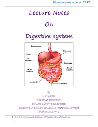
  • 1. Digestive system notes 2017 1 By, K. P. Komal, Asst. Professor of Biochemistry, chitradurga. Lecture Notes On Digestive system By, K. P. KOMAL ASSISTANT PROFESSOR DEPARTMENT OF BIOCHEMISTRY GOVERNMENT SCIENCE COLLEGE, CHITRADURGA. 577501 KARNATAKA STATE.
  • 2. Digestive system notes 2017 2 By, K. P. Komal, Asst. Professor of Biochemistry, chitradurga. Human Digestive System In the human digestive system, large organic masses are broken down into smaller particles that the body can use as fuel. This is a complex process. The breakdown of the nutrients requires the coordination of several enzymes secreted from specialized cells within the mouth, stomach, intestines, and liver. The major organs or structures that coordinate digestion within the human body include the mouth, esophagus, stomach, small and large intestines, and liver. Mouth In the human body, the mouth (oral cavity) is a specialized organ for receiving food and breaking up large organic masses. In the mouth, food is changed mechanically by biting and chewing. Humans have four kinds of teeth: incisors are chisel-shaped teeth in the front of the mouth for biting; canines are pointed teeth for tearing; and premolars and molars are flattened, ridged teeth for grinding, pounding, and crushing food. In the mouth, food is moistened by saliva, a sticky fluid that binds food particles together into a soft mass. Three pairs of salivary glands—parotid, submaxillary, and sublingual—secrete saliva into the mouth. The saliva contains an enzyme called amylase, which digests starch molecules into smaller molecules of the disaccharide maltose. During chewing, the tongue moves food about and manipulates it into a mass called a bolus. The bolus is pushed back into the pharynx (throat) and is forced through the opening to the esophagus. Esophagus The esophagus is a thick-walled muscular tube located behind the windpipe that extends through the neck and chest to the stomach. The bolus of food moves through the esophagus by peristalsis: a rhythmic series of muscular contractions that propels the bolus along. The contractions are assisted by the pull of gravity.
  • 3. Digestive system notes 2017 3 By, K. P. Komal, Asst. Professor of Biochemistry, chitradurga. Stomach The esophagus joins the stomach at a point just below the diaphragm. A valvelike ring of muscle called the cardiac sphinctersurrounds the opening to the stomach. The sphincter relaxes as the bolus passes through and then quickly closes. The stomach is an expandable pouch located high in the abdominal cavity. Layers of stomach muscle contract and churn the bolus of food with gastric juices to form a soupy liquid called chyme. The stomach stores food and prepares it for further digestion. In addition, the stomach plays a role in protein digestion. Gastric glands called chief cells secrete pepsinogen. Pepsinogen is converted to the enzyme pepsin in the presence of hydrochloric acid. Hydrochloric acid is secreted by parietal cells in the stomach lining. The pepsin then digests large proteins into smaller proteins called peptides. To protect the stomach lining from the acid, a third type of cell secretes mucus that lines the stomach cavity. An overabundance of acid due to mucus failure may lead to an ulcer. Small intestine The soupy mixture called chyme spurts from the stomach through a sphincter into the small intestine. An adult’s small intestine is about 23 feet long and is divided into three sections: the first 10 to 12 inches form the duodenum; the next 10 feet form the jejunum;and the final 12 feet form the ileum. The inner surface of the small intestine contains numerous fingerlike projections called villi (the singular is villus). Each villus has projections of cells called microvillito increase the surface area. Most chemical digestion takes place in the duodenum. In this region, enzymes digest nutrients into simpler forms that can be absorbed. Intestinal enzymes are supplemented by enzymes from the pancreas, a large, glandular organ near the stomach. In addition, bile enters the small intestine from the gallbladder to assist in fat digestion.
  • 4. Digestive system notes 2017 4 By, K. P. Komal, Asst. Professor of Biochemistry, chitradurga. The enzymes functioning in carbohydrate digestion include amylase (for starch), maltase (for maltose), sucrase (for sucrose), and lactase (for lactose). For fats, the principal enzyme is lipase. Before lipase can act, the large globules of fat must be broken into smaller droplets by bile. Bile is a mixture of salts, pigments, and cholesterol that is produced by the liver and stored in the gallbladder, a saclike structure underneath the liver. Protein digestion is accomplished by several enzymes, including two pancreatic enzymes: trypsin and chymotrypsin. Peptides are broken into smaller peptides, and peptidases reduce the enzymes to amino acids. Nucleases digest nucleic acids into nucleotides in the small intestine also. Most absorption in the small intestine occurs in the jejunum. The products of digestion enter cells of the villi, move across the cells, and enter blood vessels called capillaries. Diffusion accounts for the movement of many nutrients, but facilitated diffusion is responsible for the movement of glucose and amino acids. The products of fat digestion pass as small droplets of fat into lacteals, which are branches of the lymphatic system. Absorption is completed in the final part of the small intestine, theileum. Substances that have not been digested or absorbed then pass into the large intestine. Large intestine The small intestine joins the large intestine in the lower-right abdomen of the body. The two organs meet at a blind sac called thececum and a small fingerlike organ called the appendix.Evolutionary biologists believe the cecum and appendix are vestiges of larger organs that may have been functional in human ancestors. The large intestine is also known as the colon. It is divided into ascending, transverse, and descending portions, each about one foot in length. The colon’s chief functions are to absorb water and to store, process, and eliminate the residue following
  • 5. Digestive system notes 2017 5 By, K. P. Komal, Asst. Professor of Biochemistry, chitradurga. digestion and absorption. The intestinal matter remaining after water has been reclaimed is known as feces. Feces consist of nondigested food (such as cellulose), billions of mostly harmless bacteria, bile pigments, and other materials. The feces are stored in the rectum and passed out through the anus to complete the digestion process. Liver The liver has an important function in processing the products of human digestion. For example, cells of the liver remove excess glucose from the bloodstream and convert the glucose to a polymer called glycogen for storage. The liver also functions in amino acid metabolism. In a process called deamination, it converts some amino acids to compounds that can be used in energy metabolism. In doing so, the liver removes the amino groups from amino acids and uses the amino groups to produce urea. Urea is removed from the body in the urine. Fats are processed into two-carbon units that can enter the Kreb’s cycle for energy metabolism. The liver also stores vitamins and minerals, forms many blood proteins, synthesizes cholesterol, and produces bile for fat digestion. The metabolism of carbohydrates is the process of getting the carbohydrates in the foods we eat into the right format to provide fuel to our body's cells. This process involves digestion, absorption and transportation. Most commonly, carbohydrate metabolism results in the production of glucose molecules which are the most efficient source of energy (ATP) for our muscles and our brains. Energy or fuel from our food is used for cell growth, repair and normal cell functioning. Carbohydrate Digestion Carbohydrates are most commonly consumed as polysaccharides (e.g. starch, fibre or cellulose) or disaccharides (e.g. lactose, sucrose, galactose) and therefore need to be broken down into their simpler monosaccharide forms which the body can utilise.
  • 6. Digestive system notes 2017 6 By, K. P. Komal, Asst. Professor of Biochemistry, chitradurga. The digestion process of polysaccharides such as starch will begin in the mouth where it is hydrolysed by salivary amylase. The amount of starch hydrolysed in this environment is often quite small as most food does not stay in the mouth long. Once the food bolus reaches the stomach the salivary enzymes are denatured. As a result, digestion predominantly occurs in the small intestine with pancreatic amylase hydrolysing the starch to dextrin and maltose. Enzymes classed as glucosidases on the brush border of the small intestine break down the dextrin and maltase, lactase and sucrase convert the other disaccharides into their two monosaccharide units. Absorption & transport The monosaccharide units, glucose, galactose and fructose are transported through the wall of the small intestine into the portal vein which then takes them straight to the liver. The mode of transport varies between the monosaccharides. Both glucose and fructose are absorbed relatively quickly, depending on what other nutrients are eaten at the same time. For example a meal or food containing protein and fat causes the sugars to be absorbed slower than when consumed on their own.  Glucose, at low concentrations is transported through the mucosal lining into the epithelial cells of the intestine by active transport, via a sodium dependant transporter. At higher concentrations, a second facilitative transporter becomes involved. From the epithelial cells glucose is moved into the surrounding capillaries by facilitated diffusion.  Galactose is transported in the same way as glucose, utilising the same transporters. As galactose is not found as a monosaccharide in nature, absorbed galactose primarily comes from the breakdown of lactose.  Fructose moves entirely via facilitated diffusion. The process utilises a different transporter to glucose when entering the enterocytes, however both fructose and glucose utilise the same transporter to exit the enterocyte into the capillaries. The
  • 7. Digestive system notes 2017 7 By, K. P. Komal, Asst. Professor of Biochemistry, chitradurga. absorption of fructose is much slower than that of glucose and is quantitatively limited. Consumption of large amounts of fructose has been shown to produce a level of fructose malabsorption in almost all cases. Co-ingestion of glucose with fructose has been shown to facilitate fructose absorption. Once in the liver galactose and fructose are removed from the blood and converted into other metabolites. When eaten in moderate quantities, most fructose is taken up by the liver and converted to glucose, glycogen and lactate. A fraction may also be oxidised or converted into fatty acids and uric acid. Only a small amount of fructose reaches the bloodstream, so blood fructose concentrations are always low. Galactose is primarily converted into glucose and stored as glycogen. On the other hand most of the glucose derived from food is transported via the blood stream to the peripheral tissues where, under normal circumstances, the hormone insulin enables it to be taken up by the cells and used as an energy source via the glycolysis pathway. As glucose is the most important fuel source for the body and in particular the brain the body attempts to keep a basal circulating blood glucose of around 4-5mmol/L. This homeostasis mechanism is predominantly controlled by the actions of glycogen and insulin. Storage Surplus glucose is initially stored as glycogen in the liver or muscles. The liver can store approximately 100g of glycogen which is used to maintain basal blood glucose levels between meals, whilst the muscles typically store 400-500g often used during movement. Once these reserves are saturated, excess glucose is converted to fat for longer term storage.
  • 8. Digestive system notes 2017 8 By, K. P. Komal, Asst. Professor of Biochemistry, chitradurga. Proteins Digestion and Absorption: The major constituents of the food are carbohydrates, proteins and lipids. They are digested and absorbed in the stomach and intestine. Some of the digested/degraded components of the food stuffs may either be reutilized or may be excreted out. Chewing of food, movements of the stomach and intestine facilitate the grinding of the food materials and bring them in contact with gastric secretions. Proteolytic enzymes are absent in the salivary secretions, hence there is no digestion of proteins in the mouth. Proteolysis takes place in the gastro-intestinal tract (i.e. stomach and intestine). When the proteins enter the stomach they stimulate the secretion of the hormone called gastrin which in turn stimulates the secretion of HCl by parietal cells of the stomach and pepsinogen from the chief cells. Gastric juice is acidic i.e. the pH is 1.5—2.5. Acidic pH of the stomach has an antiseptic action that kills the bacteria and other microorganisms. At this pH the dietary proteins also get denatured. In presence of HCl, pepsinogen is converted to
  • 9. Digestive system notes 2017 9 By, K. P. Komal, Asst. Professor of Biochemistry, chitradurga. pepsin by autocatalysis resulting in removal of some of the amino acids from the amino terminal end. Pepsin is an endopeptidase for tyr, phe, trp. In the stomach the proteins are converted as follows: Protein → Metaprotein → Proteone → Peptone → Peptide As the food passes from the stomach to small intestine the low pH of the food triggers the secretion of the hormone ‘secretin’ into the blood. It stimulates the pancreas to secrete HCO3 into the small intestine in order to neutralize HCl. The secretion of HCO3 into the intestine abruptly raises the pH from 2.5 to 7.0. The entry of amino acids into the duodenum releases the hormone ‘cholecystokinin’ which in turn triggers the release of pancreatic juice (that contains many pancreatic enzymes like trypsinogen, chymotrypsinogen, procarboxypeptidases) by the exocrine cells of the pancreas (ecbolic and hydrolatic). Most of these enzymes are produced as zymogens (inactive enzymes) by the pancreas in order to protect the exocrine cells from being digested. Subsequent to the entry of trypsinogen into the small intestine it is activated to trypsin by enterokinase secreted by the intestinal cells. Trypsin is formed from trypsinogen by the removal of hexapeptide from the N-terminal end. The newly formed trypsin activates the remaining trypsinogen, Trypsin is an endopeptidase, specific for (acts on) the peptide bonds contributed by the basic amino acids like arginine, histidine and lysine. Chymotrypsin is secreted in an inactive from called chymotrypsinogen which is activated by trypsin. Chymotrypsin is an endopeptidase specific to aromatic amino acids i.e. phenylalanine, tyrosine, tryptophan.
  • 10. Digestive system notes 2017 10 By, K. P. Komal, Asst. Professor of Biochemistry, chitradurga. Carboxypeptidase secreted as procarboxypeptidase is activated again by trypsin. It is an exopeptidase that cleaves the amino acids from the carboxy terminal end. Amino peptidase secreted as pro-aminopeptidase is once again activated by trypsin. It is an exopeptidase that cleaves the amino acids from the free amino terminal end. Dipeptides acts only on dipeptides and hydrolyses it into 2 amino acids. Even after the action of all these enzymes most of the proteins remain undigested. Protein like collagen, fibrin etc., escape digestion and are excreted out. Absorption of the Digested Proteins: There are four distinct carrier systems in the intestinal epithelium for the absorption of the digested proteins. These are: (1) Carrier system for neutral amino acids (2) Carrier system for basic amino acids (3) Carrier system for acidic amino acids (4) Carrier system for glycine and imino acid (proline)
  • 11. Digestive system notes 2017 11 By, K. P. Komal, Asst. Professor of Biochemistry, chitradurga. The digested amino acids are carried across the mucosal cell membrane from the intestinal lumen to the cytoplasm of the cell by one of the above carrier systems specific to that particular amino acid. Absorption of amino acids is an up-hill process (i.e. against gradient as compared to the Na+ absorption which is downhill i.e. along the gradient). There are four systems that operate to absorb amino acids from the mucosal cells into the blood. They are: (1) A — system (alanine system) (2) L — system (leucine system) (3) Ly – system (lysine system) (4) Ala-ser-cyst – system Amino acids are taken up by the blood capillaries of the mucosa and are transported in the plasma to the liver. Some amounts of amino acids are also absorbed through the lymph. Glucagon stimulates the absorption of amino acids through ‘A’ system mediated by cAMP. Insulin stimulates the trans cellular transport of amino acids to minimize the loss in the urine. The proximal tubule cells reabsorb and return them to the blood stream. It is done by glutathione, a tripeptide. Three ATPs are utilized for the absorption of each amino acid. Absorbed amino acids do not stimulate antibody production whereas an intact protein absorbed becomes antigenic. Intestinal membranes allow the transport of proteins across them ex.—In a neonate the intestinal mucosa is permeable to y-globulin (immunoglobulin) of colostrum. The immune system in a neonate is not well developed thus absorption of intact y-globulin into the blood does not elicit any immune response instead it results in the defence of the neonate against infections. In adults, some proteins may be absorbed intact through the intestinal mucosa leading to the formation of antibodies and
  • 12. Digestive system notes 2017 12 By, K. P. Komal, Asst. Professor of Biochemistry, chitradurga. anaphylactic reactions or other such immunological phenomena after the absorption of those proteins. Thus in such cases allergies to food proteins occur. Digestion of Fats/ lipids: Stomach: Lipase present in the stomach is unable to hydrolyze fats owing to the high acidity of the gastric contents. Therefore, the major part of the ingested fat is digested in the small intestine. Small Intestine: The ingested fat reaching the duodenum is mixed with the bile and pancreatic juice which contains lipase. The bile salts emulsify the fat before the action of lipase. The emulsification is also brought about by monoglycerides, phospholipid and lysolecithin. The secreted inactive pancreatic lipase is activated by bile and Ca. The surface area of the emulsified fat becomes increased for which the rate of reaction of lipase is increased. Pancreatic lipase hydrolyzes 1- and 3-positions of the triglycerides leaving a mixture of 2- monoglycerides, 1, 2- and 2, 3-diglycerides as well as the soaps of the free fatty acids. The pancreatic juice also contains phospholipase and cholesterol-esterase which hydrolyze phospholipid and esterified cholesterol. Intestinal juice also contains a lipase whose action is not of much importance as most of the fat is hydrolyzed by the pancreatic lipase. Absorption of Fats: Several theories have been proposed for the mechanism of absorption of fats after digestion. The important theories are: A. Lipolytic hypothesis.
  • 13. Digestive system notes 2017 13 By, K. P. Komal, Asst. Professor of Biochemistry, chitradurga. B. Partition theory. C. More recent theory. A. Lipolytic Hypothesis: 1. According to this theory, fat is completely hydrolyzed to fatty acids and glycerol which are absorbed. 2. The fatty acids combining with bile salts form a miscible complex which is ab- sorbed into the intestinal mucosa. 3. The fatty acids are then separated from bile acids and converted into triglycerides by combining with glycerol. 4. The triglycerides are passed to the lacteals. They then enter the lymphatic’s and reach the systemic circulation via thoracic duct. B. Partition Theory: 1. According to this theory, 30 per cent of the triglycerides are hydrolyzed to fatty acids and glycerol while 70 per cent remain un-hydrolyzed. 2. The un-hydrolyzed triglycerides are emulsified by monoglycerides and diglycerides in combination with bile salts to form minute particles known as “micelles” of size about 0.1 to 0.5µ. 3. The resulting mixture is absorbed into the intestines, passed on to the lacteals and then to the lymphatic’s. The mixture then reaches the systemic circulation via thoracic duct. 4. The free fatty acids are absorbed as bile salt-fatty acid complex into the intestinal mucosa. The fatty acids are absorbed into the portal blood to reach the liver.
  • 14. Digestive system notes 2017 14 By, K. P. Komal, Asst. Professor of Biochemistry, chitradurga. C. Recent Theory: 1. The removal of the ester group of 2- mono-acylglycerol requires isomerization to a primary ester linkage. This is a slow process. As a result, monoacylglycerols are the major end products of fat digestion and less than one-fourth of the ingested fat is completely broken down to glycerol and fatty acids. 2. Within the intestinal wall, 2-monoacylglycerols are converted to triacylglycerol’s and l-monoacylglycerols are further hydrolyzed to form free glycerol and fatty acids. 3. The fatty acids are then activated by thiokinase in presence of ATP and coenzyme A for the resynthesize of triacylglycerol’s. 4. The free glycerol in the intestinal lumen is about 22 per cent of total amount of triacylglycerol originally present. This passes directly to the portal vein.
  • 15. Digestive system notes 2017 15 By, K. P. Komal, Asst. Professor of Biochemistry, chitradurga. 5. The glycerol within the intestinal wall is activated by glycerokinase in presence of ATP to form glycerol-3-phosphate for the synthesis of triacylglycerol followed by the combination with acyl-CoA present in the intestinal wall. 6. All long chain fatty acids present in the intestinal wall are reincorporated into triacylglycerol’s which are transported to the lymphatic vessels of the abdominal region (the so-called lacteals) for distribution to the rest of the body. 7. The great majority of absorbed fat appears in the form of chylomicrons which appear first at the lymphatic vessels of the abdominal region and later in the systemic blood. The chylomicrons contain triacylglycerol, free and esterified cholesterol, phospholipid and 0.5 per cent protein. All of the factors relating to digestion and absorption of fat are mentioned in Fig. 16.8.
  • 16. Digestive system notes 2017 16 By, K. P. Komal, Asst. Professor of Biochemistry, chitradurga. Absorption of Phospholipids: Phospholipids are split by phospholipases and their acyl chains are incorporated into chylomicrons, choline, the hydrophilic component, may be transported directly to the liver via the hepatic portal vein. Absorption of Cholesterol: It is absorbed into the lymphatic’s and recovered mainly as cholesteryl esters.
  • 17. Digestive system notes 2017 17 By, K. P. Komal, Asst. Professor of Biochemistry, chitradurga. Composition and Functions of Various Digestive Juice: There are five digestive juices; saliva, gastric juice, pancreatic juice, succus entericus (intestinal juice) and bile. The necessity for so many digestive juices is that – (a) One juice does not contain all the enzymes necessary for digesting all the different types of foodstuffs. For instance, saliva contains only carbohydrate-splitting enzymes; whereas gastric juice contains both fat- and protein -splitting enzymes but none acting on carbohydrates, and (b) The second reason is that, one particular digestive juice cannot digest a particular type of food up to completion. It will digest only up to a certain stage and then, the products will be handed over to the next digestive juice for further digestion. In this way digestion is completed. For instance, gastric juice digests protein up to the stage of peptone, pancreatic juice carries the digestion of peptone further up to lower peptide. The latter is digested completely up to amino acids by succus entericus. One more interesting fact about the digestive juices is that, their reactions are not all same. Saliva is slightly acid, gastric juice is strongly acid, but pancreatic juice is strongly alkaline. This, more or less, alternate acid and alkaline reaction, prevents any serious alteration of blood reaction. This is a special device for maintaining blood reaction constant. 1. Saliva: Characteristics:  Total Amount – 1,200 -1,500 ml in 24 hours. A large proportion of this 24- hour volume is secreted at meal time, when secretory rate is highest.  Consistency – Slightly cloudy, due to the presence of cells and mucin.
  • 18. Digestive system notes 2017 18 By, K. P. Komal, Asst. Professor of Biochemistry, chitradurga.  Reaction – Usually slightly acid (pH 6.02 – 7.05). On standing or boiling it loses CO2 and becomes alkaline. This alkaline reaction causes precipitation of salivary constituents, as tartar on the teeth or calculus in salivary duct.  Specific Gravity – 1.002 – 1.012.  Freezing Point – 0.07 – 0.34°C. Composition: 1. Water – 99.5%. 2. Solids – 0.5%. i. Cellular Constituents: Yeast cells, bacteria, protozoa, polymorphonuclear leucocytes, desquamated epithelial cells, etc. ii. Inorganic Salts: About 0.2% consists of NaCI, KCI, acid and alkaline sodium phosphate, CaCO3, calcium phosphate, potassium thiocyanate. Smoker’s saliva is rich in thiocyanate. In case of poisoning with metals like lead, mercury, etc., they are secreted in saliva. iii. Orgainc – 0.3%. a. Enzymes P tyalin (salivary amylase) lipase, carbonic anhydrase, phosphatase and a bacteriolytic enzyme, lysozyme. b. Mucin. c. Urea, amino acids, cholesterol and vitamins (in small amounts). d. The soluble specific blood group substances also form part of the organic constituents of saliva and they have the same characteristics as the agglutinogen on the erythrocyte. In human beings, A, B, O and Lea substances have been demonstrated. Their concentration in saliva is from 10 to 20 mgm per litre. 3. Gases: Saliva contains about 1 ml oxygen, 2.5 ml nitrogen and 50 ml of CO2 per 100 ml. Bicarbonates, phosphates and the proteins act as buffers. Chlorides activate amylase.
  • 19. Digestive system notes 2017 19 By, K. P. Komal, Asst. Professor of Biochemistry, chitradurga. The thiocyanate (KCNS) is a product of excretion. It is formed in the body from the cyanogen radicle (-CN) derived from proteins. Its formation is a process of detoxication of the poisonous cyanides and hence is an example of protective synthesis. With ferric chloride it gives a rich brown colour. An enzyme kallikrein is present in saliva which acts upon plasma protein to produce a substance known as kallidin or bradykinin. This produces vasodilatation of salivary gland during secretion and is also responsible for the sudden fall and consequent rise in blood pressure observed after injection of saliva in animals. Functions: 1. Mechanical Functions: a. It keeps the mouth moist and helps speech. Decrease in salivary secretion as occurs after nervousness, causes impairment of speech. b. It helps in the process of mastication of the foodstuff and in preparing it into a bolus, suitable for deglutition. Here, saliva also acts as a lubricant. c. Constant flow of saliva washes down the food debris and thereby does not allow the bacteria to grow. In acute fevers, where the salivary secretion is inhibited, the food debris is not properly washed away and the bacteria multiply. These collect as the sordes at the root of the teeth and upon the tongue. 2. Digestive Functions: Saliva contains two enzymes: a. Ptyalin: Splits starch up to maltose in the following manner [Fig. 9.24]
  • 20. Digestive system notes 2017 20 By, K. P. Komal, Asst. Professor of Biochemistry, chitradurga. b. Maltase: Maltase (in traces) converts maltose into glucose. 3. Excretory Functions: Saliva excretes urea, heavy metals (Hg, Pb, Bi, As, etc.), thiocyanates, certain drugs like iodide, etc. Alkaloids, such as morphine, antibiotics, such as penicillin, streptomycin, etc. are also excreted in the saliva. The excretion of ethyl alcohol by the salivary gland has prompted the recommendation that such a test should be used for medico-legal purpose. [It also excretes certain virulent micro-organisms, such as the virus of hydrophoboea, acute anterior poliomyelitis, mumps, etc.] 4. Helps in the Sensation of Taste: Taste is a chemical sensation. Unless the substances are in solution, the taste buds cannot be stimulated. Saliva acts as a solvent and is thus essential for taste. 5. Helps Water Balance: Saliva keeps the mouth moist. When moisture is reduced in the mouth, certain nerve endings at the back of the tongue are stimulated and the sensation of thirst arises. When body water is lost (sweating, diarrhoea, etc.) – saliva is reduced and thirst is felt. The subject feels the necessity of drinking water and thus water balance is restored. 6. Helps Heat Loss: This is mainly found in animals (dog, sheep, etc.). When they become hot or excited more saliva is secreted causing greater heat loss. 7. Buffering Action: Mainly bicarbonate and to a lesser extent phosphate and mucin present in saliva act as buffers. There is an increase in bicarbonate concentration during food intake. 8. Bacteriolytic Action:
  • 21. Digestive system notes 2017 21 By, K. P. Komal, Asst. Professor of Biochemistry, chitradurga. Cell membrane of different varieties of bacteria contains polysaccharides, lysozyme, the enzyme present in the saliva is polysaccharides, and thus it dissolves the cell wall of many bacteria and finally kills them. 2. Gastric Juice: Composition: The average composition of human gastric juice is as follows: 1. Water – 99.45%. 2. Total Solids – 0.55%. a. Inorganic – 0.15% (NaCI, KCI, CaCI,, calcium phosphate, Mag. Phosphate, bicarbonate, etc.). b. Organic – 0.40%. i. Mucin. ii. Intrinsic factor, iii. Enzymes. a. Pepsin. i. Other proteolytic enzymes of the gastric juice are; cathepsin, gastricin, parapepsin I and II. ii. Gastric rennin. iii. Gastric lipase. iv. Other gastric enzymes are present in minute amounts and are; lysozyme, gelatinase, urease, carbonic anhydrase. Characteristics: i. Total Quantity – About 500 -1,000 ml per meal (1,200 ml -1,500 ml per day). ii. Reaction – Strongly acid. iii. Free HCI – -0.4 -0.5% iv. Total Acidity – – 0.45 – 0.6%. It includes free HCI, as well as HCI combined with proteins. It also includes other acids, such as lactic acid. As ordinarily examined, the
  • 22. Digestive system notes 2017 22 By, K. P. Komal, Asst. Professor of Biochemistry, chitradurga. gastric contents show a lower acidity (0.15% to 0.25% HCI), because the HCI is partly neutralised by mucin and other substances. v. pH – 0.9-1.5 vi. Specific Gravity – 1.002 -1.004. vii. Freezing Point – 0.59°C. Functions: i. Pepsin – The enzyme pepsin, with HCI, digests proteins up to the stage of peptone. ii. Rennin – Rennin coagulates caseinogen of milk. iii. Gastric Lipase – Gastric lipase digests fat to some degree. iv. HCI Acts as an Antiseptic – HCI acts as an antiseptic and causes some hydrolysis of all the foodstuffs. v. Excretion – Toxins, heavy metals, certain alkaloids, etc., are excreted through gastric juice. 3. Pancreatic Juice: Characteristics: i. Total Quantity – About 500 ml per meal. About 1,500 ml in 24 hours. ii. Reaction – Alkaline. iii. Specific Gravity – 1.010 to 1.030 iv. pH – 8.0 – 8.3 (in dog). Constituents: i. Inorganic Constituents: The distinguishing chemical characteristic is its high bicarbonate content. The principal bases are sodium and potassium. Small amounts of calcium, magnesium and zinc are also present. ii. Organic Constituents: Enzymes:
  • 23. Digestive system notes 2017 23 By, K. P. Komal, Asst. Professor of Biochemistry, chitradurga. The enzymes of pancreatic juice are trypsinogen, chymotrypsinogen, procar- boxypeptidase, nucleotidases (ribonuclease and deoxyribonuclease), elastase, collagenase, pancreatic lipase, lecithinase, cholesterol esterase and amylase. Its composition varies according to the means used to cause Secretion. 4. Succus Entericus (Intestinal Juice): Intestinal juice, in pure form, is difficult to collect because it is mixed up with bile and pancreatic juice. It can be collected from fistula preparations, such as Thiry fistula, Thiry-Vella modification, and Mann Bollman fistula. Characteristics: i. Total Quantity – Roughly about 1-2 litres in 24 hours. [Accurate measurement is not possible due to the great length of the small intestine.] ii. Specific Gravity – Specific gravity -1.010. iii. Reaction – Faintly acid to faintly alkaline. iv. pH – Varies from 6.3 – 9.0 average 8.3. Composition: 1. Water – 98.5% 2. Solids – 1.5% i. Inorganic – 0.8% salts of sodium, potassium, calcium and magnesium with that of chloride, bicarbonate and phosphate. The bicarbonate concentration is higher than it is in the blood or interstitial fluids. ii. Organic – 0.7%. a. Activator- Enteropeptidase (previously known as enterokinase). It activates trypsinogen into trypsin. b. Enzymes. c. Mucen. Intestinal Juice Enzymes: 1. Proteolytic:
  • 24. Digestive system notes 2017 24 By, K. P. Komal, Asst. Professor of Biochemistry, chitradurga. i. Erepsin – A mixture of enzymes containing dipeptidases (break down dipeptides into amino acids) and amino peptidases (remove terminal amino acid containing free NH2 group from polypeptides). ii. Enzymes – Several enzymes acting on the different fractions of nucleic acid, such as nuclease, nucleotidase and nucleosidase. iii. Arginase Acts on arginine producing urea and ornithine. 2. Carbohydrate Splitting: i. Amylase – Found in traces, acts on starch and dextrin. ii. Sucrase (Invertase) – Digests cane sugar. iii. Maltase – Acts on maltose. iv. Isomaltase. v. Lactase – Breaks down lactose. 3. Fat-Splitting – Lipase. 4. Other Enzymes: Alkaline phosphatase, cholesterol esterase, lecithinase, etc. The small intestine does not secrete enzymes in the sense that secretion occurs in the gastric mucosa or in the pancreas. Most of these digestive enzymes are actually intracellular and are present in the juice only because cells desquamate. Enteropeptidase and amylase are highly soluble and diffusible and are present in the succus entericus. As regards other enzymes they are mostly present in the epithelial cells. Peptidases (erepsin), lactase, maltase, sucrose (invertase) and lipase are found in the intestinal epithelium as well as in the shed cells present in the juice. Proteases, nuclease, phosphatase and arginase are present in the scrapings of the mucous membrane only. These scrapings also show the presence of all the enzymes mentioned above. From this it can be concluded that the enzymes discussed above digest the foodstuffs in three ways:
  • 25. Digestive system notes 2017 25 By, K. P. Komal, Asst. Professor of Biochemistry, chitradurga. 1. Soluble enzymes- Enteropeptidase and amylase, freely exert their action on trypsinogen and starch respectively. 2. The shed cells break down in the succus entericus, set free their insoluble enzymes which digest polypeptides, disaccharides and fats. 3. Those insoluble enzymes which remain in the intestinal mucosa and found only in the scrapings, exert their actions of the corresponding substrates during their transit through the epithelium, in the course of absorption. 5. Bile: Introduction: Bile is both a product of secretion as well as of excretion of the liver. Minute droplets of bile collect inside the tiny vacuoles of the liver cells and are discharged into the bile capillaries through the intracellular canaliculi. The primary bile capillaries start between hepatic cells as blind tubules. They join together repeatedly and form bigger channels and ultimately come out of liver as the right and left hepatic ducts. The two ducts unite and form the common bile duct, which enter into the ducodenum, through the ampulla of Vater. Through the same ampulla also the pancreatic duct opens. From the upper part of the common bile duct commences the cystic duct, which ends in the gall-bladder (Fig. 9.25).
  • 26. Digestive system notes 2017 26 By, K. P. Komal, Asst. Professor of Biochemistry, chitradurga. Formation of bile by the liver is an active process, but entry of bile into the duodenum is intermittent and takes place only after meal. This necessarily indicates that bile must be stored somewhere. Gall-bladder acts as the chief storehouse. The common bile duct also stores some bile. Composition of Bile: Bile is a complex fluid containing various substances, some of which are merely waste products undergoing excretion, whereas others are products of secretion serving important physiological functions. In the gall-bladder bile is concentrated five to ten times and its alkalinity is reduced (vide ‘Gall-bladder’). The composition, as estimated by different observers, varies widely. The usual composition and the characters are as follows: Characteristics: i. Total Quantity – 500 -1,000 ml daily. On the average about 700 ml. ii. Sp. Gravity – 1.010 -1.011 (Gall-bladder bile-1.026 -1.040). iii. Colour – Human bile is yellowish-green. [In the carnivore, it is golden-yellow, due to the presence of more bilirubin. In herbivora, the colour is green, due to more biliverdin.] iv. Taste – Bitter. v. Consistency – Viscid, mucoid liquid. vi. Reaction – Liver bile is definitely alkaline, pH 7.7. [Some hold pH 8.0 – 8.6] Gall-bladder bile is neutral or slightly alkaline (pH 7.0 – 7.6) or slightly acid (pH 6.8). That of dog and cat, definitely acid (pH 5.6). Composition: Total solids, 2 -11%. The chief constituents are: i. Inorganic Salts:
  • 27. Digestive system notes 2017 27 By, K. P. Komal, Asst. Professor of Biochemistry, chitradurga. Chlorides, carbonates and phosphates of Na, K and Ca and NaHCO3. The total base is equivalent to about 170 ml of (N/10) NaOH per 100 ml of liver bile (300 ml % in gall-bladder bile). ii. Bile Salts: Sodium taurocholate and sodium glycocholate. These are the most important constituents of bile and are synthesised by the liver (secretion). iii. Bile Pigments: Of which bilirubin and biliverdin are the chief . iv. Cholesterol, Lecithin: Cholesterol, lecithin and traces of fatty acids, soaps, etc. Cholesterol is probably an excretory product, because its amount in bile varies with its level in blood. It is kept in solution by the hydrotropic action of bile salts. Average composition of human bile is given in Table 9.2. Structure of villi  The wall of the small intestine contains millions of fingerlike projections called villi; their fingerlike shape greatly increases the surface area of the intestine thereby speeding up the overall rate of absorption. Absorption is the process by which the nutrients of food move from the small intestine into the bloodstream. Villus
  • 28. Digestive system notes 2017 28 By, K. P. Komal, Asst. Professor of Biochemistry, chitradurga.  Each villus contains a lymphatic vessel called a lacteal, which is adapted to receive fatty acids from the microvilli.  And each villus contains a network of capillaries that delivers oxygen to the microvilli so they can perform cell respiration. Microvilli are hundreds of microscopic cells that project into the intestinal lumen from the villus. Like the villus, each microvillus is finger-shaped to further increase the surface area of the intestine for faster absorption.  Microvilli are extremely small cells, a feature that allows nutrients to pass through them quickly.  Microvilli contain many mitochondria, which allow them to make lots of ATP for the active transport of nutrients into the bloodstream.  Microvilli have protein pumps that move monosaccharides and amino acids into the bloodstream.  Absorption requires the nutrients to cross four cell membranes: 1) out of the small intestine lumen and into a microvillus cell; 2) out of the microvillus cell; 3) into a capillary cell; 4) out of a capillary cell and into the capillary lumen. Role of Portal vein in transport of nutrients: The portal vein or hepatic portal vein is a blood vessel that carries blood from the gastrointestinal tract, gallbladder, pancreas and spleen to the liver.
  • 29. Digestive system notes 2017 29 By, K. P. Komal, Asst. Professor of Biochemistry, chitradurga. This blood contains nutrients and toxins extracted from digested contents. Approximately 75% of total liver blood flow is through the portal vein, with the remainder coming from the hepatic artery proper. The blood leaves the liver to the heart in the hepatic veins. The portal vein is not a true vein, because it conducts blood to capillary beds in the liver and not directly to the heart. It is a major component of the hepatic portal system, one of only two portal venous systems in the body – with thehypophyseal portal system being the other. The portal vein is usually formed by the confluence of the superior mesentericand splenic veins and also receives blood from the inferior mesenteric, left andright gastric veins, and cystic veins. Functions The portal vein and hepatic arteries form the liver's dual blood supply. Approximately 75% of hepatic blood flow is derived from the portal vein, while the remainder is from the hepatic arteries.
  • 30. Digestive system notes 2017 30 By, K. P. Komal, Asst. Professor of Biochemistry, chitradurga. Unlike most veins, the portal vein does not drain into the heart. Rather, it is part of a portal venous system that delivers venous blood into another capillary system, the hepatic sinusoids of the liver. In carrying venous blood from the gastrointestinal tract to the liver, the portal vein accomplishes two tasks: it supplies the liver with metabolic substrates and it ensures that substances ingested are first processed by the liver before reaching the systemic circulation. This accomplishes two things. First, possible toxins that may be ingested can be detoxified by the hepatocytes before they are released into the systemic circulation. Second, the liver is the first organ to absorb nutrients just taken in by the intestines. After draining into the liver sinusoids, blood from the liver is drained by the hepatic vein.