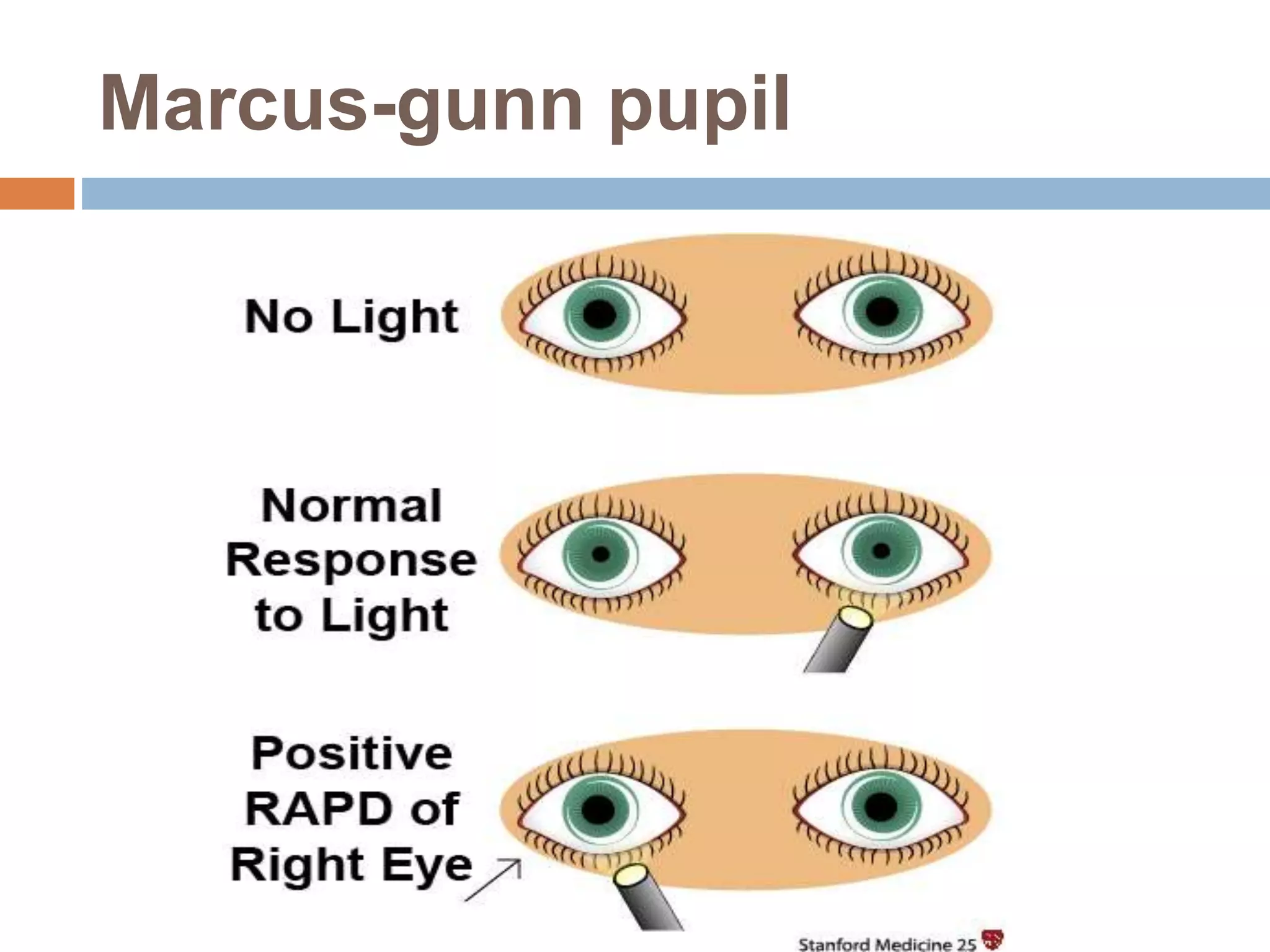
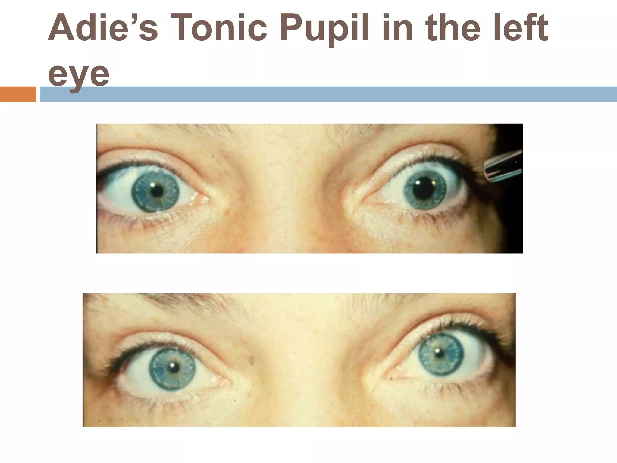
ABNORMALITIES OF
PUPIL
Kh.Md.Imrul Morshed
B.Optom( 6th Batch )
Institute of Community Ophthalmology,
Chittagong
Pupil
 It is a circular aperture at the centre of the iris
that allows light to enter the retina.
 The size of pupil normally varies between 2 to
4 mm .
Abnormal pupils
 Anisocoria:
 Anisocoria means unequal pupils.
 If there is a poor reaction to light in one eye and
the anisocoria is more evident in a well lit room,
then the affected pupil is abnormally large.
 If there is a good reaction to light in both eyes
but a poor dilation in the dark then the affected
pupil is abnormally small.
The abnormally large pupil
 Features - there is poor constriction in a
well lit room.
 Differential diagnosis –
 traumatic iris damage
 third cranial nerve palsy
 pharmacological dilation (i.e. dilating
drops)
 Adie's pupil
 iris rubeosis.
The abnormally small pupil
 Features - there is poor dilation in a dim
room.
 Differential diagnosis –
 physiologically small pupil
 pilocarpine drops
 uvietis with synaechiae
 Horner's syndrome.
Dilated pupil of right eye
Anomalies of pupillary
reflexes
 Marcus-gunn pupil:
 It is also known as Related afferent pupillary
defect (RAPD) or pupillary escape
phenomenon.
 It occurs in defect of visual pathway anterior to
the chaisma.
 It consists of –
1. A diminished amplitude to pupillary reaction
2. Pupillary dilation with continuous light
stimulation.
Marcus-gunn pupil
 Detection : The test for its detection is called
swinging flash light test . Normally, if a
illuminated pen light is alternately directed to
each eye the pupils constrict and do not vary
as the light alternate between the eyes.
 In afferent pupillary defect both pupils dilate
when the light is moved from the unaffected
eye to the affected eye. But they constrict
when the light is directed to the normal eye.
Examination of RAPD
Causes of Marcus-Gunn pupil
 Optic neuritis
 Ischemic optical disease
 Optic nerve damage
(Trauma,Radiation,Tumor)
 Retinal detachment
 Severe macular degeneration
Diseases affecting the
pupils
 Congenital Abnormalities:
 Aniridia:
 The term Aniridia implies an absence of Iris.
 In the majority of cases a small stump of iris
tissue exists at the iris root.
Aniridia with a small stump of
iris tissue at the iris root.
Etiology of Aniridia
 Aniridia is an congenital anomaly that
develops at approximately twelfth week of
gestation .
 Two-thirds of the cases are inherited in the
autosomal dominant fashion. The remaining
one-third of the cases appear as a result of
spontaneous mutation .
Ocular Signs and Symptoms
 Photophobia
 Corneal opacification
 Congenital polar cataracts
 Lens dislocations
 Progressive Glaucoma
Management
 Colored contact lens.
 Early goniotomy can be performed to prevent
blockage to trabecular meshwork.
 Filtering surgery.
 Cyclocryotherapy
Iris Coloboma
 This is an uncommon
congenital condition
characterized by a
unilateral or bilateral
partial iris defect.
 It is a condition where
normal tissues of iris is
missing from birth.
 It creates “Key-hole” or
“Cats-eye” appearance
to the iris.
Iris Coloboma With a
Key-hole appearance
Etiology of Iris coloboma
 The eye develops quickly during the first three
months of growth of a fetus. A gap , known as
the choroidal fissure, appears at the bottom of
the stalks that eventually form the eye.This
fissure generally closes by the seventh week
of gestation. If it doesnot close , a coloboma or
space forms.
Signs and Symptoms
 Missing iris tissue
 Reduced vision
 Key-hole or cat-eye shaped pupil
 photophobia
Management
 Usually there is no cure for coloboma.
 Colored contact lens
 Surgery
leukocoria
 Refers to a white pupillary reflex from the retina of
the eye.
Leukocoria in the left eye
Etiology of Leukocoria
 Many conditions cause leukocoria that are-
 Congenital cataract
 Retinal detachment
 Retinoblastoma
 Endophthalmitis
 Retinopathy of prematurity
Signs and Symptoms
 Vision problems.
 Eye pain.
 Redness of the white part of the eye.
 Bleeding in the front part of the eye.
 Bulging of the eye.
 A pupil that doesn’t constrict when exposure to
the bright light.
 A different color in each iris.
Management
 Leukocoria is managed by treating any other
underlying conditions ( Retinal detachment ,
cataract e.t.c ) which are responsible for the
appearance of the white pupil.
Acquired Structural
Abnormalities
 Pseudoexfoliation Syndrome:
 This is a condition characterized by deposition of
a grey-white fibro granular extracellular matrix
material deposited on the anterior lens.
 It is seen on the slit-lamp examination , as a fine
grey dusting around the pupil .
 Pupil shape and function are not affected
 It is clinically significant due to its association with
glaucoma and its potential to make cataract
surgery more tricky.
Pseudoexfoliation Syndrome
Signs and Symptoms
 White flaky materials on pupillary border of the
iris.
Management
 IOP lowering medications as first line therapy.
 Argon Laser Trabeculoplasty (ALT)
 Selective Laser Trabeculoplasty (SLT)
Sphincter Tear
 Iris tear can occur as a
result of blunt or
penetrating trauma and
can also occur during
intraocular surgery.
 Tears may be
associated with
Glaucoma and visual
problems. Tear in the spincter
muscle
Neurological Abnormalities
 Horner’s Syndrome:
 It is a syndrome that consists of miosis, partial
ptosis and endophthalmitis.
 Sometimes associated with unilateral absence of
sweating of the face of the affected side
(Anhydrosis).
 In congenital form there may be associated
heterochromia of iris .
Horner’s Syndrome
Etiology
 Horner’s syndrome can be caused by any
interruption in a set of nerve fibers that start in
the part of brain called hypothalamus and
travel to the face and eyes .
 Injury to the carotid artery of the brain.
 Injury to the nerves at the base of the neck
called the brachial plexus.
 Migraine
 Tumor in the top of the lung.
Management
 If cosmetically unaccepted , ptosis may be
corrected surgically.
Adie’s Tonic Pupil
 It is a neurological disorder characterized by a
tonically dilated pupil that reacts slowly to light
but shows a more definite response to
accommodation.
 Cause:
 Caused by Postganglionic parasympathetic
pupillomotor damage.
Adie’s Tonic Pupil in the left
eye
Signs and Symptoms
 Abnormally dilated pupil which doesn’t
constrict in response to light .
 Hyperopia due to accommodation paresis .
 Photophobia
 Blurry vision
 Facial pain
 Headache
Management
 There is no effective treatment.
 Weak miotics may be helpful
 Near correction may be given to the affected
eye.
Argyll-Robertson’s Pupil
 It is a bilateral abnormality characterized by
failure of the pupils to constrict with light , but
retention of constriction present with
accommodation.
Argyll Robertson pupil
Causes
 Neuro-syphilis- here all signs are present.
 Non-syphilitic causes :
 Diabetes
 Multiple sclerosis
 Hemorrhage and tumors involving the pretectal
region .
Management
 Syphilis is the major cause of Argyll Robertson
pupil. Administration of penicillin intravenously
is the method to treat almost all stages of
syphilis.
Abnormalities of pupil

Abnormalities of pupil

  • 1.
    ABNORMALITIES OF PUPIL Kh.Md.Imrul Morshed B.Optom(6th Batch ) Institute of Community Ophthalmology, Chittagong
  • 2.
    Pupil  It isa circular aperture at the centre of the iris that allows light to enter the retina.  The size of pupil normally varies between 2 to 4 mm .
  • 3.
    Abnormal pupils  Anisocoria: Anisocoria means unequal pupils.  If there is a poor reaction to light in one eye and the anisocoria is more evident in a well lit room, then the affected pupil is abnormally large.  If there is a good reaction to light in both eyes but a poor dilation in the dark then the affected pupil is abnormally small.
  • 4.
    The abnormally largepupil  Features - there is poor constriction in a well lit room.  Differential diagnosis –  traumatic iris damage  third cranial nerve palsy  pharmacological dilation (i.e. dilating drops)  Adie's pupil  iris rubeosis.
  • 5.
    The abnormally smallpupil  Features - there is poor dilation in a dim room.  Differential diagnosis –  physiologically small pupil  pilocarpine drops  uvietis with synaechiae  Horner's syndrome.
  • 6.
  • 7.
    Anomalies of pupillary reflexes Marcus-gunn pupil:  It is also known as Related afferent pupillary defect (RAPD) or pupillary escape phenomenon.  It occurs in defect of visual pathway anterior to the chaisma.  It consists of – 1. A diminished amplitude to pupillary reaction 2. Pupillary dilation with continuous light stimulation.
  • 8.
  • 9.
     Detection :The test for its detection is called swinging flash light test . Normally, if a illuminated pen light is alternately directed to each eye the pupils constrict and do not vary as the light alternate between the eyes.  In afferent pupillary defect both pupils dilate when the light is moved from the unaffected eye to the affected eye. But they constrict when the light is directed to the normal eye.
  • 10.
  • 11.
    Causes of Marcus-Gunnpupil  Optic neuritis  Ischemic optical disease  Optic nerve damage (Trauma,Radiation,Tumor)  Retinal detachment  Severe macular degeneration
  • 12.
    Diseases affecting the pupils Congenital Abnormalities:  Aniridia:  The term Aniridia implies an absence of Iris.  In the majority of cases a small stump of iris tissue exists at the iris root.
  • 13.
    Aniridia with asmall stump of iris tissue at the iris root.
  • 14.
    Etiology of Aniridia Aniridia is an congenital anomaly that develops at approximately twelfth week of gestation .  Two-thirds of the cases are inherited in the autosomal dominant fashion. The remaining one-third of the cases appear as a result of spontaneous mutation .
  • 15.
    Ocular Signs andSymptoms  Photophobia  Corneal opacification  Congenital polar cataracts  Lens dislocations  Progressive Glaucoma
  • 16.
    Management  Colored contactlens.  Early goniotomy can be performed to prevent blockage to trabecular meshwork.  Filtering surgery.  Cyclocryotherapy
  • 17.
    Iris Coloboma  Thisis an uncommon congenital condition characterized by a unilateral or bilateral partial iris defect.  It is a condition where normal tissues of iris is missing from birth.  It creates “Key-hole” or “Cats-eye” appearance to the iris. Iris Coloboma With a Key-hole appearance
  • 18.
    Etiology of Iriscoloboma  The eye develops quickly during the first three months of growth of a fetus. A gap , known as the choroidal fissure, appears at the bottom of the stalks that eventually form the eye.This fissure generally closes by the seventh week of gestation. If it doesnot close , a coloboma or space forms.
  • 19.
    Signs and Symptoms Missing iris tissue  Reduced vision  Key-hole or cat-eye shaped pupil  photophobia
  • 20.
    Management  Usually thereis no cure for coloboma.  Colored contact lens  Surgery
  • 21.
    leukocoria  Refers toa white pupillary reflex from the retina of the eye. Leukocoria in the left eye
  • 22.
    Etiology of Leukocoria Many conditions cause leukocoria that are-  Congenital cataract  Retinal detachment  Retinoblastoma  Endophthalmitis  Retinopathy of prematurity
  • 23.
    Signs and Symptoms Vision problems.  Eye pain.  Redness of the white part of the eye.  Bleeding in the front part of the eye.  Bulging of the eye.  A pupil that doesn’t constrict when exposure to the bright light.  A different color in each iris.
  • 24.
    Management  Leukocoria ismanaged by treating any other underlying conditions ( Retinal detachment , cataract e.t.c ) which are responsible for the appearance of the white pupil.
  • 25.
    Acquired Structural Abnormalities  PseudoexfoliationSyndrome:  This is a condition characterized by deposition of a grey-white fibro granular extracellular matrix material deposited on the anterior lens.  It is seen on the slit-lamp examination , as a fine grey dusting around the pupil .  Pupil shape and function are not affected  It is clinically significant due to its association with glaucoma and its potential to make cataract surgery more tricky.
  • 26.
  • 27.
    Signs and Symptoms White flaky materials on pupillary border of the iris. Management  IOP lowering medications as first line therapy.  Argon Laser Trabeculoplasty (ALT)  Selective Laser Trabeculoplasty (SLT)
  • 28.
    Sphincter Tear  Iristear can occur as a result of blunt or penetrating trauma and can also occur during intraocular surgery.  Tears may be associated with Glaucoma and visual problems. Tear in the spincter muscle
  • 29.
    Neurological Abnormalities  Horner’sSyndrome:  It is a syndrome that consists of miosis, partial ptosis and endophthalmitis.  Sometimes associated with unilateral absence of sweating of the face of the affected side (Anhydrosis).  In congenital form there may be associated heterochromia of iris .
  • 30.
  • 31.
    Etiology  Horner’s syndromecan be caused by any interruption in a set of nerve fibers that start in the part of brain called hypothalamus and travel to the face and eyes .  Injury to the carotid artery of the brain.  Injury to the nerves at the base of the neck called the brachial plexus.  Migraine  Tumor in the top of the lung.
  • 32.
    Management  If cosmeticallyunaccepted , ptosis may be corrected surgically.
  • 33.
    Adie’s Tonic Pupil It is a neurological disorder characterized by a tonically dilated pupil that reacts slowly to light but shows a more definite response to accommodation.  Cause:  Caused by Postganglionic parasympathetic pupillomotor damage.
  • 34.
    Adie’s Tonic Pupilin the left eye
  • 35.
    Signs and Symptoms Abnormally dilated pupil which doesn’t constrict in response to light .  Hyperopia due to accommodation paresis .  Photophobia  Blurry vision  Facial pain  Headache
  • 36.
    Management  There isno effective treatment.  Weak miotics may be helpful  Near correction may be given to the affected eye.
  • 37.
    Argyll-Robertson’s Pupil  Itis a bilateral abnormality characterized by failure of the pupils to constrict with light , but retention of constriction present with accommodation.
  • 38.
  • 39.
    Causes  Neuro-syphilis- hereall signs are present.  Non-syphilitic causes :  Diabetes  Multiple sclerosis  Hemorrhage and tumors involving the pretectal region .
  • 40.
    Management  Syphilis isthe major cause of Argyll Robertson pupil. Administration of penicillin intravenously is the method to treat almost all stages of syphilis.